Human Anatomy Part 1 - K. A. Dyubenko, A. K. Kolomiysev, Yu. B. Chaykovsky 2002
Special Part
Respiratory System, systema respiratorium [respiratory apparatus, apparatus respiratorius] - General Overview
External Nose
The external Nose, nasus externus (Greek rhinos, whence inflammation of the nasal mucosa is termed rhinitis), comprises the ROOT of the nose, radix nasi, the dorsum of the nose, dorsum nasi, and the apex of the nose, apex nasi. In addition, the alae of the nose, alae nasi, and the nasal openings, i.e., the nostrils or nares, are distinguished.
The framework of the external nose is formed by the nasal PARTS OF THE Frontal bone, the frontal processes of the Maxilla, the nasal bones, and the nasal cartilages, cartilagines nasi (Fig. 281):
1. Lateral nasal Cartilage, cartilago nasi lateralis, - a paired, irregular triangular cartilage located in the lateral part of the external nose.
2. Greater alar cartilage, cartilago alaris major, - a paired cartilage consisting of two thin plate-like crura joined at an angle. The lateral crus, crus laterale, forms the foundation of the nasal ala, while the medial crus, crus mediale, connects with the cartilage of the nasal septum.
3. Lesser alar cartilages, cartilagines alares minores, - small cartilages situated in the posterior parts of the alae of the nose.
4. Accessory nasal cartilages, cartilagines nasi accessoriae, - one to two small cartilages located between the lateral nasal cartilage and the greater alar cartilage.
5. Vomeronasal cartilage, cartilago vomeronasalis, - an unpaired cartilage positioned near the anterosuperior margin of the Vomer.
6. Cartilage of the nasal septum, cartilago septi nasi, - an unpaired cartilage forming the anteroinferior part of the nasal septum; its posterior margin articulates with the vomer, and its superior margin connects with the perpendicular plate of the Ethmoid bone.
Class="center">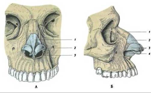
Fig. 281. External nose, nasus externus:
A - anterior view: 1 - lateral nasal cartilage; 2 - greater alar cartilage of the nose; 3 - septal nasal cartilage; B - lateral view: 1 - Nasal bone; 2 - lateral nasal cartilage; 3 - greater alar cartilage; 4 - cartilage of the nasal septum
The aforementioned cartilages are connected to one another and to the margin of the piriform aperture by Connective Tissue. Externally, they are covered by facial Muscles and Skin, and internally, by the mucous membrane.
Last update: 08/08/2026
Editorial and Educational Adaptation: This material has been compiled based on the primary/original source text. The project team performed an editorial review, corrected technical inaccuracies, structured sections, and adapted the content for an educational format.
What was processed:
- elimination of formatting defects (OCR errors, structural breaks, corrupted characters);
- editorial organization of content;
- standardization of terminology in accordance with academic sources;
- verification of factual statements against the original source text.
All mentions of the author, publication year, and origin of the primary text have been preserved in accordance with the source.