HUMAN ANATOMY ATLAS - G.L. Bilich - 2014
APPARATUS OF LOCOMOTION
SKELETAL SYSTEM
TOPOGRAPHY OF THE SKULL. THE SKULL AS A WHOLE
The individual BONES OF THE Skull articulate with one another to form the skull, which encloses the Brain, the Organs of Vision, Hearing, equilibrium, and Olfaction within its cavities. The skull also serves as the bony framework for the initial segments of the digestive and respiratory systems. All the bones of the skull, with the exception of the Mandible, are firmly and practically immovably connected to one another by sutures. Only the mandible is movably articulated with the remaining bones of the skull via the paired temporomandibular joint.
Class="center">
Fig. 64. Mandible (A — anterior view, B — posterior view, C — lateral view, right):
1 — Mcntal protuberance; 2 — Body of mandible; 3 — Mental foramen; 4 — Dental alveoli; 5 — Oblique line; 6 — Coronoid process; 7— Condylar process; 8 — Alveolar pari; 9 —Ramus of mandible; 10— Mandibular foramen; 11 — Mylohyoid line; 12 — Anglc of mandible; 13 — Pterygoid fovea; 14 — Mandibular notch; 15 — Mcntal tubercle
The Structure of the skull is complex; it features several surfaces with distinct relief, encompassing cavities and fossae that house vital organs. Blood Vessels and nerves pass through foramina within the cranial bones. For the convenience of studying the intact skull, K. Baer proposed back in the mid-19th century viewing it from five different Perspectives (five norma). From above (vertical aspect, norma verticalis), the cranial vault or roof is visible; from below (basilar aspect, norma basilaris), the external Base of the skull is seen; from the front, the facial aspect (norma facialis) allows The Study of the facial surface of the skull; from the side, the lateral aspect (norma lateralis). The lateral surface of the skull features fossae and foramina. Posteriorly lies the occipital aspect (norma occipitalis), corresponding to the rear surface of the skull.
To characterize the dimensions and shape of the neurocranium, measurements of its three principal diameters—longitudinal, transverse, and vertical—along with their ratios (indices) are determined. For this purpose, craniology (the Study of the skull) and anthropology (the study of humans) utilize craniometric points.
Glabella — the most anteriorly projecting point in the interciliary region, where the Frontal bone forms a more or less pronounced prominence (this prominence is absent in infant skulls).
Gnathion — the point on the inferior margin of the mandible in the midline.
Metopion — the point located at the intersection of the line connecting the apices of the frontal eminences with the sagittal plane (the line of the sagittal suture).
Bregma — the point at the junction of the sagittal and coronal sutures.
Lambda — the point located at the intersection of the lambdoid and sagittal sutures.
Basion — the point at the midpoint of the anterior margin of the foramen magnum (occipital foramen).
Nasion — the point of intersection of the frontonasal suture with the sagittal plane.
Inion — the external occipital protuberance.
The longitudinal dimension (diameter)—the maximum length of the skull—represents the distance between the glabella and the most distant point of the occiput in the sagittal plane (inion). In modern humans, this dimension ranges from 167 to 198 mm. The transverse diameter is measured at the point of maximum cranial width in the frontal plane between the most laterally projecting points of the lateral surface of the skull located on the Parietal bone (euryon). This dimension ranges between 123 and 160 mm. The vertical diameter is measured on The Skull as the distance between the basion and bregma points, varying from 124 to 145 mm. The ratio of these specified dimensions (diameters) is expressed using indices, calculated as the percentage of one value relative to another. The cephalic, or cranial (transverse-longitudinal) index is the ratio of the transverse diameter to the longitudinal diameter; the vertico-longitudinal index is the ratio of the vertical dimension to the longitudinal dimension; the vertico-transverse index is the ratio of the vertical dimension to the transverse dimension.
Topinard's line — the line connecting the glabella and gnathion, used in craniometry (Topinard, Paul, 1830–1912 — French anthropologist).
Neurocranium. The upper part of the neurocranium is the vault, or roof of the skull, and the lower part is the cranial base. The boundary between the vault and the base on the external surface of the skull is an imaginary line that runs through the external occipital protuberance, then along the superior nuchal line to the base of the mastoid process, above the external acoustic meatus, along the base of the zygomatic process of the Temporal bone, and along the infratemporal crest of the greater wing of the Sphenoid bone. This line then ascends to the zygomatic process of the frontal bone and reaches the frontonasal suture along the supraorbital margin.

Fig. 65. Vault (roof) of the skull (A — superior view, B — internal view, from the cranial cavity side):
1 — Lambdoid suture; 2 — Occipital bone; 3 — Parietal foramen; 4 — Frontal bone; 5 — Coronal suture; 6 — Parietal bone; 7— Sagittal suture; 8 — Frontal sinus; 9 — Frontal crest; 10— Groove for superior sagittal sinus; 11 — Granular foveolae; 12 — Arterial grooves
The cranial vault (calvaria) is formed by the squamous part of the frontal bone, the parietal bones, the squamous PARTS OF THE occipital and temporal bones, and the lateral parts of the greater wings of the sphenoid bone (Fig. 65). On the external surface of the cranial vault along the midline lies the sagittal suture (sutura sagittalis), formed by the Articulation of the medial margins of the parietal bones. Perpendicular to this suture, at the boundary between the frontal squama and the parietal bones in the frontal plane, runs the coronal suture (sutura coronalis). Between the parietal bones and the squamous part of the occipital bone lies the lambdoid suture (sutura lambdoidea), which resembles the Greek letter "lambda" in shape. On the lateral surface of the cranial vault on each side, between the squamous part of the temporal bone and the parietal bone, there is the squamous suture (sutura squamosa), as well as serrated sutures (suturae serratae) between the lateral part of the greater wing of the sphenoid bone and adjacent bones (temporal, parietal, and frontal). Serrated sutures are visible between the mastoid process of the temporal bone, the parietal bone, and the occipital bone. Along the course of the sutures and within the fontanelles, accessory cranial bones—Andernach's bones (syn.: Wormian bones, sutural bones)—may form (Andernach, Günther Johann, 1487–1574 — French anatomist and surgeon). For example, small, inconstant, isolated bones may be located in the petro-occipital suture—Riolan's bones (Riolan, Jean, 1577–1657 — French physician and anatomist). In the region of the pterion—the convergence point of the frontal, parietal, and temporal bones and the greater wing of the sphenoid bone—accessory cranial bones—Flower's bones—may form (Flower, William Henry, 1831–1899 — English surgeon and anatomist). Frequently, the upper section of the occipital squama separates into Goethe's bone (syn.: interparietal bone, os interparietale) (Goethe — see p. 95).
In the anterior Regions of the cranial vault lies the convex part—the forehead (frons), formed by the squama of the frontal bone. On the frontal squama (squama frontalis), frontal bumps, or frontal eminences (tuber frontale; eminentia frontalis), are visible on the sides, superciliary arches (arcus superciliares) are located above the orbits, and a small flat area—the glabella—lies in the middle. Parietal bumps, or parietal eminences (tuber parietale; eminentia parietalis), project on the lateral surfaces of the cranial vault. Below each bump, extending from the base of the zygomatic process of the frontal bone to the junction of the parietal and occipital bones, runs an arch-shaped superior temporal line (linea temporalis superior), to which the temporal fascia attaches. Below this line, the more distinctly defined inferior temporal line (linea temporalis inferior) is visible, marking the Water/144.html">Origin of the temporal Muscle.
The anterolateral region of the cranial vault, bounded superiorly by the inferior temporal line and inferiorly by the infratemporal crest of the greater wing of the sphenoid bone, is the temporal fossa (fossa temporalis). The infratemporal crest separates it from the infratemporal fossa (fossa infratemporalis). Laterally, the temporal fossa is bounded by the zygomatic arch (arcus zygomaticus), and anteriorly by the temporal surface of the Zygomatic bone (facies temporalis ossis zygomatici).
On the internal (cerebral) surface of the cranial vault, cranial sutures (sagittal, coronal, lambdoid, squamous) and digital impressions (impressiones digitatae)—the imprints of cerebral gyri—are visible, alongside narrow, sometimes quite deep arterial and venous sulci (sulci arteriosi et venosi), which mark the paths of adjacent Arteries and Veins. Near the sagittal suture lie the granular foveolae (foveolae granulares)—Pacchionian depressions—rounded pits of varying size on the inner table of the cranial vault bones on both sides of the sagittal sulcus, containing arachnoid granulations that communicate with the diploic channels of the cranial vault bones (Antonio Pacchioni [1665–1726], Italian anatomist and physician).
Within the spongy substance of the cranial vault bones are bony channels for the diploic veins—Breschet's canals (syn.: Dupuytren's canals, diploic canals, canales diploici) (Gilbert Breschet [1784–1860], French anatomist; Guillaume Dupuytren [1777–1835], French surgeon).
Like the cranial vault, the cranial base can be examined from two perspectives: externally (from below)—the external cranial base, and internally—the internal cranial base, following a horizontal section made at the level of the boundary with the vault or a sagittal section of the skull. In this case, the cerebral surface of the base, or internal cranial base, is examined. The boundary between the vault and the internal cranial base on the cerebral surface is indistinct; only in the posterior part can it be approximated by the groove for the transverse sinus, which corresponds to the superior nuchal line on the external surface of the occipital bone.
The external cranial base (basis cranii externa) is not visible anteriorly because it is covered by the bones of the facial Skeleton. The posterior region of the cranial base is formed by the external surfaces of the occipital, temporal, and sphenoid bones. Numerous foramina are visible here, through which arteries, veins, and nerves pass in the living human (Fig. 66, Table 12). Located almost in the center of this region is the foramen magnum, flanked by oval prominences—the occipital condyles (condyli occipitales). Posterior to each condyle lies a poorly defined condylar fossa (fossa condylaris) with an inconstant opening leading to the condylar canal (canalis condylaris). The base of each condyle is pierced transversely by the hypoglossal canal (canalis nervi hypoglossi). The posterior region of the cranial base terminates in the external occipital protuberance (protuberantia occipitalis externa), with the superior nuchal line extending laterally to the right and left. Anterior to the foramen magnum is the basilar part of the occipital bone (pars basilaris ossis occipitalis) featuring a well-defined pharyngeal tubercle (tuberculum pharyngeum). The basilar part transitions into the body of the sphenoid bone (corpus ossis sphenoidalis).

Fig. 66. External cranial base:
1 — Highest nuchal line; 2 — Superior nuchal line; 3 — Inferior nuchal line; 4 — Foramen magnum; 5 — Hypoglossal canal; 6 — Foramen lacerum; 7— Carotid canal; 8 — Stylomastoid foramen; 9— Foramen spinosum; 10 — Foramen ovale; 11 — Vomer; 12 — Pterygoid process, medial plate; 13 — Pterygoid process, lateral plate; 14 — Lesser palatine foramina; 15 — Greater palatine foramen; 16 — Palatine bone; 17 — Transverse palatine suture; 18 — Median palatine suture; 19 — Incisive foramina; 20 — Maxilla, palatine process; 21 — Teeth; 22 — Choana; Posterior nasal aperture; 23 — Maxilla, zygomatic process; 24 — Inferior orbital fissure; 25 — Zygomatic bone, temporal surface; 26 — Pharyngeal tubercle; 27 — Zygomatic arch; 28 — Temporal bone; 29 — Mandibular fossa; 30 — Styloid process; 31 — Mastoid process; 32 — Mastoid notch; 33 — Mastoid foramen; 34 — Occipital condyle; 35 — Parietal bone; 36 — Condylar canal; 37 — External occipital protuberance
Flanking the occipital bone on each side is the Inferior surface of the petrous part of the temporal bone, which houses vital structures: the external openings of the carotid canal and musculotubal canal, the jugular fossa, and the jugular notch. The latter, together with the jugular notch of the occipital bone, forms the jugular foramen, the styloid process, and the mastoid process. Between the styloid and mastoid processes lies the stylomastoid foramen, which marks the termination of Fallopian canal (syn.: facial canal, canalis nervi facialis) within the petrous part of the temporal bone; originating at the bottom of the internal acoustic meatus, the facial canal transmits the facial and intermediate nerves and contains the geniculate ganglion. Laterally, the tympanic part of the temporal bone, which surrounds the external acoustic opening, adjoins the petrous part of the temporal bone (Gabriele Falloppio [1523–1562], Italian anatomist).
The greater wing of the sphenoid bone may feature Vesalius' foramen (syn.: venous foramen, foramen venosum)—an inconstant aperture located between the foramen rotundum and foramen ovale, through which an emissary vein passes (Andreas Vesalius [1515–1564], prominent Renaissance scientist and founder of modern anatomy).
An important topographical and anatomical landmark for determining the PROJECTION OF THE cranial base is Reid's line—a horizontal line drawn through the centers of the external acoustic meatuses (Robert William Reid [1851–1939], Scottish anatomist).
Posteriorly, the tympanic part of the temporal bone is separated from the mastoid process by the tympanomastoid fissure (fissura tympanomastoidea). On the posteromedial aspect of the mastoid process lie the mastoid notch (incisura mastoidea) and the sulcus for the occipital artery (sulcus arteriae occipitalis).
Between the tympanic part of the temporal bone and the projecting outer margin of the tympanic cavity roof lies a narrow space—the exit site of the chorda tympani—Glaserian fissure (syn.: Huguier's canal, Civinini's canal, petrotympanic fissure, fissura petrotympanica) (Johann Heinrich Glaser [1629–1675], Swiss physician and anatomist; Filippo Civinini [1805–1854], Italian anatomist).
On the horizontally positioned segment of the squamous part of the temporal bone (pars squamosa ossis temporalis) lies the mandibular fossa, which articulates with the condylar process of the mandible. Anterior to this fossa is the articular tubercle (tuberculum articulare). In the intact skull, the posterior part of the greater wing of the sphenoid bone (ala major ossis sphenoidalis)—in which the foramen spinosum and foramen ovale are clearly visible—wedges into the gap between the petrous and squamous parts of the temporal bone. Above the acoustic meatus lies the petrosquamous canaliculus—Verga's canal (canaliculus), which is consistently present in the fetus but not always in adults (Andreas Verga [1811–1895], Italian neuropathologist and anatomist). This canaliculus runs toward and opens onto the surface via the petrosquamous fissure (Otto's aperture). Otto's aperture (fissura petrosquamosa) is a narrow space between the squamous part of the temporal bone and the projecting outer margin of the petrous pyramid, where the petrosquamous canaliculus opens (Adolph W. Otto [1786–1845], German surgeon and anatomist). On the external surface of the temporal bone, above the upper margin of the external acoustic opening, is a projection known as Zuckerkandl's spine (syn.: Henle's spine [Friedrich Gustav Jacob Henle (1809–1885), German anatomist and pathologist], suprameatal spine of the temporal bone, spina suprameatica) (Emil Zuckerkandl [1849–1910], Austrian anatomist). On the lateral surface of the mastoid process is Chassaignac's triangle—a triangle bounded superiorly by the continuation of the superior temporal line of the parietal bone onto the temporal squama, anteriorly by a line extending from the apex of the mastoid process to the suprameatal spine, and posteriorly by the attachment line of the sternocleidomastoid muscle on the mastoid process.
Table 12. Foramina of the external cranial base and their contents
Cranial region |
Foramen name |
Location on the skull |
Structures passing through the foramen |
||
Arteries |
Veins |
Nerves |
|||
Hard palate |
Incisive foramen |
Posterior to the medial incisors, within the median palatine suture |
- |
- |
Nasopalatine nerve - from a branch of the Trigeminal nerve (V) |
Greater palatine foramen |
In the posterolateral region of the hard palate |
Greater palatine artery - branch of the descending palatine artery (from the maxillary artery) |
Greater palatine vein draining into the palatine veins - tributaries of the facial vein |
Greater palatine nerve - from the second branch of the trigeminal nerve (V) |
|
Lesser palatine foramina |
In the posterolateral region of the hard palate |
Lesser palatine arteries - Branches of the descending palatine artery (from the maxillary artery) |
Lesser palatine veins draining into the palatine veins - tributaries of the facial vein |
Lesser palatine nerves - from the second branch of the trigeminal nerve (V) |
|
Middle part of the external cranial base, between the choanae and the foramen magnum |
Foramen ovale |
In the posterior part of the base of the greater wing of the sphenoid bone |
Meningeal branch of the middle meningeal artery (from the maxillary artery) |
Emissary sphenoidal sinus (venous plexus of foramen ovale) connects the cavernous sinus with the pterygoid venous plexus |
Mandibular nerve - third branch of the trigeminal nerve (V) |
Foramen spinosum |
Cerebral surface of the greater wing of the sphenoid bone, posterior and lateral to the foramen ovale |
Middle meningeal artery - branch of the maxillary artery |
Middle meningeal veins draining into the retromandibular vein |
Meningeal branch - from the third branch of the trigeminal nerve (V) |
|
Foramen lacerum |
Between the apex of the petrous part of the temporal bone, the lateral margin of the occipital bone base, and the posterior margin of the pterygoid process base of the sphenoid bone |
- |
- |
Lesser petrosal nerve - terminal branch of the tympanic nerve from the Glossopharyngeal nerve (IX) |
|
External opening of the carotid canal |
On the inferior surface of the petrous part of the temporal bone, anterior to the jugular fossa |
Internal carotid venous plexus |
Internal carotid (sympathetic) plexus |
||
Inferior opening of the tympanic |
At the floor of the petrosal fossa, between the external opening of the carotid canal and the jugular fossa |
Inferior tympanic artery - branch of the ascending pharyngeal artery |
- |
Tympanic nerve - branch of the glossopharyngeal nerve (IX) |
|
External opening of the mastoid canaliculus |
Floor of the jugular fossa |
- |
- |
Auricular branch of the Vagus nerve (X) |
|
Petrotympanic fissure |
Between the anterior margin of the tympanic part of the temporal bone and the margin of the tympanic cavity roof (posterior to the petrosquamous fissure) |
Anterior tympanic artery - branch of the maxillary artery |
Tympanic tributaries of the retromandibular vein |
Chorda tympani - branch of the Facial Nerve (VII) |
|
Tympanosquamous fissure |
Between the anterior margin of the tympanic part and the posterior margin of the inferior part of the temporal squama |
Deep auricular artery - branch of the maxillary artery |
- |
- |
|
Jugular foramen |
Between the petrous part of the temporal bone anteriorly and the jugular notch of the occipital bone posteriorly |
Posterior meningeal artery - branch of the ascending pharyngeal artery |
Glossopharyngeal (IX), vagus (X), accessory (XI) nerves, meningeal branch of the vagus nerve (X) |
||
At the level of the foramen magnum |
Stylomastoid foramen |
At the base of the styloid process, between it and the mastoid process of the temporal bone |
Stylomastoid artery - branch of the posterior auricular artery |
Stylomastoid vein draining into the retromandibular vein |
Facial nerve (VII) |
Mastoid foramen |
Base of the mastoid process, closer to the posterior margin of the temporal bone |
Meningeal branch of the occipital artery |
Mastoid emissary vein (connects the sigmoid sinus and occipital vein) |
- |
|
Condylar canal |
In the condylar fossa, posterior to the occipital condyle |
- |
Condylar emissary vein (connects the sigmoid sinus with the vertebral venous plexus) |
- |
|
External opening of the hypoglossal canal |
At the base of the occipital condyle, on its lateral side |
- |
Hypoglossal venous plexus (draining into the internal jugular vein) |
Hypoglossal nerve (XII) |
|
Foramen magnum |
In the center of the occipital bone |
Vertebral, anterior and posterior spinal arteries (branches of the vertebral arteries) |
Basilar venous plexus |
||
The petrous part of the temporal bone is separated from the occipital bone by the petro-occipital fissure (fissura petrooccipitalis), and from the greater wing of the sphenoid bone by the sphenopetrosal fissure (fissura sphenopetrosa). On the inferior surface of the external cranial base, an irregularly shaped opening is also visible—the foramen lacerum (foramen lacerum), bounded laterally and posteriorly by the apex of the petrous part (apex partis petrosae) of the temporal bone, which wedges between the body of the occipital bone and the greater wing of the sphenoid bone. On the anterosuperior area of the mastoid process stands out MacEwen's triangle (or Chassaignac's area), which is bounded by a horizontal line drawn through the midpoint of the posterior wall of the external acoustic meatus and a vertical line corresponding to the bony crest On the surface of the mastoid process. This region serves as the pathway for vessels connecting the mucous membrane of the mastoid antrum with the periosteum of the mastoid process, housing perivascular Connective Tissue that can facilitate the spread of pus during mastoiditis.
When describing skull shapes across various races in anthropology, the term Serres' angle (syn. metafacial angle) is widely used—the angle formed by the pterygoid process of the sphenoid bone and the cranial base (Antoine Étienne Renaud Augustin Serres [1786–1868], French biologist and anatomist).
The internal cranial base (basis cranii interna) features a concave, irregular surface that conforms to the shape of the inferior surface of the brain. It contains three cranial fossae: anterior, middle, and posterior (Fig. 67). The posterior margins of the lesser wings (ala minor) and the tuberculum sellae of the sphenoid bone (tuberculum sellae turcicae ossis sphenoidalis) separate the anterior cranial fossa (fossa cranii anterior) from the middle cranial fossa (fossa cranii media). The boundary between the middle and posterior cranial fossae (fossa cranii posterior) is formed by the superior borders of the petrous parts of the temporal bones (margines superiores partis petrosae) and the dorsum sellae of the sphenoid bone. Upon inspection of the internal cranial base, numerous foramina for the passage of arteries, veins, and nerves are visible (Table 13).
The anterior cranial fossa (fossa cranii anterior) is formed by the orbital parts of the frontal bone (pars orbitalis ossis frontalis)—featuring prominent cerebral eminences and digital impressions—and the cribriform plate of the Ethmoid bone (lamina cribrosa ossis ethmoidalis), through the foramina of which numerous bundles of Olfactory nerve fibers pass. In the center of the cribriform plate rises the crista galli, anterior to which lie the foramen caecum (Morand's foramen), bounded by the alar processes of the crista galli of the ethmoid bone and the crura of the frontal crest (Sauveur François Morand [1697–1773], French surgeon and anatomist), and the frontal crest. Near the crista galli of the ethmoid bone is Palfyn's sinus—a space communicating with the frontal and ethmoidal Cells (Jean Palfyn [1650–1730], French physician and anatomist).
The middle cranial fossa (fossa cranii media) is significantly deeper than the anterior fossa. The walls of the middle fossa are formed by the body and greater wings of the sphenoid bone (corpus et alae majores ossis sphenoidalis), the anterior surface of the petrous pyramids, and the squamous part of the temporal bones (facies anterior partis petrosae et pars squamosa ossis temporalis). The middle cranial fossa can be divided into a central part and lateral regions. The central part is occupied by the sella turcica with its hypophysial fossa. At the floor of the hypophysial fossa in the body of the sphenoid bone, an inconstant structure may occur (found in 0.3% of adults)—Landuzent's canal (syn.: craniopharyngeal canal, canalis craniofaryngealis). It pierces the body of the sphenoid bone and opens on its inferior surface (near the junction of the vomer wings) via a "pharyngeal" aperture. The canal contains a continuation of the dura mater in the form of a fibrous sleeve enclosing connective tissue and Blood Vessels (veins) (Feodor Pavlovich Landuzent [1833–1889], professor at the St. Petersburg Medico-Surgical Academy).

Fig. 67. Internal cranial base:
1 — Groove for transverse sinus; 2 — Groove for sigmoid sinus; 3 — Hypoglossal canal; 4 — Clivus; 5 — Foramen lacerum; 6 — Arterial grooves; 7 — Foramen spinosum; 8 — Foramen ovale; 9 — Anterior clinoid process; 10 — Optic canal; 11 — Cribriform plate; 12 — Frontal crest; 13 — Frontal sinus; 14 — Ethmoid: Ethmoidal bone, crista galli; 15 — Frontal bone; 16 — Sphenoid; Sphenoidal bone, lesser wing; 17 — Sphenoid; Sphenoidal bone, greater wing; 18 — Sphenoid; Sphenoidal bone, hypophysial fossa; 19 — Posterior clinoid process; 20 — Temporal bone, petrous part; 21 — Internal acoustic meatus; 22 — Jugular foramen; 23 — Foramen magnum; 24 - Cerebellar fossa; 25 — Cerebral fossa
Fig. 67. Cranial fossae
The internal base of the skull is depressed and features three cranial fossae: anterior, middle, and posterior. These depressions slope downward from the frontal region to the occipital bone, creating a terraced structure.
✵ The anterior cranial fossa is formed by the orbital parts of the frontal bones, the cribriform plate of the ethmoid bone, and the greater wings of the sphenoid bone.
✵ The middle cranial fossa is formed by the body and greater wings of the sphenoid bone, the anterior surfaces of the petrous parts, and the squamous parts of the temporal bone.
✵ The posterior cranial fossa is formed by the occipital bone, the posterior surface of the petrous parts and the internal surfaces of the mastoid processes of the temporal bones, and the posterior part of the body of the sphenoid bone (the sella turcica back).
Table 13. Foramina of the internal cranial base and their contents
Cranial region |
Foramen name |
Location on the skull |
Passing structures |
||
Arteries |
Veins |
Nerves |
|||
Anterior cranial fossa |
Cribriform foramina |
In the middle parts of the anterior cranial fossa, flanking the crista galli |
Anterior ethmoidal artery - a branch of the ophthalmic artery |
Ethmoidal veins (tributaries of the superior ophthalmic vein) |
Olfactory nerves (I) |
Optic canal |
At the base of the lesser wing of the sphenoid bone |
Ophthalmic artery |
- |
Optic nerve (II) |
|
Middle cranial fossa |
Superior orbital fissure |
Between the greater and lesser wings of the sphenoid bone (in the anterior part of the middle cranial fossa) |
Anterior meningeal artery (a branch of the anterior ethmoidal artery) |
Superior ophthalmic vein, draining into the cavernous sinus |
Oculomotor (III), trochlear (IV), and abducens (VI) nerves, ophthalmic nerve - 1st branch of the trigeminal nerve (V) |
Carotid canal (internal opening) |
At the apex of the petrous part of the temporal bone |
Internal carotid artery |
Internal carotid venous plexus |
Internal carotid sympathetic plexus |
|
Foramen rotundum |
At the base of the greater wing of the sphenoid bone, posterior to the superior orbital fissure |
Maxillary nerve - 2nd branch of the trigeminal nerve (V) |
|||
Foramen ovale |
At the base of the greater wing of the sphenoid bone, posterior and lateral to the foramen rotundum |
Accessory branch of the middle meningeal artery |
Emissary vein of the foramen ovale, connecting the cavernous sinus and the pterygoid venous plexus |
Mandibular nerve - 3rd branch of the trigeminal nerve (V) |
|
Foramen spinosum |
In the region of the posterior angle of the greater wing of the sphenoid bone (posterior and lateral to the foramen ovale) |
Middle meningeal artery - a branch of the maxillary artery |
Middle meningeal veins |
Meningeal branch - from the 3rd branch of the trigeminal nerve (V) |
|
Hiatus for the greater petrosal nerve (canalicular opening of the facial nerve) |
On the anterior surface of the petrous part of the temporal bone, lateral to the trigeminal impression |
Superficial petrosal branch - a branch of the middle meningeal artery |
Tympanic vein draining into the superior petrosal sinus |
Greater petrosal nerve - a branch of the facial nerve (intermediate nerve) (VII) |
|
Hiatus for the lesser petrosal nerve (superior aperture of the tympanic canaliculus) |
Lateral to the previous opening (hiatus for the greater petrosal nerve) |
Superior tympanic artery - a branch of the middle meningeal artery |
Lesser petrosal nerve - a branch of the tympanic nerve (from the glossopharyngeal nerve - IX) |
||
Posterior cranial fossa |
Internal acoustic meatus |
On the posterior surface of the petrous part of the temporal bone |
Labyrinthine artery - a branch of the Basilar artery |
Labyrinthine veins, draining into the inferior petrosal sinus |
Facial nerve (VII), Vestibulocochlear nerve (VIII) |
External aperture of the vestibular aqueduct |
On the posterior surface of the petrous part of the temporal bone, lateral to the internal acoustic meatus |
Endolymphatic duct and sac |
|||
External aperture of the cochlear canaliculus |
On the posterior surface of the petrous part of the temporal bone, inferior to the internal acoustic meatus |
Perilymphatic duct, draining into the superior bulb of the internal jugular vein, vein of the cochlear canaliculus |
|||
Mastoid foramen |
At the lateral margin of the sigmoid sulcus |
Meningeal branch of the occipital artery |
Mastoid emissary vein, connecting the sigmoid sinus and the occipital vein |
||
Jugular foramen |
Posterior to the petrous part of the temporal bone, inferior to the internal acoustic meatus |
Posterior meningeal artery - a branch of the ascending pharyngeal artery |
Internal jugular vein |
Glossopharyngeal (IX), vagus (X), and accessory (XI) nerves, meningeal branch of the vagus nerve (X) |
|
Foramen magnum |
In the floor of the posterior cranial fossa, posterior to the clivus |
Vertebral, anterior, and posterior spinal arteries |
Basilar venous plexus |
Medulla oblongata |
|
Hypoglossal canal |
On the internal surface of the lateral part of the occipital bone, lateral to the foramen magnum |
Venous network of the hypoglossal canal, draining into the internal jugular vein |
Hypoglossal nerve (XII) |
||
Condylar canal |
In the floor of the condylar fossa |
Condylar emissary vein, connecting the sigmoid sinus with the vertebral venous plexus |
|||
Anterior to the hypophyseal fossa lies the optic chiasm sulcus (sulcus chiasmatis), leading to the right and left optic canals (canalis opticus), through which the optic nerves pass. On the lateral surface of the body of the sphenoid bone is the prominent carotid sulcus (sulcus caroticus), and near the apex of the petrous part lies the irregularly shaped foramen lacerum. Also situated here, between the greater and lesser wings and the body of the sphenoid bone, is the superior orbital fissure (fissura orbitalis superior), through which the oculomotor, trochlear, and ophthalmic nerves enter the Orbit. Posterior to the superior orbital fissure is the foramen rotundum, which transmits the maxillary nerve, followed by the foramen ovale for the mandibular nerve. Near the posterior margin of the greater wing of the sphenoid bone lies the foramen spinosum, transmitting the middle meningeal artery into the cranial cavity. On the anterior surface of the petrous part of the temporal bone is the trigeminal impression (impressio trigemini) — Meckel's cave (Johann Friedrich Meckel the Elder (1724–1774) — German anatomist); lateral to it is the hiatus for the greater petrosal nerve (hiatus canalis nervi petrosi majoris) — Tarin's opening, through which the greater petrosal nerve exits, along with the groove for the petrosal nerve (Pierre Tarin (1725–1761) — French physician and anatomist). Further lateral and anterior are the hiatus and groove for the lesser petrosal nerve. Lateral and posterior to these structures lie the tegmen tympani and the arcuate eminence (eminentia arcuata). Between the carotid canal and the trigeminal ganglion — Gasserian ganglion (syn. ganglion trigeminale) on the petrous part of the temporal bone is Gruber's notch (syn. jugular notch, incisura jugularis), covered by a thin bony plate (Johann Laurentius Gasser (1723–1769) — Austrian physician and anatomist; Wenzel Leopold Gruber (1814–1890) — Austrian anatomist working in Russia). Within the petrous part of the temporal bone, beneath the dura mater, lies Dorello's canal formed by the dura and the groove for the inferior petrosal sinus — a canal transmitting the inferior petrosal sinus, vessels, and the Abducens nerve on its course to the cavernous sinus (Paolo Dorello (born 1872) — Italian anatomist). Near the apex of the petrous part of the temporal bone is Princeteau's tubercle — an elevation adjacent to the superior petrosal sinus (Laurent Princeteau (1858–1932) — French physician and anatomist). A crucial topographical landmark for surgical interventions on the labyrinth, and less commonly on the Cerebellum, is Trautmann's triangle — a cranial region bounded posteriorly by the sigmoid sinus of the dura mater, anteriorly by the posterior semicircular canal of the Inner ear, and superiorly by the superior border of the petrous part of the temporal bone (Moritz Trautmann (1832–1902) — German surgeon).
The posterior cranial fossa (fossa cranii posterior) is the deepest of all. It is formed by the occipital bone, the posterior surfaces of the petrous parts and the internal surfaces of the mastoid processes of the right and left temporal bones, as well as the posterior part of the body of the sphenoid bone and the posteroinferior angles of the parietal bones. In the center of the fossa lies the foramen magnum, anterior to which is the clivus (Blumenbach's clivus) formed by the fused bodies of the sphenoid and occipital bones in adults, supporting the Pons and medulla oblongata (Johann Friedrich Blumenbach (1752–1840) — German physician, anatomist, zoologist, and anthropologist). An accessory bone, Albrecht's ossicle, may occasionally be found between the bodies of the occipital and sphenoid bones (Karl Martin Paul Albrecht (1851–1894) — German anatomist). Along the posterior margin of the foramen magnum, in the course of development, Kerckring's ossicle appears as an ossification center of the occipital bone (Theodor Kerckring (1640–1693) — Dutch physician and anatomist).
Posterior to the foramen magnum in the midline lie the internal occipital crest (crista occipitalis interna) and the cruciate eminence (eminentia cruciformis). On the posterior surface of the petrous part on each side is the internal acoustic opening (porus acusticus internus), leading into the internal acoustic meatus (meatus acusticus internus). Deep within it begins the facial canal, transmitting the facial nerve. The vestibulocochlear nerve exits from the internal acoustic opening. On the floor of the posterior cranial fossa posterior to the petrous parts lies the paired jugular foramen (foramen jugulare), through which the glossopharyngeal, vagus, and accessory nerves pass, with the hypoglossal canal for the nerve of the same name located medially to it. The internal jugular vein also exits the cranial cavity through the jugular funnel as a continuation of the sigmoid sinus, which lies within the groove of the same name.
On The surface of the cranial vault, 3 cm posterior and superior to the upper margin of the external acoustic meatus lies Keen's point, which serves as a topographical landmark for puncture of the inferior horn of the lateral ventricle of the brain (William Williams Keen (1837–1932) — American surgeon).
On the internal base of the skull, within the posterior cranial fossa, lies Moure's area — a region bounded superiorly by the inferior petrosal sinus of the dura mater, posteriorly by the transverse sinus, and anteriorly and medially by the internal acoustic meatus on the petrous part of the temporal bone; this area is a frequent site for cerebellar abscesses.
The boundary between the cranial vault and the internal base in the region of cases involving the posterior cranial fossa is the transverse sinus sulcus (sulcus sinus transversi), which continues on each side into the sigmoid sinus sulcus (sulcus sinus sigmoidei).
Facial skeleton. Anterior view of the facial skeleton (norma facialis) reveals structures that house vital organs: the orbits, Nasal cavity, and Oral Cavity. LATERAL VIEW OF the skull (norma lateralis) displays the temporal, infratemporal, and pterygopalatine fossae, as well as the foramina and canals connecting them to adjacent cavities (Table 14, see Figs. 40, 41).
The orbit (orbita) is a paired cavity resembling a quadrilateral pyramid with rounded edges. The base of the orbit faces anteriorly, forming the orbital opening (aditus orbitae). The apex of the orbit is directed posteriorly and medially toward the optic canal (canalis opticus). The orbital cavity contains the Eyeball, its extrinsic Muscles, the lacrimal gland, and other Accessory structures of the eye. The orbit has four walls: superior, medial, inferior, and lateral (Figs. 68, 69).
Table 14. Fossae (cavities) of the facial skeleton
Fossa (cavity) name |
Walls of the fossa (cavity) |
Communications with other cavities (fossae) |
|||||
Superior |
Inferior |
Lateral |
Medial |
Anterior |
Posterior |
||
Orbit |
Orbital plate of the frontal bone; posteriorly, the lesser wing of the sphenoid bone |
Orbital surfaces of the maxilla, zygomatic bone, and orbital process of the palatine bone |
Orbital surfaces of the greater wing of the sphenoid bone, frontal process of the zygomatic bone, and zygomatic process of the frontal bone |
Frontal process of the maxilla, Lacrimal bone, orbital plate of the ethmoid bone, body of the sphenoid bone (posteriorly), and the most medial part of the orbital plate of the frontal bone (superiorly) |
Wall absent |
Wall absent |
Nasolacrimal canal - with the nasal cavity; superior orbital fissure and optic canal - with the middle cranial fossa; inferior orbital fissure - with the pterygopalatine and infratemporal fossae |
Nasal cavity |
Nasal bones, nasal part of the frontal bone, cribriform plate of the ethmoid bone, and inferior surface of the body of the sphenoid bone |
Palatine processes of the maxillae and horizontal plates of the palatine bones |
Nasal surface of the body and frontal process of the maxilla, lacrimal bone, ethmoidal labyrinth, perpendicular plate of the palatine bone, medial plate of the pterygoid process of the sphenoid bone (posteriorly) |
Absent (the osseous nasal septum, formed by the perpendicular plate of the ethmoid bone and vomer, divides the nasal cavity into two halves) |
Piriform aperture, bounded by the nasal notches (right and left) of the maxillae and the inferior margins of the nasal bones |
Choanae, bounded by the medial plate of the pterygoid process, vomer, body of the sphenoid bone, and horizontal plate of the palatine bones |
Choanae - with the nasopharynx, ethmoidal cells, and apertures of the frontal, maxillary, and sphenoid sinuses. Sphenopalatine foramen - with the pterygopalatine fossa; nasolacrimal canal - with the orbit |
Pterygopalatine fossa |
Base of the greater wing of the sphenoid bone |
Wall absent, continues into the greater palatine canal |
Wall absent, continues into the infratemporal fossa |
Perpendicular plate of the palatine bone |
Maxillary tuberosity |
Base of the pterygoid process of the sphenoid bone |
Sphenopalatine foramen - with the nasal cavity; foramen rotundum - with the middle cranial fossa; pterygoid canal - with the region of the foramen lacerum; greater palatine canal - with the oral cavity; inferior orbital fissure - with the orbit |
Infratemporal fossa |
Temporal bone and base of the greater wing of the sphenoid bone |
Absent |
Partially the zygomatic arch and the ramus of the mandible |
Lateral plate of the pterygoid process of the sphenoid bone |
Maxillary tuberosity and partially the zygomatic bone |
Absent |
Inferior orbital fissure - with the orbit; pterygomaxillary fissure - with the pterygopalatine fossa |
Numerous foramina in the orbital walls transmit various blood vessels and nerves (Table 15).
The superior wall (paries superior) forms the roof of the orbit; it is smooth, slightly concave, and lies nearly horizontally. It is formed by the orbital part of the frontal bone (pars orbitalis ossis frontalis), with the lesser wing of the sphenoid bone (ala minor ossis sphenoidalis) completing it posteriorly. At the junction of the superior and lateral walls lies the shallow lacrimal gland fossa (fossa glandulae lacrimalis). Near the medial margin of the superior wall, close to the supraorbital notch, is an inconspicuous depression — the trochlear fossa, next to which a small bony projection, the trochlear spine, is sometimes found.
The medial wall (paries medialis) is oriented sagittally. It is formed by the orbital plate of the ethmoid bone, the frontal process of the maxilla, the lacrimal bone, the body of the sphenoid bone (posteriorly), and the most medial part of the orbital plate of the frontal bone (superiorly). As an anatomical variation of facial bones, an accessory lacrimal bone, Rousseau's bone, may be located anterior to the lacrimal bone (Louis François Emmanuel Rousseau (1788–1868) — French anatomist and histologist).
In the anterior part of the medial wall lies the lacrimal sac fossa (fossa sacci lacrimalis), which leads inferiorly into the nasolacrimal canal (canalis nasolacrimalis) opening into the inferior nasal meatus of the nasal cavity. Slightly posterior and superior to the lacrimal sac fossa, in the suture between the frontal bone and the orbital plate of the ethmoid bone, two foramina are visible: the anterior ethmoidal foramen (foramen ethmoidale anterius) and the posterior ethmoidal foramen (foramen ethmoidale posterius), which transmit the corresponding nerves and vessels.
Table 15. Foramina in the orbital walls and their transmitted structures
Foramina |
Structures |
|
Optic canal |
✵ Optic nerve (II) ✵ Ophthalmic artery |
|
Superior orbital fissure |
✵ Oculomotor nerve (III) ✵ Trochlear nerve (IV) ✵ Ophthalmic nerve - 1st branch of the trigeminal nerve (V) - Lacrimal nerve - Frontal nerve - Nasociliary nerve ✵ Abducens nerve (VI) ✵ Anterior meningeal artery - branch of the anterior ethmoidal artery ✵ Superior ophthalmic vein |
|
Inferior orbital fissure |
✵ Zygomatic nerve - branch of the maxillary nerve (V) ✵ Inferior ophthalmic vein ✵ Infraorbital artery, vein, and nerve - branch of the maxillary nerve (V) |
|
Nasolacrimal canal |
✵ Nasolacrimal duct |
|
Infraorbital canal |
✵ Infraorbital artery, vein, and nerve - branch of the maxillary nerve (V) |
|
Supraorbital foramen |
✵ Supraorbital artery and vein ✵ Supraorbital vein ✵ Supraorbital nerve (lateral branch) - branch of the frontal nerve (V) |
|
Frontal notch |
✵ Supratrochlear artery ✵ Supratrochlear nerve (medial branch) - branch of the frontal nerve (V) |
|
Anterior ethmoidal foramen |
✵ Anterior ethmoidal artery, vein, and nerve - branch of the nasociliary nerve (V) |
|
Posterior ethmoidal foramen |
✵ Posterior ethmoidal artery, vein, and nerve - branch of the nasociliary nerve (V) |
|
Clinically important structures surrounding the orbit |
||
Direction |
Structures adjacent to the orbit |
|
Inferiorly |
Maxillary sinus |
|
Superiorly |
Frontal sinus Anterior cranial fossa (containing the frontal lobes of the brain) |
|
Medially |
Ethmoidal air cells |
|
Deeply |
Sphenoid sinus Middle cranial fossa Optic chiasm Cavernous sinus Pterygopalatine fossa |
|
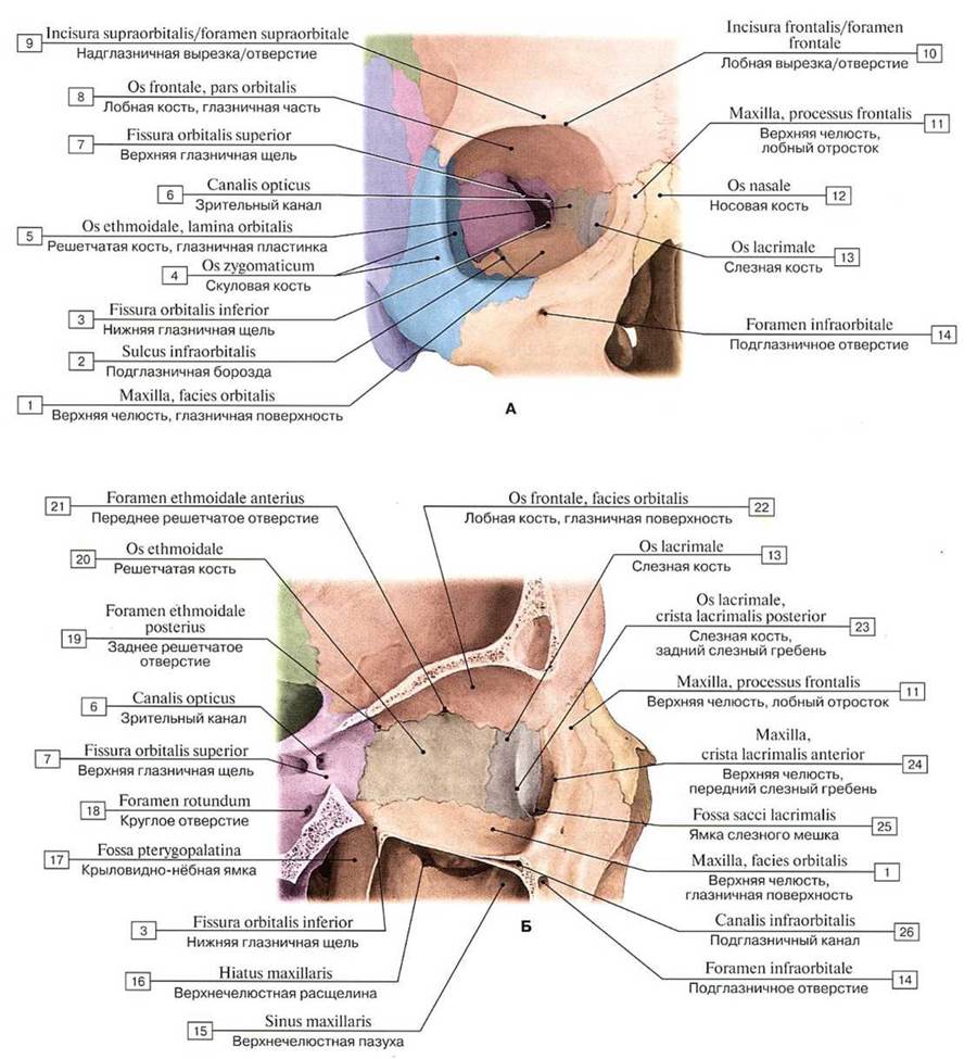
Fig. 68. Right orbit (A - anterior view, B - lateral view from the outside, sectioned through the orbit, showing the medial wall):
1 — Maxilla, orbital surface; 2 — Infra-orbital groove; 3 — Inferior orbital fissure; 4 — Zygomatic bone; 5 — Ethmoid; Ethmoidal bone, orbital plate; 6 — Optic canal; 7 — Superior orbital fissure; 8 — Frontal bone, orbital part; 9 — Supra-orbital notch/foramen; 10 — Frontal notch/foramen; 11 — Maxilla, frontal process; 12 — Nasal bone; 13 — Lacrimal bone; 14 — Infra-orbital foramen; 15 — Maxillary sinus; 16 — Maxillary hiatus; 17 — Pterygopalatine fossa; 18 — Foramen rotundum; 19 — Posterior ethmoidal foramen; 20 — Ethmoid; Ethmoidal bone; 21 — Anterior ethmoidal foramen; 22 — Frontal bone, orbital surface; 23 — Lacrimal bone, posterior lacrimal crest; 24 — Maxilla, anterior lacrimal crest; 25 — Fossa for lacrimal sac; 26 — Infra-orbital canal
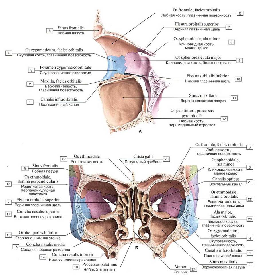
Fig. 69. Left orbit (A — lateral view from the inside, section passing through the orbit, showing the lateral wall; B — orbits and nasal cavity with surrounding air sinuses of the skull):
1 — Infra-orbital canal; 2 — Maxilla, orbital surface; 3 — Zygomatico-orbital foramen; 4 — Zygomatic bone, orbital surface; 5 — Frontal sinus; 6 — Frontal bone, orbital surface; 7 — Superior orbital fissure; 8 — Sphenoid; Sphenoidal bone, lesser wing; 9 — Sphenoid; Sphenoidal bone, greater wing; 10 — Inferior orbital fissure; 11 — Maxillary sinus; 12 — Palatine bone, pyramidal process; 13 — Palatine process; 14 — Inferior nasal concha; 15 — Middle nasal concha; 16 — Orbit, floor; 17 — Superior nasal concha; 18 — Ethmoid; Ethmoidal bone, perpendicular plate; 19 — Ethmoid; Ethmoidal bone; 20 — Crista galli; 21 — Optic canal; 22 — Ethmoid; Ethmoidal bone, orbital plate; 23 — Greater wing, orbital surface; 24 — Vomer
The inferior wall (paries inferior), or orbital floor, is formed by the orbital surfaces of the maxilla and zygomatic bone. Posteriorly, the wall is supplemented by the orbital process of the palatine bone (processus orbitalis ossis palatini). The infra-orbital groove (sulcus infraorbitalis) lies in the inferior orbital wall, continuing anteriorly into the canal of the same name, which opens on the anterior surface of the maxillary body via the infra-orbital foramen (foramen infraorbitale). The infra-orbital nerve is housed within this groove and canal.
The lateral wall (paries lateralis) is formed by the orbital surfaces of the greater wing of the sphenoid bone (facies orbitalis alae majoris ossis sphenoidalis) and the frontal process of the zygomatic bone (processus frontalis ossis zygomatici), as well as a section of the zygomatic process of the frontal bone (processus zygomaticus ossis frontalis). On the orbital surface of the frontal process of the zygomatic bone is the tubercle of Whitnall (syn.: marginal tubercle, tuberculum marginale) (Samuel Ernst Whitnall, 1876–1950, English anatomist).
The zygomaticoorbitale foramen (foramen zygomaticoorbitale) for the zygomatic nerve is located on the lateral wall of the orbit, leading into a canal that divides deep within the bone into two branch canaliculi. One of them opens on the lateral surface of the zygomatic bone as the zygomaticofacial foramen (foramen zygomaticofaciale), while the other opens on the temporal surface as the zygomaticotemporal foramen (foramen zygomaticotemporale).
Between the lateral and superior walls deep in the orbit lies the superior orbital fissure (fissura orbitalis superior), which leads from the orbit into the middle cranial fossa and is bounded by the lesser and greater wings of the sphenoid bone. Between the lateral and inferior walls is the inferior orbital fissure (fissura orbitalis inferior), formed by the posterior margin of the orbital surface of the maxillary body and the orbital process of the palatine bone on one side, and the inferior margin of the orbital surface of the greater wing of the sphenoid bone on the other. Through this fissure, the orbit communicates with the pterygopalatine and infratemporalis fossae (fossae pterygopalatina et infratemporalis). Blood vessels and nerves pass through the superior and inferior orbital fissures. The orbit is surrounded by numerous structures of high clinical significance (see Table 15).
Two important topographical-anatomical landmarks are distinguished in this region: Camper's plane (line), a line connecting the anterior nasal spine to the upper margin of the external acoustic meatus; and Camper's facial angle (syn.: Topinard's angle, general facial angle), the angle between the orbitomeatal horizontal and the line connecting the nasion (located in the medio-sagittal plane approximately at the level of the frontonasal suture) and prosthion, the most anterior point of the alveolar arch of the maxilla in the midline. This is an anthropometric index (Peter Camper, 1722–1789, Dutch physician, anthropologist, paleontologist, and artist; Paul Topinard, 1830–1912, French anthropologist).
The nasal cavity (cavum nasi), centrally located in the facial skeleton, opens anteriorly via the piriform aperture (apertura piriformis), which is bounded by the nasal notches (right and left) of the maxillae and the inferior margin of the nasal bones. In the lower part of the piriform aperture, the anterior nasal spine (spina nasalis anterior) projects forward, continuing posteriorly into the bony nasal septum. The bony nasal septum (septum nasi osseum), formed by the perpendicular plate of the ethmoid bone (lamina perpendicularis ossis ethmoidalis) and the vomer situated inferiorly on the nasal crest, divides the nasal cavity into two halves (Fig. 70). The posterior apertures of the nasal cavity, or choanae (choanae), connect the nasal cavity to the pharyngeal cavity. Each choana is bounded laterally by the medial plate of the pterygoid process (lamina medialis processus pterygoidei), medially by the vomer, superiorly by the body of the sphenoid bone (corpus ossis sphenoidalis), and inferiorly by the horizontal plate of the palatine bone (lamina horizontalis ossis palatini).
The nasal cavity has three walls: superior, inferior, and lateral (Fig. 71).
The superior wall of the nasal cavity (paries superior) is formed anteriorly by the nasal bones and the nasal part of the frontal bone, and posteriorly by the cribriform plate of the ethmoid bone and the inferior surface of the sphenoid body. The ethmoidal cells project downward into the nasal cavity from above (Fig. 72).

Fig. 70. Bony nasal septum, right view from the inside:
1 — Maxilla, palatine process; 2 — Palatine bone, horizontal plate; 3 — Posterior process; Sphenoid process; 4 — Vomer; 5 — Sphenoidal crest; 6 — Hypophyseal fossa; 7 — Sphenoidal sinus; 8 — Cribriform plate; 9 — Anterior cranial fossa; 10 — Frontal sinus; 11 — Crista galli; 12 — Nasal bone; 13 — Ethmoid; Ethmoidal bone, perpendicular plate; 14 — Septal nasal Cartilage; 15 — Major alar cartilage, medial crus; 16 — Nasal crest; 17 — Incisive canal; 18 — Oral cavity

Fig. 71. Skeleton of the nasal cavity and hard palate, posterior view:
1 — Median palatine suture; 2 — Pterygoid process, lateral plate; 3 — Pterygoid process, medial plate; 4 — Choana; Posterior nasal aperture; 5 — Inferior orbital fissure; 6 — Pterygoid fossa; 7 — Opening of sphenoidal sinus; 8 — Anterior clinoid process; 9 — Septum of sphenoidal sinuses; 10 — Optic canal; 11 — Superior orbital fissure; 12 — Middle nasal concha; 13 — Ethmoid; Ethmoidal bone, perpendicular plate; 14 — Inferior nasal concha; 15 — Palatine bone, pyramidal process; 16 — Vomer; 17 — Maxilla, palatine process; 18 — Incisive foramina

Fig. 72. Skeleton of the nasal cavity and orbits, inferior view (horizontal section through the middle parts of the orbital entrance):
1 — Pterygoid canal; 2 — Pterygospinous process; 3 — Ethmoidal cells; 4 — Posterior ethmoidal foramen; 5 — Greater wing; 6 — Orbital plate, ethmoidal labyrinth; 7 — Fossa for lacrimal gland; Lacrimal fossa; 8 — Anterior ethmoidal foramen; 9 — Nasal bone; 10 — Ethmoid; Ethmoidal bone, perpendicular plate; 11 — Frontal bone, orbital part; 12 — Optic canal; 13 — Superior orbital fissure; 14 — probe in Opening of sphenoidal sinus; 15 — Sphenoidal sinus
The inferior wall of the nasal cavity (paries inferior) is formed by the palatine processes of the maxillae and the horizontal plates of the palatine bones. Along the midline, these bones form the nasal crest (crista nasalis), to which the bony nasal septum attaches, serving as the medial wall for the right and left halves of the nasal cavity (Fig. 73).
The lateral wall of the nasal cavity (paries lateralis) is formed by the nasal surface of the body and frontal process of the maxilla, the nasal bone, lacrimal bone, ethmoidal labyrinth of the ethmoid bone, perpendicular plate of the palatine bone, and medial plate of the pterygoid process of the sphenoid bone. Three curved bony plates project from the lateral wall of the nasal cavity as nasal conchae, positioned one above the other (Fig. 74). The superior and middle nasal conchae belong to the ethmoidal labyrinth, whereas the inferior nasal concha (concha nasalis inferior) is an independent bone (see Fig. 58). Above the superior nasal concha, the supreme nasal concha (concha nasalis suprema), also known as Santorini's concha, may sometimes be present as an inconstant thin bony plate located on the medial wall of the ethmoidal labyrinth (Giovanni Domenico Santorini, 1681–1737, Italian anatomist).
The nasal conchae divide the lateral region of the nasal cavity into three narrow longitudinal passages known as nasal meatuses: superior, middle, and inferior (Table 16). These structures are of great clinical significance because inflammatory processes (such as sinusitis and ethmoiditis) frequently develop within them. The superior nasal meatus (meatus nasi superior) lies between the superior nasal concha (concha nasalis superior), also known as Morgagni's concha, superiorly and the middle nasal concha (concha nasalis media) inferiorly (Giovanni Battista Morgagni, 1682–1771, Italian anatomist and physician). The posterior-inferior and anterior ethmoidal cells open into this short nasal meatus, which is located in the posterior part of the nasal cavity. The sinuses of the pneumatic bones open into the nasal cavity (Figs. 75, 76).
Table 16. Communication of the Paranasal Sinuses and nasolacrimal duct with the nasal cavity
Nasal cavity |
Opening structures |
Superior nasal meatus |
Posterior ethmoidal cells, Sphenoidal sinus |
Middle nasal meatus |
Frontal sinus, Maxillary sinus, Anterior ethmoidal cells, Middle ethmoidal cells |
Inferior nasal meatus |
Nasolacrimal duct |
Sphenoethmoidal recess |
Sphenoidal sinus |

Fig. 73. Inferior wall of the nasal cavity (bony palate), superior view (horizontal section through the zygomatic processes of the maxillae):
1 — Lesser palatine foramina; 2 — Posterior nasal spine; 3 — Pyramidal process; 4 — Palatine bone, liorizontal piate; 5 — Transverse palatine suture; 6 — Maxillary sinus; 7 — Nasal crest; 8 — Incisive foramina; 9 — Anterior nasal spine; 10 — Maxilla, palatine process; 11 — Maxilla, zygomatic process; 12 — Greater palatine canal; 13 — Pterygoid process, lateral piate; 14 — Pterygoid process, medial piate

Fig. 74. Lateral wall of the nasal cavity, left, internal view:
1 — Palatine bone, liorizontal piate; 2 — Pterygoid process, lateral piate; 3 — Choana; Posterior nasal aperture; 4 — Pterygoid process, medial piate; 5 — Sphenoid; Sphenoidal bone, body; 6 — Superior nasal concha; 7 — Sphenoidal sinus; 8 — Hypophysial fossa; 9 — Middle cranial fossa; 10 — Sphenoid; Sphenoidal bone, lesser wing; 11 — Superior nasal meatus; 12 — Cribriform piate; 13 — Frontal sinus; 14 — Anterior cranial fossa; 15 — Crista galli; 16 — Frontal bone; 17— Nasal bone; 18 — Maxilla, frontal process; 19 — Piriform aperture; 20— Middle nasal meatus; 21 — Inferior nasal concha; 22 — Maxilla, palatine process; 23 — Inferior nasal meatus; 24— Middle nasal concha
The middle nasal meatus (meatus nasi medius) lies between the middle and inferior nasal conchae. It is significantly longer and wider than the superior nasal meatus. The anterior and middle ethmoidal cells, the opening of the frontal sinus via the ethmoidal infundibulum, and the semilunar hiatus (hiatus semilunaris), which leads into the maxillary sinus (sinus maxillaris) — also known as Highmore's antrum (Nathaniel Highmore, 1613–1685, English physician and anatomist) — all open into the middle nasal meatus.
Through the sphenopalatine foramen (foramen sphenopalatinum), located posterior to the middle nasal concha, the middle nasal meatus communicates with the pterygopalatine fossa, from which the sphenopalatine artery and nerves (nasal branches of the pterygopalatine ganglion) enter the nasal cavity.
The inferior nasal meatus (meatus nasi inferior) is the longest and widest nasal passage. It is bounded superiorly by the inferior nasal concha and inferiorly by the hard palate (the nasal surface of the palatine process of the maxilla and the horizontal plate of the palatine bone). The lateral wall of the inferior nasal meatus is formed by the lower part of the maxillary sinus wall. The nasolacrimal canal, which originates in the orbit (lacrimal sac fossa) and transmits the nasolacrimal duct, opens into the anterior part of the inferior nasal meatus.

Fig. 75. Sinuses of the pneumatic bones of the skull (paranasal sinuses) (highlighted in color) (A — anterior view, B — lateral view, left side, C — age-related changes of the frontal and maxillary sinuses, D — projections of the skull's paranasal sinuses):
1 — Frontal sinus: 2 — Ethmoidal labyrinth; 3 — Maxillary sinus; 4 — Sphenoidal sinus

Fig. 76. Nasal cavity (A — lateral wall (left), right internal view, nasal conchae removed, B — nasal cavity and right orbit, sagittal section):
1 — Palatine bone, perpendicular piate; 2 — Pterygoid process, medial piate; 3 — Maxillary hiatus: 4 — Middle nasal concha; 5 — Sphenoid; Sphenoidal bone; 6 — Sphenopalatine foramen; 7 — Sphenoidal sinus; 8 — Hypophysial fossa; 9 — Superior nasal concha; 10 — Cribriform piate; 11 — Anterior cranial fossa; 12 — Frontal sinus; 13 — Crista galli; 14 — Frontal bone; 15 — Ethmoidal bulla; 16 — Frontal process; 17 — Inferior nasal concha; 18 - Lacrimal bone; 19 — Uncinate process; 20 — Palatine process; 21 — Oral cavity; 22 — Maxillary sinus; 23 — Ethmoidal cells; 24 — Orbit; 25 - Nasal cavity; 26 — Nasal septum
A narrow sagittal cleft bounded medially by the nasal septum and laterally by the nasal conchae forms the common nasal meatus (meatus nasi communis).
In the region of the niche of the round window of the tympanic cavity of the Middle ear, there is Hyrtl's fissure (Joseph Hyrtl, 1811–1894, Austrian anatomist), running parallel to the cochlear canaliculus. This fissure may remain patent During the first months of an infant's life, and in cases of purulent otitis media, it can serve as a pathway for pus to spread into the lateral triangle of the neck, leading to abscess formation.
An important topographical landmark is Hyrtl's line (syn.: facial line, linea facialis), a vertical line connecting the supraorbital, infraorbital, and mental foramina, which serve as the exit points for branches of the trigeminal nerve on the anterior face.
The rudiment of the frontal sinus appears during the first year of life, and the sphenoidal sinus begins to form in the third year. The maxillary sinus starts developing during the fifth to sixth month of intrauterine life (at birth, only this sinus is present, roughly the size of a pea) — Table 17.
The bony (hard) palate (palatum osseum), which separates the nasal and oral cavities, is formed by the palatine processes of the right and left maxillae joined by the median palatine suture, as well as the horizontal plates of the palatine bones (Fig. 77). Anteriorly and laterally, the hard palate is bounded by the alveolar processes of the maxillae, which together form the upper dental arcade. The posterior border of the hard palate is smooth, and the median suture terminates in a projection known as the posterior nasal spine. The inferior (palatine) surface of the hard palate facing downward is concave, uneven, and rough, bearing impressions from adjacent glands, vessels, and nerves. Running along the midline is the median palatine suture (sutura palatina mediana), at the anterior end of which lies the incisive canal (canalis incisivus), transmitting the nasopalatine nerve. The transverse palatine suture (sutura palatina transversa) marks the junction between the posterior margins of the maxillary palatine processes and the horizontal plates of the palatine bones. Just behind the lateral ends of this suture, at the base of each horizontal plate, are the greater palatine foramen (foramen palatinum majus) and two to three lesser palatine foramina (foramina palatina minora), through which the oral cavity communicates with the pterygopalatine fossa. The greater and lesser palatine nerves pass through these foramina from the pterygopalatine fossa into the oral cavity.

Fig. 77. Hard palate (A — position of the hard palate on the skull, inferior view, B — inferior view, choanae, posterior apertures of the nasal cavity originating at the posterior border of the hard palate):
1— Pterygoid canal; 2 — Pyramidal process; 3 — Inferior orbilal fissure; 4 — Lcsser palatine foramen; 5 — Greater palatine foramen; 6 — Posterior nasal spine; 7— Choana; Posterior nasal aperture; 8 — Transverse palatine suture; 9 — Incisive canals; 10 — Mcdian palatine suture; 11 — Maxilla, palatine process; 12 — Pterygoid process, ntedial piale; 13 —- Pterygoid fossa; 14 — Pterygoid process, latcral piate; 15 — Scaphoid fossa; 16— Foramen ovale: 17— Vomer
Table 17. Timing of appearance of cavities (sinuses and cells) in the pneumatic bones of the skull
Air cavity |
Time of appearance |
|
in the fetus |
postnatal |
|
Maxillary sinus |
5-6 mos |
- |
Mastoid cells |
- |
5-8 mos |
Ethmoidal cells |
- |
9-12 mos |
Frontal sinus |
- |
12 mos |
Sphenoidal sinus |
- |
3 years |
In the anteroinferior part of the maxilla during Embryogenesis, the bone of Vicq d'Azyr (syn.: Goethe's bone, Kölliker's bone, incisive bone, os incisivum) develops as an independent ossification center. It bears the upper incisors; sometimes the bone remains separate even after birth (Vicq-d'Azyr Felix, 1748–1794, French anatomist; Kölliker Hans Theodor Alfons, 1852–1937, German physiologist and surgeon; Goethe Johann Wolfgang, 1749–1832, German poet and natural scientist. He developed METABOLISM/2.html">THE CONCEPT OF a general morphological type, considering the skeleton as The basis of animal body form; he raised questions about the relationship between the Organism and the environment, and the interrelationship of organs within the organism. He authored the vertebral theory of skull structure, according to which the skull evolved from corresponding cervical vertebrae in parallel with The Development of the brain).
On the inferior surface of the hard palate lies Stensen's duct (syn.: incisive canal, canalis incisivus), a single bony canal that begins with the incisive foramen (foramen incisivum) — Stensen's foramen — an opening in the anterior part of the inferior wall of the nasal cavity that leads into the incisive canal and opens onto the inferior surface of the hard palate; the nasopalatine nerve passes through this canal (Stenon (Stensen) Niels, 1638–1686, Danish anatomist, geologist, and paleontologist).
The bony palate serves as the hard (bony) foundation of the superior wall of the Oral Cavity and the inferior wall of the nasal cavity. The superior and inferior alveolar arches along with the teeth, as well as the body and rami of the mandible, form the skeleton of the anterior and lateral walls of the oral cavity.
When examining the skull from the sides (lateral norma) at the border between the facial and cranial parts of the skull, posterior to the maxilla, lies the infratemporal fossa (fossa infratemporalis), which is separated superiorly from the temporal fossa by the infratemporal crest of the greater wing of the sphenoid bone (Fig. 78). The superior wall of the infratemporal fossa is formed by the temporal bone and the greater wing of the sphenoid bone (infratemporal crest). The medial wall is formed by the lateral plate of the pterygoid process of the sphenoid bone. The anterior wall is formed by the maxillary tuber (tuber maxillae; eminentia maxillae) and partially by the zygomatic bone. The infratemporal fossa lacks lateral and inferior walls; it is only partially covered by the ramus of the mandible. Anteriorly, the infratemporal fossa communicates with the orbit via the inferior orbital fissure, and medially with the pterygopalatine fossa via the pterygomaxillary fissure (fissura pterygomaxillaris).

Fig. 78. Temporal fossa, infratemporal fossa, and pterygopalatine fossa, right view, zygomatic arch removed:
1 — Pterygoid hamulus; 2 — Palatine bone, pyramidal process; 3 — Pterygoid process, lateral plate; 4 — Pterygopalatine fossa; 5 — Infratemporal fossa; 6 — Infratemporal crest; 7 — Temporal fossa; 8 — Sphenosquamous suture; 9 — Sphenoid; Sphenoidal bone, greater wing; 10 — Sphenozygomatic suture; 11 — Sphenopalatine foramen; 12 — Inferior orbital fissure; 13 — Alveolar foramina
The pterygopalatine fossa communicates with the middle cranial fossa, the orbit, and the nasal cavity. It contains the pterygopalatine (parasympathetic) ganglion and transmits numerous nerves and blood vessels that innervate and supply blood to adjacent organs. Laterally, the pterygopalatine fossa communicates with the infratemporal fossa. During surgical interventions in this region for tumors, the pterygopalatine fossa is accessed through the infratemporal fossa.
The pterygopalatine fossa (fossa pterygopalatina) — Bichat's fossa (Fig. 79, see Fig. 78) — is a narrow cavity whose entrance is located deep within the infratemporal fossa (Bichat Marie François Xavier, 1771–1802, French anatomist and physiologist). The pterygopalatine fossa has four walls: anterior, superior, posterior, and medial (Table 18). The anterior wall of the fossa is the maxillary tuber, the superior is the inferolateral surface of the body and the base of the greater wing of the sphenoid bone, the posterior is the anterior surface of the base of the pterygoid process of the sphenoid bone, and the medial is the external surface of the perpendicular plate of the palatine bone. Laterally, the pterygopalatine fossa has no bony wall and communicates with the infratemporal fossa via a narrow fissure. Inferiorly, the pterygopalatine fossa gradually narrows and transitions into the greater palatine canal (canalis palatinus major), which inferiorly lies between the posterior part of the maxilla (laterally) and the perpendicular plate of the palatine bone (medially).

Fig. 79. Pterygopalatine fossa, inferior view. The arrows in the diagram indicate the approach to the pterygopalatine fossa through the cranial base. The fossa itself (not shown in the figure) is located lateral to the lateral plate of the pterygoid process of the sphenoid bone
Table 18. Walls of the pterygopalatine fossa
Walls |
Structures |
Anterior |
Maxillary tuber |
Posterior |
Anterior surface of the base of the pterygoid process of the sphenoid bone |
Medial |
External surface of the perpendicular plate of the palatine bone |
Lateral |
No bony wall; communicates with the infratemporal fossa through the pterygomaxillary fissure |
Superior |
Base of the greater wing and inferolateral surface of the body of the sphenoid bone |
Inferior |
Retropharyngeal space |
The pterygopalatine fossa has five openings (Table 19). Superiorly, the fossa communicates with the orbit via the inferior orbital fissure; medially, with the nasal cavity via the sphenopalatine foramen; posteriorly, with the middle cranial fossa via the foramen rotundum; superiorly and posteriorly, via the pterygoid canal (canalis pterygoideus) — Vidian canal — at the base of the eponymous process with the area of the foramen lacerum; and inferiorly, via the greater palatine canal with the oral cavity (Vidius Vidus (Guido Guidi), 1500–1569, Italian anatomist). Table 20 presents the openings in the walls of the facial skull cavities and the arteries, veins, and nerves passing through them.
Table 19. Communications of the pterygopalatine fossa
Openings, canals |
Structure communicating with the fossa |
Vessels and nerves |
Foramen rotundum |
Middle cranial fossa |
Maxillary nerve (branch of the trigeminal nerve, V) |
Pterygoid canal |
External cranial base |
Nerve of the pterygoid canal (greater petrosal nerve [parasympathetic branch of the facial nerve, VII]; deep petrosal nerve [sympathetic fibers of the carotid plexus]; artery of the pterygoid canal and accompanying veins) |
Greater palatine canal |
Palate |
Greater palatine nerve (branch of the trigeminal nerve, V); descending palatine artery; greater palatine artery; greater palatine vein |
Lesser palatine canal |
Palate |
Lesser palatine nerves (branches of the trigeminal nerve, V); lesser palatine arteries (terminal branches of the descending palatine artery); lesser palatine veins |
Sphenopalatine foramen |
Nasal cavity |
Sphenopalatine artery and accompanying vein; posterior superior lateral and medial nasal branches of the nasopalatine nerve (branches of the trigeminal nerve, V) |
Inferior orbital fissure |
Orbit |
Infraorbital nerve (branch of the trigeminal nerve, V); zygomatic nerve (branch of the trigeminal nerve, V); orbital branches (branch of the trigeminal nerve, V); infraorbital artery with accompanying veins; inferior ophthalmic vein |
Figure 80 shows a computed tomography scan of the skull (paranasal sinuses). On radiographs taken in various projections (Fig. 81), one can observe the cranial vault and base, as well as the facial bones. The contours of the cranial vault on radiographs appear as a double line (lighter in tone) formed by compact Bone tissue. The sharper and smoother outer line corresponds to the outer table of the cranial vault bones, while the inner line of varying thickness represents the inner table. The narrow radiolucent ("darker") band between them reflects the spongy bone tissue, the diploë. In the anterior parts of the cranial vault, this narrow dark radiolucent band transitions into a noticeable oval- or triangular-shaped widening corresponding to the frontal sinus. Posteriorly, the outer contour of the cranial vault terminates in a more or less pronounced external occipital protuberance. Internal to the protuberance is a thickening of the cruciform eminence with a small depression belonging to the transverse sinus sulcus.

Fig. 80. Paranasal sinuses. Axial projection (computed tomography):
1 — nasal part of the frontal bone; 2 — bony nasal septum; 3 — zygomatic bone; 4 — sphenoidal sinus; 5 — carotid artery; 6 — clivus; 7 — external acoustic meatus; 8 — sphenoid bone; 9 — temporalis muscle; 10 — ethmoidal cells (after S. K. Ternovoy)
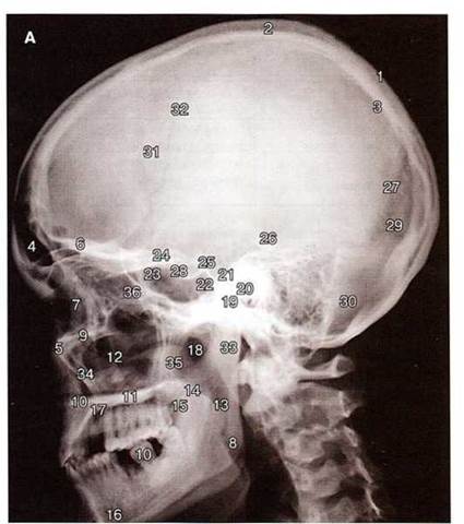
Fig. 81-A. Skull radiograph, lateral projection:
1 — outer table of the cranial vault bones; 2 — diploë; 3 — inner table of the cranial vault bones; 4 — frontal sinus; 5 — nasal bone; 6 — frontal bone, orbital part; 7 — orbit; 8 — angle of the mandible; 9 — maxilla, zygomatic process; 10 — inferior wall of the nasal cavity (bony palate); 11 — bony palate; 12 — maxillary sinus; 13 — ramus of the mandible; 14 — soft palate; 15 — ROOT of the Tongue; 16 — body of the mandible; 17 — hard palate; 18 — mandibular notch; 19 — external acoustic meatus; 20 — internal acoustic meatus; 21 — petrous part of the temporal bone (pyramid); 22, 23 — sphenoidal sinus; 24 — anterior clinoid process of the sphenoid bone; 25 — posterior clinoid process of the sphenoid bone; 26 — LOCATION OF THE auricle; 27 — lambdoid suture; 28 — hypophyseal fossa (sella turcica); 29 — transverse sinus sulcus; 30 — occipitomastoid suture; 31 — coronal suture; 32 — arterial sulcus; 33 — condylar process of the mandible; 34 — anterior wall of the maxillary sinus; 35 — coronoid process of the mandible; 36 — ethmoidal cells (after A. Yu. Vasilyev)
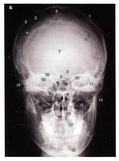
Fig. 81-B. Skull X-ray, nasomental (occipitomental) projection:
1 — outer table of the parietal bone; 2 — inner table of the parietal bone; 3 — parietal bone; 4 — coronal suture; 5 — lambdoid suture; 6 — sagittal suture; 7 — metopic suture; 8 — frontal sinuses; 9 — crista galli; 10 — sphenoid bone, lesser wing; 11 — petrous part of the temporal bone; 12 — sphenoid bone, greater wing; 13 — superior wall of the orbit; 14 — zygomatic bone; 15 — temporal bone, mastoid process; 16 — mandible; 17 — alveolar process of the maxilla; 18 — maxillary sinus; 19 — bony nasal septum; 20 — inferior nasal meatus; 21 — ethmoidal labyrinth (after A.Yu. Vasiliev)
Table 20. Openings in the walls of the facial skull cavities and their contents
Skull region |
Foramen |
Location on the skull |
Passing through the foramen |
||
arteries |
veins |
nerves |
|||
Optic canal |
In the region of the orbital apex |
Ophthalmic artery |
Optic nerve |
||
Anterior ethmoidal |
In the upper part of the medial wall, within the suture between the frontal bone and the orbital plate of the ethmoid bone |
Anterior ethmoidal artery - branch of the supraorbital artery |
Anterior ethmoidal vein - tributary of the superior ophthalmic vein |
Anterior ethmoidal nerve - branch of the nasociliary nerve (from the ophthalmic nerve) |
|
Superior orbital fissure |
Between the superior and lateral walls of the orbit |
Anterior meningeal artery |
Superior ophthalmic vein (draining into the cavernous sinus) |
Oculomotor, trochlear, abducens, ophthalmic (1st branch of the trigeminal nerve) |
|
Inferior orbital fissure |
Between the lateral and inferior walls of the orbit |
Infraorbital artery |
Inferior ophthalmic vein - tributary of the pterygoid venous plexus |
Infraorbital and zygomatic nerves - branches of the maxillary nerve |
|
Orbit |
Nasolacrimal canal |
Lacrimal sac fossa in the anterior part of the medial orbital wall, continuing into a canal that connects the orbit with the nasal cavity and opens into the inferior nasal meatus |
The nasolacrimal duct passes through the canal |
||
Supraorbital (or supraorbital notch) |
In the anterior part of the superior orbital wall (above the supraorbital margin of the frontal bone) |
Supraorbital artery - branch of the ophthalmic artery |
Supraorbital vein - tributary of the facial vein |
Supraorbital nerve - branch of the frontal nerve |
|
Posterior ethmoidal |
In the upper part of the medial orbital wall, within the suture between the frontal bone and the orbital plate of the ethmoid bone |
Posterior ethmoidal artery - branch of the supraorbital artery |
Posterior ethmoidal vein - tributary of the superior ophthalmic vein |
Posterior ethmoidal nerve - branch of the nasociliary nerve |
|
Infraorbital |
On the anterior surface of the maxilla as the anterior continuation of the infraorbital canal |
Infraorbital artery - branch of the maxillary artery |
Infraorbital vein |
Infraorbital nerve |
|
Alveolar (anterior and middle) |
On the inferior wall of the infraorbital sulcus and canal |
Anterior superior alveolar arteries |
Superior alveolar veins - tributaries of the maxillary vein |
Anterior and middle superior alveolar branches of the infraorbital nerve |
|
Zygomaticoorbital |
On the orbital surface of the zygomatic bone |
Zygomaticoorbital artery - branch of the superficial temporal artery |
Zygomatic nerve - branch of the maxillary nerve |
||
Piriform aperture - anterior opening of the nasal cavity |
On the anterior surface of the facial skull, in its central part |
||||
Choanae - posterior openings of the nasal cavity |
Posterior to the nasal cavity, beneath the external base of the neurocranium |
||||
Nasal cavity |
Ethmoidal |
In the superior wall of the nasal cavity |
Anterior ethmoidal artery |
Ethmoidal veins (draining into the superior ophthalmic vein) |
Olfactory (cranial nerve I) |
Nasolacrimal canal |
Inferior nasal meatus (in the anterior section) |
||||
Sphenopalatine |
Posterior to the middle nasal concha |
Sphenopalatine artery - branch of the maxillary artery |
Sphenopalatine vein; draining into the pterygoid venous plexus |
Posterior superior nasal branches (lateral and medial) - branches of the pterygopalatine ganglion |
|
Incisive canal |
In the anterior part of the median palatine suture |
Posterior septal nasal branch of the sphenopalatine artery; branch of the greater palatine artery (from the descending palatine artery) |
Nasopalatine nerve - from the pterygopalatine ganglion |
||
Greater palatine |
In the posterior part of the lateral |
Descending palatine ar- |
Greater palatine vein - |
Greater palatine |
|
canal |
wall of the nasal cavity |
tery and greater palatine artery |
draining into the palatine veins |
nerve |
|
Greater palatine foramen |
In the posterior part of the lateral region of the hard palate, between the greater palatine groove of the perpendicular plate of the palatine bone and the corresponding groove of the maxilla |
Greater palatine artery and descending palatine artery |
Greater palatine vein - draining into the palatine veins |
Greater palatine nerve - from the pterygopalatine ganglion |
|
Lesser palatine foramina |
In the posterior part of the lateral region of the hard palate, posterior to the greater palatine foramen, passing through the pyramidal process of the palatine bone |
Lesser palatine arteries - branches of the descending palatine artery |
Lesser palatine veins - draining into the palatine veins, which are tributaries of the facial vein |
Lesser palatine nerves - from the pterygopalatine ganglion |
|
Against the Background of the cranial bones, lighter lines ("radiopacities") corresponding to cerebral juga (prominences) and darker areas of various shapes ("radiolucencies") representing digital impressions are visible. The coronal and lambdoid sutures can be distinguished against the background of the cranial vault bones, along with the occipitomastoid suture as a posterior and inferior continuation of the lambdoid suture. Other cranial sutures in the lateral projection are poorly defined or entirely invisible. Sutures should be distinguished from wavy dark bands indicating the course of diploic veins, as well as the grooves for meningeal arteries. Within the cranial base, overlapping dense "shadows" of the petrous parts of the temporal bones are visible. Anterior to them lies the body of the sphenoid bone containing the sella turcica, the walls of which have clear margins. Within the body of the bone, beneath the sella turcica, There is a large dark patch ("radiolucency") corresponding to the sphenoid sinus.
Posterior to the sella turcica, the clivus begins as a line extending toward the anterior margin of the foramen magnum, while posterior to the "shadows" of the petrous pyramids of the temporal bones, one can observe the radiolucencies of the mastoid air cells and the broad radiolucent (dark) groove of the sigmoid sinus.
In the facial skeleton, the orbits appear as cones with their bases directed anteriorly and their apices posteriorly. The pattern of the ethmoidal labyrinth air cells is superimposed upon the orbits. Anterior to the orbits, the outlines of the nasal bones are visible, with their bases directed superiorly and posteriorly, and their apices inferiorly and anteriorly. The nasal cavity is superimposed on the orbits and the maxillary sinuses located below the orbits, which appear on the radiograph as quadrilateral or irregularly shaped dark areas. Against this quadrilateral background, the "shadows" of the nasal conchae can be distinguished as elongated, semi-oval radiopaque bands, with the nasal meatuses lying between them. Inferior to the overlapping images of the nasal cavity and maxillary sinuses, a horizontally positioned radiopaque strip ("shadow") is visible, representing the bones of the hard palate. Inferior and anterior to it lie the alveolar arch of the maxilla and the upper teeth. The outlines of the superimposed right and left halves of the mandible and the teeth are clearly visible on the lateral radiograph. Along the body and the lower part of the mandibular ramus, a darker strip corresponding to the mandibular canal can be traced.
The posteroanterior (PA) radiograph reveals both halves of the skull, outlining the cranial vault where the pattern of the frontal bone is superimposed on that of the occipital bone. The orbital margins are clearly defined, with the nasal cavity—divided by its septum—lying between them and slightly inferiorly. Intense "shadows" of the petrous pyramids of the temporal bones are superimposed on the lower parts of the orbits lateral to the nasal cavity. Projected onto the upper part of the nasal cavity between the orbits are the body of the sphenoid bone with the sphenoid sinus, the ethmoid air cells, and the outlines of the nasal conchae. Lateral to the nasal cavity and inferior to the orbits, dark areas ("radiolucencies") corresponding to the maxillary sinuses stand out. In the lower part of the facial skeleton, the upper and lower teeth are visible, along with the mandible showing its right and left rami.
Last update: 08/08/2026
Editorial and Educational Adaptation: This material has been compiled based on the primary/original source text. The project team performed an editorial review, corrected technical inaccuracies, structured sections, and adapted the content for an educational format.
What was processed:
- elimination of formatting defects (OCR errors, structural breaks, corrupted characters);
- editorial organization of content;
- standardization of terminology in accordance with academic sources;
- verification of factual statements against the original source text.
All mentions of the author, publication year, and origin of the primary text have been preserved in accordance with the source.