LEHNINGER PRINCIPLES OF BIOCHEMISTRY - VOL. 1. THE FOUNDATIONS OF BIOCHEMISTRY: STRUCTURE AND CATALYSIS - 2011
PART I. STRUCTURE AND CATALYSIS
6. ENZYMES
6.5. Regulatory Enzymes
In The Cell, groups of Enzymes work together in sequential pathways of enzyme-catalyzed reactions—such as the multistep breakdown of glucose to lactate or the multistep synthesis of Amino Acids FROM SIMPLE MOLECULES—in which the product of one reaction becomes the substrate for the next.
Most enzymes in metabolic pathways follow the kinetic patterns discussed thus far. However, each metabolic pathway has one or more enzymes that exert a greater influence on the overall rate of the pathway than do any of the Other Enzymes. These regulatory enzymes exhibit increased or decreased catalytic activity in response to certain signals. Adjusting the rates of these regulatory reactions allows the cell to adapt its METABOLISM to changing conditions of energy demand and to the needs for Biomolecules for growth and repair.
In most multienzyme systems, the first enzyme in the sequence is the regulatory enzyme. This is a very efficient strategy for regulating a pathway, since investing energy and metabolites into making the first few intermediates of a pathway whose end product is already in oversupply wastes both energy and starting Materials. Other enzymes in the pathway may also play subtle regulatory roles in fine-tuning flux through the pathway, as we will discuss in Chapter 15.
The activity of regulatory enzymes is modulated in a variety of ways. In allosteric enzymes, activity is regulated by the reversible, noncovalent binding of small molecules called allosteric modulators or effectors, which are generally small metabolites or Cofactors. Other enzymes are regulated by reversible covalent modification. In both cases, regulatory Proteins are typically multi-subunit proteins, and the regulatory sites and active sites are frequently located on different subunits. Metabolic systems also employ at least two other MECHANISMS OF ENZYME regulation. The activity of certain enzymes is stimulated or inhibited by binding to other regulatory proteins. Yet other enzymes are activated by proteolytic Cleavage of a peptide fragment; unlike regulation by effectors, proteolytic cleavage is irreversible. Both mechanisms are widely used in physiological processes such as Digestion, Blood clotting, hormone action, and Vision.
Cellular growth and survival depend on the efficient use of energy and nutrients, and this efficiency is governed by regulatory enzymes. There is no single rule determining which type of regulation operates in a given system. To some degree, allosteric (noncovalent) mechanisms allow fine-tuning of metabolic pathways that must operate continuously, but at fluctuating rates depending on cellular conditions. Regulation by covalent modification can operate either in an all-or-none fashion (as in proteolytic cleavage) or as a subtle rheostat-like adjustment. A single regulatory enzyme may utilize several types of regulation. The concluding section of this chapter examines these Methods of enzymatic regulation in greater detail.
Allosteric Enzymes Undergo Conformational Changes in Response to Modulator Binding
As discussed in Chapter 5, allosteric proteins are those having "other shapes," or Conformations, induced by the binding of a modulator. The same is true of certain regulatory enzymes: Conformational changes induced by one or more modulators interconvert less active and more active forms of the enzyme. Modulators of allosteric enzymes may be inhibitory or stimulatory. Often, the modulator is the substrate itself; regulatory enzymes for which substrate and modulator are identical are called homotropic. This behavior is analogous to the binding of oxygen to Hemoglobin (Chapter 5): the binding of a Ligand (in this case, the substrate) induces a conformational change that alters the affinity of remaining sites for the ligand. When the modulator is a molecule other than the substrate, the enzyme is heterotropic. Note that allosteric modulators should not be confused with uncompetitive or mixed inhibitors. Although the latter bind outside the Active Site, they do not necessarily induce conformational transitions between active and inactive states, and their kinetic manifestations are distinct.
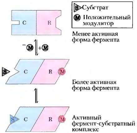
The kinetic and structural properties of allosteric enzymes differ markedly from those of simple, nonregulatory enzymes. Structurally, allosteric enzymes generally have one or more regulatory, or allosteric, sites for binding the modulator, In addition to the active site (Fig. 6–31). Just as the active site is specific for its substrate, each regulatory site is specific for its modulator. Enzymes with multiple modulators usually have separate binding sites for each. In homotropic enzymes, the active site and the regulatory site are the same.
Class="center">Figure 6–31. Model of interaction between subunits of an allosteric protein, showing binding sites for an inhibitor and an activator. In many allosteric enzymes, the substrate-binding site and the modulator-binding site(s) are on separate subunits—the catalytic (C) subunit and regulatory (R) subunit, respectively. The binding of a positive modulator (activator, M) to a specific site on the regulatory subunit induces a conformational change in the catalytic subunit. As a result, the catalytic subunit becomes active and can bind the substrate (S) with higher affinity. Dissociation of the modulator from the regulatory site returns the enzyme to the less active state.
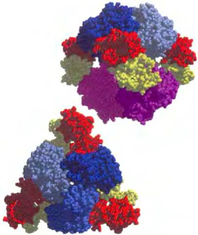
Allosteric enzymes are generally larger and more complex than nonregulatory enzymes. Most consist of two or more polypeptide chains (subunits). Aspartate transcarbamoylase, which catalyzes an early reaction in the Biosynthesis OF PYRIMIDINE NUCLEOTIDES (Fig. 22–36), consists of 12 polypeptide chains organized into catalytic and Regulatory Subunits. The quaternary Structure of this protein, determined by X-ray crystallography, is shown in Figure 6–32.
Figure 6–32. Model of the regulatory enzyme aspartate transcarbamoylase, viewed from different angles (PDB ID 2AT2). This allosteric enzyme has two catalytic clusters, each consisting of three catalytic polypeptide chains (different shades of blue and pink), and three regulatory clusters, each consisting of two regulatory polypeptide chains (red and yellow). The regulatory clusters form the vertices of a triangle surrounding the catalytic subunits. The binding sites for allosteric modulators are located on the regulatory subunits. Modulator binding causes substantial Changes in the structure and activity of the enzyme. The Role of this protein in nucleotide synthesis and the details of its regulation are discussed in Chapter 22.
In Many Metabolic Pathways, the Regulatory Step Is Catalyzed by an Allosteric Enzyme
In many multienzyme systems, the regulatory enzyme is specifically inhibited by the end product of the pathway whenever the concentration of that product exceeds the cell's needs. As the reaction catalyzed by the regulatory enzyme slows down, all subsequent enzyme-catalyzed steps slow down as well, as their substrates are depleted. Consequently, The rate of production of the end product is brought into balance with cellular needs. This type of regulation is called feedback inhibition. The accumulation of the end product of a metabolic pathway thus slows down the entire pathway.
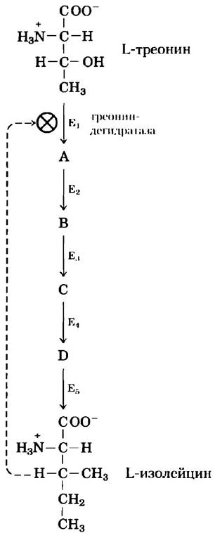
One of the first systems in which this mechanism was discovered is the bacterial enzyme system that catalyzes the five-step conversion of L-Threonine to L-isoleucine (Fig. 6–33). The first enzyme in this system, Threonine dehydratase, is inhibited by isoleucine, the end product of the pathway. This is an example of heterotropic allosteric inhibition. Isoleucine is a remarkably specific inhibitor: none of the pathway intermediates inhibit threonine dehydratase, and no other enzyme in the pathway is inhibited by isoleucine. Isoleucine binds not at the active site, but at a distinct regulatory site on the enzyme molecule. This binding is noncovalent and reversible; if the concentration of isoleucine drops, the rate of threonine dehydration increases. Thus, the activity of threonine dehydratase responds rapidly and reversibly to fluctuations in the cellular concentration of isoleucine. As we will see in Part II of this book, The regulatory mechanisms of many other Metabolic pathways are far more complex.
Figure 6–33. Feedback inhibition. The conversion of L-threonine to L-isoleucine proceeds via a sequence of five enzymes (E1 through E5). The first enzyme, threonine dehydratase, is subject to allosteric inhibition by the end product of the series (isoleucine) but is not inhibited by any of the intermediates (A through D). Feedback inhibition is indicated by the dashed line and the symbol  crossing the arrow for the reaction catalyzed by threonine dehydratase. This convention for representing feedback inhibition is used throughout the book.
Allosteric Enzymes Do Not Follow Michaelis-Menten Kinetics
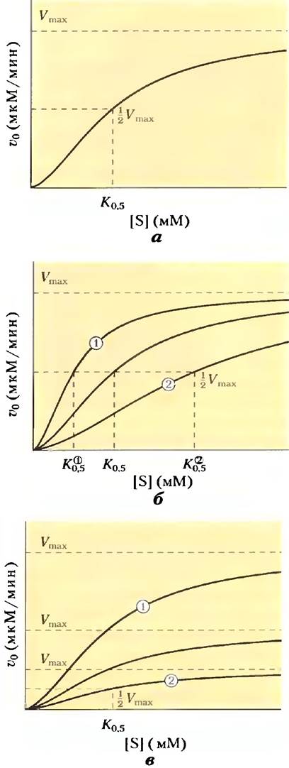
The plot of v0 versus [S] for allosteric enzymes does not follow Michaelis-Menten kinetics. Although these enzymes show substrate saturation at sufficiently high [S], the plot of v0 versus [S] (Fig. 6–34) is sigmoidal rather than the hyperbolic curve characteristic of nonregulatory enzymes. On a sigmoidal curve, we can still find the [S] corresponding to 1/2 Vmax, but this value is not designated as Km because the enzyme does not obey Michaelis-Menten kinetics. Instead, the kinetic behavior of allosteric enzymes is described by the symbols [S]0.5 or K0.5, representing the concentration of substrate that yields half-maximal velocity (Fig. 6–34).
Figure 6–34. Substrate-activity curves for Different types of allosteric enzymes, illustrating complex responses to modulators. (a) Sigmoidal curve for a homotropic enzyme, in which the substrate also acts as a positive (activating) modulator. Note the similarity to the oxygen-saturation curve of hemoglobin (Fig. 5–12). (b) Effect of a positive (+) and a negative (-) modulator on The behavior of an allosteric enzyme: K0.5 changes, whereas Vmax remains constant. The central curve represents the behavior in the absence of a modulator. (c) A less common type of modulator action in which Vmax changes while K0.5 remains nearly constant.
A sigmoidal curve for v0 versus [S] typically indicates cooperative interactions between protein subunits. In other words, a structural change in one subunit, driven by noncovalent interactions at the subunit interface, induces structural changes in the neighboring subunit. This principle is particularly evident in non-enzymatic proteins, such as the binding of oxygen to hemoglobin. The sigmoidal nature of this relationship is explained by two models—concerted (Symmetry) and sequential—that describe subunit interactions (Fig. 5-15).
Homotropic allosteric enzymes are typically multimeric proteins. As mentioned earlier, the binding site on each subunit of such a Protein Functions simultaneously as both an active site and a regulatory site. Most commonly, the substrate acts as a positive modulator (activator) because the subunits act cooperatively: the binding of a substrate molecule at one site induces a conformational change in the protein that facilitates the binding of subsequent substrate molecules. This type of interaction is precisely what gives rise to the sigmoidal profile of the v0 versus [S] curve. A key feature of sigmoidal kinetics is that small changes in modulator concentration can dramatically affect enzyme activity. As shown in Fig. 6-34a, a relatively small increase in [S] along the steep region of the curve causes a significant increase in v0.
For heterotropic allosteric enzymes, which are modulated by metabolites other than their normal substrate, the shape of the v0 versus [S] curve can vary. The action of an activator may convert the curve to a nearly hyperbolic profile with a decreased K0,5 value but an unchanged Vmax, which manifests as an increased reaction rate at a fixed Substrate Concentration (v0 values lie higher across all [S]; Fig. 6-34b, upper curve). Other heterotropic allosteric enzymes respond to activators with an increased Vmax without significant changes in K0,5 (Fig. 6-34c). Conversely, the action of a negative modulator (inhibitor) can produce a more pronounced sigmoidal curve characterized by an increased K0,5 (Fig. 6-34b, lower curve). Thus, the dependence of heterotropic allosteric enzyme activity on substrate concentration can be modulated in various ways, as some of these enzymes are regulated by inhibitory modulators, others by activators, and still others by both.
Some enzymes are regulated by reversible covalent modification
In another major class of regulatory enzymes, activity is controlled by the Covalent Modification of one or more amino acid residues within the enzyme molecule. Over 500 distinct types of covalent modification have been documented in proteins. Common modifying groups include phosphoryl, acetyl, adenylyl, uridylyl, methyl, amidyl, carboxyl, myristoyl, palmitoyl, prenyl, hydroxyl, sulfate, and ADP-ribosyl groups (Fig. 6-35). There are even entire proteins that serve as specialized modifying groups, such as ubiquitin and SUMO proteins (small ubiquitin-like modifiers). The respective groups are typically attached to and removed from the regulatory protein molecule by distinct sets of enzymes.
Fig. 6-35. Examples of enzyme modification reactions (E denotes the enzyme).
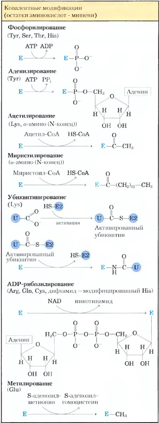
During amino acid modification, one amino acid is replaced by another with different properties. The Introduction of a charged group can locally alter The properties of the enzyme and induce conformational changes. The incorporation of a hydrophobic group may promote the association of the protein with a membrane. These alterations are often profound and can be critical for the proper function of the modified enzyme.
The number of possible enzyme modifications is too vast to discuss in detail; we will examine only a few representative examples.
An example of an enzyme regulated by methylation is the methyl-accepting chemotaxis protein in Bacteria. This protein is part of a system that allows motile bacteria to move toward attractants (such as sugars) and away from repellents. S-Adenosylmethionine (adoMet) serves as the methylating agent (Fig. 18-18b). Acetylation is a very common modification affecting about 80% of all soluble eukaryotic proteins, including many N-terminally acetylated enzymes. Ubiquitin is attached to proteins as a "tag" signaling their targeted proteolytic degradation (see Fig. 27-47). Ubiquitination can also serve regulatory functions. SUMO proteins are attached to numerous Nuclear Proteins in eukaryotes and participate in Transcriptional Regulation, Chromatin Organization, and DNA Repair.
ADP-ribosylation is a fascinating reaction undergone by only a select group of proteins. ADP-ribose is a derivative of nicotinamide adenine dinucleotide (NAD) (Fig. 8-39). This type of modification regulates bacterial Nitrogenase reductase, which plays a vital role in Nitrogen Fixation. Diphtheria and cholera toxins are enzymes that catalyze ADP-ribosylation, leading to the inactivation of key cellular enzymes and proteins.
Perhaps the most important type of regulatory modification is phosphorylation. It is estimated that one-third of all proteins in Eukaryotic Cells are phosphorylated, and one or (frequently) multiple phosphorylation steps occur in virtually every regulatory pathway. In some proteins only a single residue is phosphorylated, in others several, and Some proteins contain dozens of phosphorylation sites. Because this mode of covalent modification plays a critical role in a vast number of regulatory pathways, we will examine it in detail and revisit it in Chapter 12.
All of these modification mechanisms will be discussed in subsequent chapters of the book.
Phosphorylation Affects Protein Structure and Catalytic Activity
The attachment of a phosphoryl group to amino acid residues of a protein is catalyzed by protein Kinases; the removal of a phosphoryl group is catalyzed by Phosphatases. The addition of a phosphoryl group to Ser, Thr, and Tyr residues introduces bulky, charged groups into nonpolar Regions of the protein. The oxygen atoms of the phosphoryl group can participate in hydrogen bonding with protein groups, most commonly with the amide groups of the peptide chain at THE START OF an α-helical segment or with the charged guanidino group of an Arg residue. Two negative charges on the phosphorylated side chain can repel nearby negatively charged residues (Asp and Glu). If the modified side chain is located in a region of the protein critical for maintaining its three-dimensional structure, phosphorylation can profoundly affect protein conformation and, consequently, substrate binding and catalysis.
An important example of regulation via phosphorylation is the catalysis of the following reaction in Muscle and Liver by Glycogen phosphorylase ($M_r = 94,500$) (Chapter 15):

The resulting glucose-1-phosphate can be used for ATP Synthesis in muscle or converted into free glucose in liver. Glycogen phosphorylase exists in two forms: the more active phosphorylase a and the less active phosphorylase b (Figure 6-36). Phosphorylase a consists of two subunits, each possessing a specific Ser residue that undergoes phosphorylation at its hydroxyl group. Phosphorylation of the Serine residue is a prerequisite for maximum enzyme activity. The phosphoryl groups can be removed by the action of another enzyme, phosphorylase phosphatase:

Figure 6-36. Regulation of Muscle glycogen phosphorylase activity by covalent modification. The activity of muscle glycogen phosphorylase is regulated through multiple mechanisms, including covalent modification (phosphorylation), Allosteric Regulation, and a hormone-dependent regulatory cascade that acts on the phosphorylation and dephosphorylation enzymes. In the more active phosphorylase a, specific Ser residues on both subunits are phosphorylated. It is converted to the less active phosphorylase b upon the removal of phosphoryl groups by phosphorylase phosphatase 1 (PP1). The reverse reaction is catalyzed by phosphorylase kinase. The activity of both enzyme forms is subject to allosteric regulation by an activator (AMP) and inhibitors (glucose-6-phosphate and ATP), which bind to distinct sites on the enzyme molecule. The activities of phosphorylase kinase and PP1 are also regulated by a short cascade dependent on the Hormones Glucagon and epinephrine. When blood sugar levels drop, the Pancreas and Adrenal Glands begin producing glucagon and epinephrine. Epinephrine binds to its receptor in muscle and certain other Tissues and activates adenylate cyclase. Glucagon plays a similar role by binding to its receptor in the liver, stimulating the synthesis of large amounts of a modified nucleotide, cyclic AMP (cAMP; see p. 427), which activates cAMP-dependent protein kinase (PKA). PKA phosphorylates several target proteins, among them phosphorylase kinase and the inhibitor of phosphoprotein phosphatase 1 (PPI-1). Phosphorylated phosphorylase kinase is activated and, in turn, phosphorylates and activates glycogen phosphorylase. At the same time, phosphorylated PPI-1 interacts with PP1 and inhibits its action. Additionally, PPI-1 maintains itself in the active (phosphorylated) form by inhibiting phosphoprotein phosphatase 2B (PP2B), which dephosphorylates and thereby inactivates PPI-1. As a result, the equilibrium between the a and b forms of glycogen phosphorylase is strongly shifted toward the more active glycogen phosphorylase a. Both forms of phosphorylase kinase are activated to some extent by Ca2+ ions (not shown). For further details, see Chapters 14, 15, and 23.

In accordance with this reaction, phosphorylase a is converted to phosphorylase b through the cleavage of covalent bonds between the phosphoric acid residues and the two specific serine residues on the two subunits of glycogen phosphorylase.
Phosphorylase b, in turn, can also be converted back into active phosphorylase a by the action of another enzyme, phosphorylase kinase, which catalyzes The transfer of a phosphoryl group from ATP to the hydroxyl group of specific serine residues in phosphorylase b:

Glycogen breakdown in Skeletal Muscle and liver is regulated by altering The ratio of two forms of glycogen phosphorylase. Forms a and b differ in their secondary, tertiary, and quaternary structures; their interconversion is accompanied by changes in The structure of the active site and, consequently, in catalytic activity.
The Regulation of Glycogen phosphorylase by phosphorylation illustrates The Effect of modification on both the structure and the catalytic activity of the enzyme. Each subunit of the unphosphorylated protein is folded such that 20 amino acid residues at the N-terminus (including several basic residues) are located in a region containing acidic amino acid residues. The resulting electrostatic attraction stabilizes the molecule. Phosphorylation of Ser14 disrupts this interaction and displaces the N-terminal domain from the acidic region of the protein, leading to a conformation that favors interaction between and the side chains of several Arg residues. In this conformation, the protein is much more active.
Phosphorylation of an enzyme can also affect catalysis in another way, namely by altering the enzyme's affinity for its substrate. For example, upon phosphorylation of isocitrate dehydrogenase (an enzyme of The Citric Acid Cycle, Chapter 16), the electrostatic repulsion of the phosphate groups inhibits the binding of citrate (a tricarboxylate) at the Active Site of the enzyme.
Multiple Phosphorylation Permits Fine-Tuning
The Ser, Thr, and Tyr residues subject to phosphorylation in regulatory proteins occur within common Structural motifs called consensus sequences, which are recognized by specific protein kinases (Table 6–10). Some kinases prefer to phosphorylate residues adjacent to basic amino acids; others phosphorylate residues located, for example, near a Pro residue. However, the Amino Acid Sequence is not the only factor determining whether a given residue will be phosphorylated. As a result of protein folding, residues that are far apart in the Primary Structure may be brought into close proximity; it is the three-dimensional STRUCTURE OF THE protein that determines whether a residue is accessible to a protein kinase and whether the kinase recognizes it as a substrate. Another factor influencing the substrate Specificity of certain protein kinases is the proximity of other phosphorylated residues.
Table 6–10. Consensus Sequences for Protein Kinases
Protein kinase |
Consensus sequence and phosphorylated residue* |
Protein kinase A |
-x-R-[RK]-x-[ST]-B- |
Protein kinase G |
-x-R-[RK]-x-[ST|-x- |
Protein kinase C |
-[RK](2)-x-[ST] B-[RK](2) |
Protein kinase B |
-x-R-x-[ST]-x-K- |
Ca2+/calmodulin-dependent kinase I |
-B-x-R-x(2)-[ST]-x(3)-B- |
Ca2+/calmodulin-dependent kinase II |
-B-x-[RK]-x(2)-[ST]-x(2) |
Myosin light chain kinase (smooth muscle) |
-K(2)-R-x(2)-S-x-B(2)- |
Phosphorylase b kinase |
-K- R-K- Q-I-S- V-R- |
Extracellular signal-regulated kinase (ERK) |
-P-x-[ST]-P(2)- |
Cyclin-dependent protein kinase (cdc2) |
-x-[ST]-P-x-[KR]- |
Casein kinase 1 |
-[SpTp]-x(2)-[ST]-B-** |
Casein kinase II |
-x-[ST ]-x(2)-[ED]-x- |
β-Adrenergic receptor kinase |
-[DE](n)-[ST ]-x(3)- |
Rhodopsin kinase |
-x(2)-[ST]-(E)(n)- |
Insulin Receptor kinase |
-x-E(3)-Y-M(4)-K(2)-S-R-G-D-Y-M-T-M-Q-I- G-K(3)-L-P-A-T-G-D-Y-M-N-M-S-P- V-G-D- |
Epidermal growth factor (EGF) receptor kinase |
-E(4)-Y-F-E-L-V- |
* Capital letters denote residues deduced from nucleotide sequences, and italics denote actual amino acid residues in the sequences of known substrates. The Ser (S), Thr (T), and Tyr (Y) residues subject to phosphorylation are highlighted in red (for the single-letter amino acid nomenclature, see Table 3–1). x represents any amino acid, B any hydrophobic amino acid, and Sp, Tp, and Yp are prephosphorylated residues of Ser, Thr, and Tyr, respectively.
** The optimal target contains two amino acid residues separating the phosphorylated residue and a key Ser/Thr residue. Sites with one or three intervening residues are recognized much less efficiently.
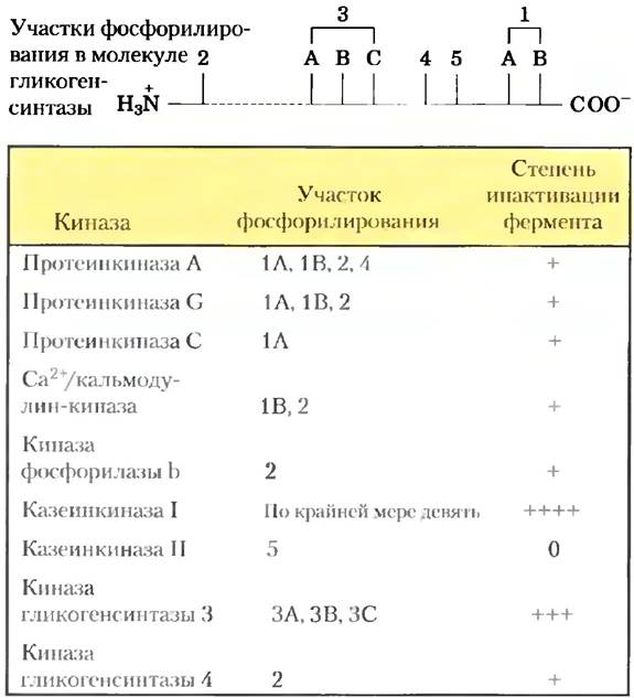
Regulation by phosphorylation is often quite complex. Some proteins have consensus sequences recognized by several different protein kinases, each of which can phosphorylate the protein and alter its enzymatic activity. In other cases, phosphorylation is a hierarchical process: a specific residue is phosphorylated only if a neighboring residue has already been phosphorylated. For example, glycogen synthase, which catalyzes the Condensation of glucose monomer units to form glycogen (Chapter 15), is inactivated by phosphorylation of specific Ser residues, and its activity is also modulated by the action of at least four other protein kinases that phosphorylate four different sites on the enzyme (Fig. 6–37). This enzyme is not a substrate for glycogen synthase kinase 3 until one site has been phosphorylated by casein kinase II. The attachment of phosphate groups at different sites inhibits glycogen synthase to varying degrees, and in some cases phosphorylation exerts a cumulative effect. Such multiple phosphorylation serves to exquisitely fine-tune enzymatic activity.
Fig. 6–37. Multiple phosphorylation. The glycogen synthase enzyme has at least nine distinct sites for phosphorylation by cellular protein kinases in five regions of the molecule indicated on the diagram. Thus, The regulation of this enzyme does not operate as a simple on/off switch, but rather through a much finer modulation in response to various signals.
For this regulatory mechanism to be effective, phosphorylation must be reversible. Phosphate groups are typically attached and removed by different enzymes, allowing these processes to be regulated independently. Cells contain a family of phosphoprotein phosphatases that hydrolyze  esters with the release of inorganic phosphate. The phosphoprotein phosphatases known to date do not act on all Phosphoproteins, but they generally exhibit lower substrate specificity than protein kinases.
Some Enzymes and Other Proteins Are Regulated by Proteolytic Cleavage of a Precursor
Certain enzymes are converted to their active form by the cleavage of an inactive precursor called a zymogen. Notably, many Proteolytic Enzymes (proteases) of the Stomach AND PANCREAS are regulated in this manner. Chymotrypsin and Trypsin are initially synthesized as chymotrypsinogen and trypsinogen (Fig. 6–38). Specific cleavage induces conformational changes that make the active sites of these enzymes accessible. Because this type of activation is irreversible, other mechanisms must exist to inactivate such proteases. Inactivation is brought about by inhibitory proteins that bind very tightly to the active site. For example, pancreatic trypsin inhibitor (Mr = 6,000) binds to and inhibits trypsin; α₁-antiproteinase (Mr = 53,000) primarily inhibits neutrophil Elastase (neutrophils are a type of WHITE BLOOD CELL; elastase degrades Elastin found in certain Connective Tissues). A deficiency of α₁-antiproteinase, which occurs in cigarette smokers, can lead to lung damage, including emphysema.
Fig. 6–38. Activation of zymogens by proteolytic cleavage. The Mechanism of formation of chymotrypsin and trypsin from their zymogens is shown. The bars represent Amino acid sequences in polypeptide chains, and the numbers indicate the relative positions of amino acid residues (the N-terminal residue is number 1). The residues at the newly formed ends of the cleaved polypeptide fragments are indicated below the bar. Note that certain residues are absent in the final active form. The three polypeptide chains of chymotrypsin (A, B, and C) are linked by Disulfide Bonds.

Proteases are not the only proteins activated by proteolysis. In other cases, however, the precursor proteins are called proenzymes or proproteins rather than zymogens. For instance, Collagen, a major component of Connective Tissue, is initially synthesized as a soluble precursor, procollagen. The blood-clotting cascade provides numerous examples of proteolytic activation. The blood clot protein fibrin is generated by the proteolysis of the inactive proprotein fibrinogen. This conversion is catalyzed by the protease Thrombin (which in many respects resembles chymotrypsin), and thrombin itself is generated by the proteolysis of prothrombin (in this case, a zymogen).
Some Regulatory Enzymes Use Multiple Regulatory Mechanisms
Glycogen Phosphorylase catalyzes the first reaction of the metabolic pathway that converts stored glucose into the energy required by the cell (Chapters 14 and 15). This is a crucial metabolic pathway, and its regulation is correspondingly complex. Although the activity of glycogen phosphorylase is primarily regulated via covalent modification, as shown in Fig. 6–36, it is also subject to allosteric regulation by AMP—an activator of phosphorylase b—as well as by glucose 6-phosphate and ATP (both of which are inhibitors). Furthermore, the enzymes that add and remove phosphoryl groups are themselves regulated by hormones that control blood glucose levels (Fig. 6–36; see also Chapters 15 and 23).
Other complex regulatory enzymes are found at the intersections of metabolic pathways. Bacterial Glutamine Synthetase, which catalyzes the reaction by which reduced nitrogen enters cellular metabolism (Chapter 22), is one of the most complex enzymes known. It is subject to allosteric regulation and reversible covalent modification (involving at least eight modulators); in addition, enzyme activity is regulated by binding to other regulatory proteins (a mechanism we will discuss in detail when covering the regulation of individual metabolic pathways).
What is the advantage of such complex REGULATION OF ENZYMATIC Activity? At the beginning of this chapter, we emphasized The Central Role of catalysis in the very existence of life. Controlling this catalysis is equally vital. If all possible reactions in a cell were to proceed simultaneously, macromolecules and metabolites would rapidly degrade into much simpler chemical components. This does not happen because cells catalyze only those reactions they currently need. When chemical resources are abundant, cells synthesize and store glucose and other metabolites. When resources are depleted, cells draw upon these reserves as fuel for metabolism. Chemical energy is expended economically, distributed across Various metabolic pathways in accordance with current demands. The presence of powerful catalysts specific to each individual reaction makes this regulation possible. All of these factors together create the complex, finely tuned network we call life.
Summary of Section 6.5 Regulatory Enzymes
■ The flux through Metabolic Pathways in the cell is regulated by controlling the activity of specific enzymes.
■ In feedback inhibition, the end product of a metabolic pathway inhibits the first enzyme unique to that pathway.
■ The activity of allosteric enzymes is controlled through the Reversible Binding of specific modulators to the regulatory site. The modulator can be the substrate itself or another metabolite; its effect may be either inhibitory or activating. The kinetic behavior of allosteric enzymes reflects the cooperative nature of subunit interactions.
■ The activity of other regulatory enzymes is modulated by the covalent modification of specific functional groups essential for catalysis. One of the most common mechanisms for regulating enzymatic activity is the phosphorylation of amino acid residues.
■ Many proteolytic enzymes are initially synthesized as inactive precursors (zymogens), which are activated by the cleavage of a small peptide fragment.
■ Enzymes operating at the intersection of Major Metabolic Pathways can be regulated through a complex combination of effects, allowing the coordination of these metabolic pathways.
Last update: 06/08/2026
Editorial and Educational Adaptation: This material has been compiled based on the primary/original source text. The project team performed an editorial review, corrected technical inaccuracies, structured sections, and adapted the content for an educational format.
What was processed:
- elimination of formatting defects (OCR errors, structural breaks, corrupted characters);
- editorial organization of content;
- standardization of terminology in accordance with academic sources;
- verification of factual statements against the original source text.
All mentions of the author, publication year, and origin of the primary text have been preserved in accordance with the source.