Human Anatomy (with the Fundamentals of Dynamic and Sports Morphology) - Ivanitsky M. F. 2008
Osteology and Arthrology
Skull
The Skull (Fig. 21) forms the skeletal framework of the HEAD, determining its external contour. It articulates with THE Vertebral Column via the atlas. The skull serves as a rigid protective case for the initial segments of the digestive and respiratory systems, as well as the Sensory Organs, and encloses the cranial cavity that houses the Brain. Anatomically, the skull is divided into two major parts: the neurocranium (braincase) and the viscerocranium (facial Skeleton).
Class="center">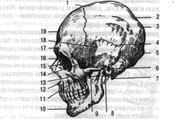
Fig. 21. Skull (lateral view):
1 - coronal suture; 2 - Parietal bone; 3 - lambdoid suture; 4 - squamous suture; 5 - Occipital bone (external occipital protuberance); 6 - external acoustic meatus; 7 - mastoid process; 8 - styloid process; 9 - zygomatic process; 10 - mental foramen of the Mandible; 11 - Zygomatic bone; 12 - anterior nasal spine of the Maxilla; 13 - infraorbital foramen of the maxilla; 14 - greater wing of the Sphenoid bone; 15 - Orbit; 16 - Nasal bone; 17 - superciliary arch; 18 - squamous part of the Temporal bone; 19 - Frontal bone (after G.F. Ivanov)
The neurocranium is formed by 8 bones: 4 unpaired (occipital, sphenoid, ethmoid, and frontal) and 2 paired (temporal and parietal). The viscerocranium comprises 6 paired bones (maxilla, zygomatic, palatine, nasal, lacrimal, and Inferior nasal concha) and 3 unpaired bones (Vomer, mandible, and Hyoid bone), along with the ethmoid and sphenoid bones, which structurally belong to the neurocranium. In addition, the facial skeleton includes 3 paired auditory ossicles (malleus, incus, and stapes), which are typically considered together with the Organ of Hearing (see p. 342).
BONES OF THE Neurocranium
Due to the evolutionary Development of the brain, the human neurocranium is larger in volume than the facial skeleton, whereas in all other vertebrate species, the facial skeleton predominates over the braincase. Structurally, the bones of the neurocranium are either flat (parietal, occipital) or pneumatized (frontal, sphenoid, ethmoid, temporal). The internal surface of the neurocranial bones exhibits prominent, fingerprint-like depressions alternating with raised elevations. These depressions and eminences correspond to the sulci and gyri of the Cerebral Cortex. Furthermore, the internal surface features grooves that lodge the Branches of the middle meningeal artery.
The occipital bone (see Fig. 21) contributes to The formation of the cranial base and the posterior region of the cranial vault. It consists of four parts arranged around the foramen magnum: anteriorly lies the basilar part, laterally are the paired lateral parts, and posteriorly forms the occipital squama. The foramen magnum transmits the Medulla Oblongata, vertebral Arteries, and accessory nerves.
The basilar part of the occipital bone fuses with the body of the sphenoid bone by the age of 18–20 years, as the intervening Cartilage is replaced by bone (transitioning from synchondrosis to synostosis). The superior surface of the basilar part faces the cranial cavity and presents a flattened area that, together with the sphenoid bone, forms the clivus, which Supports the Brainstem. Laterally, the lateral parts transition into the occipital squama. Their inferior surfaces bear the ellipsoidal occipital condyles, which articulate with the atlas. The hypoglossal canal pierces the base of each condyle. The lateral margin presents the jugular notch, which, together with the corresponding notch of the temporal bone, forms the jugular foramen transmitting the glossopharyngeal, vagus, and accessory nerves, as well as the Internal jugular vein. The superior surface of the lateral parts features the sigmoid sulcus, which lodges the sigmoid venous sinus. The occipital squama is a broad plate curved posteriorly and inferiorly; its external surface bears the external occipital protuberance, with superior and inferior nuchal lines radiating outwards for Muscle attachment.
The internal surface of the occipital squama features the internal occipital protuberance. This landmark divides the internal surface into four fossae: the two inferior fossae lodge the cerebellar hemispheres, while the two superior fossae accommodate the occipital lobes of the cerebral hemispheres. Ascending from the internal occipital protuberance is the unpaired sulcus for the superior sagittal sinus, while transversely run the grooves for the transverse sinuses, housing the corresponding venous sinuses of the cerebral dura mater.
The sphenoid bone (see Fig. 21) comprises a central body and three pairs of processes: the lesser wings projecting laterally and superiorly, the greater wings projecting laterally, and the pterygoid processes extending inferiorly.
The body of the sphenoid bone is roughly cuboidal and contains the sphenoidal sinus, which opens anteriorly into the Nasal cavity. The superior surface of the body features a deep depression known as the sella turcica, which lodges the Pituitary Gland. The lateral margins of the sella turcica are flanked by grooves that house the internal carotid arteries. The optic canal opens at the base of each lesser wing, transmitting the Optic nerve and ophthalmic artery into the orbit. The greater wings present three surfaces: a concave cerebral surface facing the cranial cavity, a flat orbital surface contributing to the orbit, and a slightly concave temporal surface forming the floor of the temporal fossa. The bases of the greater wings are perforated by several foramina: the foramen rotundum (transmitting the maxillary nerve, or cranial nerve V2), the foramen ovale (transmitting the mandibular nerve, or cranial nerve V3), and the foramen spinosum (transmitting the middle meningeal artery). The Inferior surface of the greater wings faces the infratemporal fossa. Between the greater and lesser wings lies the superior orbital fissure, through which pass the oculomotor, trochlear, ophthalmic, and abducens nerves, as well as the superior ophthalmic vein. The pterygoid processes descend vertically from the body of the sphenoid bone. Each process consists of a medial and a lateral plate, with the medial plate terminating inferiorly in a small hook-like hamulus.
The frontal bone (see Fig. 21) participates in forming both the cranial vault and the cranial base. It is divided into four parts: the frontal squama projecting superiorly, two horizontally oriented orbital parts, and a nasal part situated between the orbital parts.
The external surface of the frontal squama faces anteriorly, while its internal surface faces the cranial cavity. The external surface is smooth and bounded inferiorly by a sharp supraorbital margin, above which lie the right and left superciliary arches. Superior to the superciliary arches are the frontal eminences, while the depressed area between the superciliary arches is termed the glabella. The internal surface of the frontal squama exhibits the median groove for the superior sagittal sinus, flanked by impressions corresponding to cerebral convolutions. The lateral, temporal surface of the frontal squama articulates inferiorly with the greater wings of the sphenoid bone and posteriorly and superiorly with the parietal bones.
The orbital PARTS OF THE frontal bone form thin plates whose inferior surfaces comprise the roof of the orbit, and superior surfaces face the cranial cavity. The lateral region of the orbital part features a depression known as the lacrimal fossa. The ethmoidal notch is located between the two orbital plates.
The nasal part of the frontal bone closes the ethmoidal notch anteriorly and contains two apertures leading into the frontal sinus.
The Ethmoid bone resembles a laterally compressed cube and is characterized by its light, delicate Structure. It consists of two main plates—the cribriform and perpendicular plates—and the ethmoidal labyrinths. The cribriform plate lies horizontally within the ethmoidal notch of the frontal bone. It is perforated by numerous foramina, and along its median plane projects a superior bony crest, the crista galli, which serves for the attachment of the falx cerebri. The olfactory nerves pass from the nasal cavity into the cranial cavity through the foramina of the cribriform plate.
The perpendicular plate of the ethmoid bone lies in the median plane and descends vertically from the cribriform plate, contributing to the Formation of the nasal septum.
The right and left ethmoidal labyrinths are constructed from delicate bony lamellae oriented in various planes to enclose air-filled ethmoidal Cells that communicate with the nasal cavity. The openings of these cells facing the nasal cavity are partially closed by curved bony shelves: the superior and middle nasal conchae, between which lies the superior nasal meatus.
On its lateral aspect, the ethmoid bone presents a thin orbital plate that forms the medial wall of the orbit.
The parietal bone (Fig. 21) is a paired bone forming the central region of the cranial vault. It is a quadrilateral plate, convex externally and concave internally. Its convex surface features the parietal eminence, which is easily palpable beneath the scalp. Extending laterally and inferiorly below the eminence is the rough temporal line, which serves as one of the sites of origin for the temporalis muscle. The internal concave surface faces the cranial cavity and displays arterial sulci, digital impressions, and the groove for the superior sagittal sinus running along its sagittal margin. When apposed, the sagittal grooves of both parietal bones form a single continuous sulcus that houses the superior sagittal sinus of the dura mater.
The temporal bone (see Fig. 21) is a paired bone that contributes to the formation of the cranial base and partially the cranial vault. It consists of three distinct parts: the petrous part (or pyramid), the squamous part, and the tympanic part.
The petrous part is shaped like a three-sided pyramid, with the mastoid process attached to its posterior region. Its inferior surface faces downward toward the external cranial base, while its anterior and posterior surfaces face the cranial cavity. The anterior surface of the pyramid bears the trigeminal impression near its apex. This anterior surface forms the roof of the tympanic cavity, serving as one of the walls of the Middle ear.
The posterior surface of the petrous part features the internal acoustic meatus, through which pass the facial and vestibulocochlear nerves.
The inferior surface of the petrous part presents the external opening of the carotid canal, through which the Internal Carotid Artery enters its respective canal. At the apex of the petrous part, corresponding to the anterior end of the carotid canal, lies the internal carotid opening, where the internal carotid artery enters the cranial cavity. Near the posterior margin of the Base of the petrous part on its inferior surface is the jugular fossa. Posterior and lateral to the fossa is the stylomastoid foramen, through which the Facial Nerve exits the skull. Anterior to this foramen lies the slender styloid process.
At the top of the petrous part of the temporal bone, alongside the carotid canal, lies the musculotubular canal, which houses the pharyngotympanic (auditory) tube and the tensor tympani muscle. The facial canal, containing the facial nerve, also passes through the petrous part of the temporal bone. This canal begins deep within the internal acoustic meatus and terminates at the stylomastoid foramen.
The mastoid process is a part of the petrous temporal bone located posterior to the external acoustic meatus, which can be easily palpated beneath the Skin. It serves as the attachment site for the sternocleidomastoid muscle and contains air cells. On its medial (internal) surface lies the mastoid notch, the Water/144.html">Origin of the digastric muscle, while the cerebral surface features a wide sigmoid sulcus—a continuation of the corresponding groove on the occipital bone and the site of the sigmoid sinus of the dura mater.
The squamous part of the temporal bone resembles a semicircular, vertically oriented plate that contributes to the formation of the cranial vault (roof of the skull). Its internal surface faces the cranial cavity, while its external surface forms the floor of the temporal fossa. Projecting from it is the zygomatic process, which unites with the temporal process of the zygomatic bone to form the zygomatic arch, easily palpable beneath the skin. At the base of this process lies the mandibular fossa, which articulates with the head of the mandible. Anterior to the fossa is the articular tubercle.
The tympanic part of the temporal bone is a curved plate that forms the inferior and anterior BOUNDARIES OF THE external acoustic meatus.
Bones of the Facial Skeleton
The maxilla (upper jaw) (see Fig. 21) is a paired bone. Together with the zygomatic bones and the mandible, it forms the major portion of the facial skeleton, participating in the formation of the orbital, nasal, and oral cavities, as well as the infratemporal and pterygopalatine fossae. This bone articulates with all the bones of the facial skeleton, as well as with the frontal, ethmoid, and sphenoid bones of the neurocranium. The maxilla consists of a body and four processes: the frontal, zygomatic, alveolar, and palatine processes. The shape of the maxillary body resembles a quadrilateral prism, with its anterior surface facing the face, its superior (orbital) surface facing the orbit, its internal (nasal) surface facing the nasal cavity, and its posterior (infratemporal) surface facing the infratemporal fossa.
The body of the maxilla contains a large paranasal sinus (the maxillary sinus) that communicates directly with the nasal cavity. The orbital surface of the body features a groove that continues anteriorly into a canal, opening on the anterior surface via the infraorbital foramen above a depression known as the canine fossa. The infraorbital nerve (a branch of the Trigeminal nerve) passes through this foramen to the face. On the anteromedial margin of the maxillary body is the nasal notch, terminating anteriorly in the anterior nasal spine. Together with the nasal bone, this notch contributes to the formation of the piriform aperture. The posterior surface of the maxilla presents the maxillary tuber (tuberosity), which serves as a muscle attachment site.
The frontal process of the maxilla projects vertically upward from the body, articulating anteriorly with the nasal bone, superiorly with the frontal bone, and posteriorly with its surface abutting the Lacrimal bone. It contributes to the bony framework of the Nose.
The short and broad zygomatic process extends laterally from the body of the bone to articulate with the zygomatic bone.
The alveolar process extends inferiorly from the body of the maxilla, forming the alveolar arch, which contains dental alveoli (tooth sockets) separated from one another by interalveolar septa. In an adult, there are eight such sockets in each maxilla.
The palatine process projects from the inferior margin of the nasal surface of the maxillary body as a horizontally oriented plate. Together with the corresponding process of the opposite bone, it forms the hard palate. Its anterior medial margin features a groove that, combined with the matching groove of the contralateral side, forms the incisive canal. This canal opens onto the hard palate to transmit the nasopalatine nerve.
The zygomatic bone (see Fig. 21) is a paired, highly durable bone of irregular quadrilateral shape. It is easily palpable beneath the skin and articulates with the frontal, sphenoid, and temporal bones, as well as the maxilla. The shape of the middle third of the face depends largely on the Morphology of the zygomatic bone.
The Palatine bone is a paired bone situated between the maxilla and the sphenoid bone, forming the posterior region of the facial skeleton. It consists of two plates: a horizontal plate, which contributes to the formation of the hard palate, and a perpendicular plate, which helps form the lateral wall of the nasal cavity.
The nasal bone (see Fig. 21) is a small, paired, gutter-shaped bone located between the frontal processes of the maxilla and the nasal part of the frontal bone. It forms the bony Skeleton of the nasal bridge and participates in forming the piriform aperture.
The lacrimal bone is a paired, thin, quadrilateral plate situated in the anterior part of the medial orbital wall, positioned between the frontal process of the maxilla (anteriorly) and the orbital plate of the ethmoid bone (posteriorly). Superiorly, it contacts the frontal bone, and inferiorly, it articulates with the body of the maxilla. Its lateral surface features a vertically oriented groove that contributes to the formation of the nasolacrimal canal.
The inferior nasal concha resembles the conchae of the ethmoid bone but is considerably larger. It is an independent paired bone located on the lateral wall of the nasal cavity.
The vomer is an unpaired bone shaped like a quadrilateral plate. Together with the perpendicular plate of the ethmoid bone, it forms the bony part of the nasal septum.
The mandible (lower jaw) (see Fig. 21) is a robust, thick, unpaired bone that articulates on the right and left sides with the temporal bones. It consists of a body and right and left rami.
The body of the mandible has external and internal surfaces. It thickens inferiorly to form the base of the mandible, while its superior portion features the alveolar part, which forms the alveolar arch containing 16 tooth sockets in the adult. On its anterior surface along the midline, the mandible exhibits the mental protuberance, flanked inferiorly and laterally by the mental tubercles, which are readily palpable beneath the skin. Running through the interior of the mandible is the mandibular canal, which begins on the Medial surface of each ramus and opens anteriorly on either side of the mental protuberance via the mental foramina. Blood Vessels and nerves pass through this canal. Additionally, the inner surface of the mandibular body bears the mylohyoid line (for the attachment of the mylohyoid muscle) on both the right and left sides, as well as the digastric fossae, which serve as the origin sites for the digastric Muscles.
The ramus of the mandible forms an angle with the body that can be easily palpated beneath the skin. The outer surface of this angle features the masseteric tuberosity for the attachment of the masseter muscle, while the inner surface bears the pterygoid tuberosity for the insertion of the medial pterygoid muscle. Each ramus terminates in two processes: an anterior coronoid process and a posterior condylar process. The former serves for the insertion of the temporalis muscle, whereas the latter terminates in a head (condyle) that articulates with the temporal bone. This head is easily palpable beneath the skin immediately anterior to the external acoustic meatus.
Between the coronoid and condylar processes lies the mandibular notch.
The hyoid bone has a curved, horseshoe-like appearance. It is located posterior and slightly inferior to the mandible within the Muscles of the anterior neck. The hyoid bone comprises a body—easily palpable beneath the skin—and two pairs of processes: the greater horns and lesser horns, which serve as attachment sites for muscles and ligaments.
The Skull as a Whole. The cranial bones articulate with one another to form numerous cavities, fossae, and depressions.
The neurocranium is divided into a superior portion—the cranial roof (calvaria)—and an inferior portion—the cranial base.
The cranial roof is formed by the parietal bones and, in part, by the frontal, occipital, and temporal bones. The cranial base is formed by the orbital parts of the frontal bone, as well as the ethmoid, sphenoid, temporal, and occipital bones.
The skull features all Types of bone connections: continuous (sutures, gomphosises, and synchondroses) and discontinuous (the temporomandibular joint).
Most sutures of the cranial vault are serrated. These include the coronal suture between the frontal and parietal bones, the sagittal suture between the right and left parietal bones, and the lambdoid suture between the parietal and occipital bones. An exception is the articulation between the squamous part of the temporal bone and the parietal bone, where one margin overlaps the other to form the so-called squamous suture. The facial bones are connected by plane sutures.
Gomphosis is characteristic of The connection between the tooth ROOT* and the alveoli of the upper and lower jaws, with a thin layer of Connective Tissue in between. Synchondroses are located in children between individual bone parts, as well as between the sphenoid and occipital bones.
The temporomandibular joint is formed by the condylar process of the mandibular ramus and the mandibular fossa of the temporal bone (Fig. 22). It is a two-chambered, combined joint. An articular disc is located within it, dividing the joint into two chambers and increasing its range of motion. In shape, the joint is ellipsoidal, while functionally (due to the presence of the disc) it is spheroidal.
The following movements are possible in the temporomandibular joint: depression and elevation of the mandible, as well as forward, backward, and lateral movements. Palpating the head of the mandible during Mouth opening and closing easily confirms these movements.

Fig. 22. Right temporomandibular joint (external view, sectioned):
1 - zygomatic arch; 2 - mandibular ramus; 3 - articular capsule; 4 - external acoustic meatus; 5 - mastoid process; 6 - mandibular fossa; 7 - articular disc; 8 - articular tubercle
The joint is reinforced by the following ligaments: the lateral ligament, located on the outside of the joint and extending from the zygomatic process of the temporal bone to the neck of the mandible; and the sphenomandibular and stylomandibular ligaments, which run from their respective bones to the mandible.
Upon removing the cranial vault, one can examine the internal cranial base (Fig. 23), which is subdivided into three cranial fossae: anterior, middle, and posterior. The anterior cranial fossa is formed by the orbital part of the frontal bone, the cribriform plate of the ethmoid bone, and the lesser wings of the sphenoid bone; the middle cranial fossa is formed primarily by the cerebral surface of the greater wings of the sphenoid bone, the superior surface of its body, and the anterior surface of the petrous part of the temporal bone; the posterior cranial fossa is formed by the occipital bone and the posterior surface of the petrous part of the temporal bone.
The anterior cranial fossa houses the frontal lobes of the cerebral hemispheres, the middle fossa houses the temporal lobes, and the posterior fossa houses the Cerebellum, Pons, and medulla oblongata. Each fossa contains A number of openings. The anterior cranial fossa has the openings of the cribriform plate, connecting it to the nasal cavity. From the middle cranial fossa, the superior orbital fissure and the optic canal lead into the orbital cavity; the foramen rotundum leads into the pterygopalatine fossa and through it into the orbit; the foramen ovale and foramen spinosum connect the middle cranial fossa to the external cranial base. The posterior cranial fossa contains several openings: the foramen magnum, which connects the cranial cavity with the vertebral canal; the jugular foramen, leading to the external surface of the cranial base; and the internal acoustic meatus, leading into the Inner ear.

Fig. 23. Base of the skull (internal surface):
1 - frontal bone (orbital part); 2 - foramen caecum; 3 - frontal crest; 4 - ethmoid bone; 5 - crista galli; 6 - cribriform plate; 7 - sphenofrontal suture; 8 - tuberculum sellae of the sphenoid bone; 9 - optic canal; 10 - hypophysial fossa of the sphenoid bone; 11 - dorsum sellae; 12 - anterior clinoid process; 13 - foramen rotundum; 14 - body of the sphenoid bone; 15 - foramen ovale; 16 - foramen lacerum; 17 - foramen spinosum; 18 - petro-occipital fissure; 19 - internal acoustic opening; 20 - jugular foramen; 21 - lateral part of the occipital bone; 22 - sulcus for sigmoid sinus; 23 - mastoid foramen; 24 - parietomastoid suture; 25 - occipitomastoid suture; 26 - lambdoid suture; 27 - basilar part of the occipital bone and clivus; 28 - sulcus for transverse sinus; 29 - internal occipital protuberance; 30 - internal occipital crest; 31 - foramen magnum; 32 - squamous part of occipital bone; 33 - spheno-occipital synchondrosis; 34 - parietal bone; 35 - mastoid part of the temporal bone; 36 - petrous part of the temporal bone; 37 - sulcus for greater petrosal nerve; 38 - sulcus for lesser petrosal nerve; 39 - squamous part of the temporal bone; 40 - squamous suture; 41 - sphenoparietal suture; 42 - coronal suture; 43 - greater wing of the sphenoid bone; 44 - lesser wing of the sphenoid bone (after G. F. Ivanov)
Viewing the skull from below, one can see that the anterior part of the cranial base is covered by the facial bones, which form the hard palate consisting of the palatine processes of the maxillae and the palatine bones. In the middle and posterior parts, the cranial base is formed by the inferior surfaces of the sphenoid, occipital, and temporal bones. They feature A large number of openings, notably the jugular foramen between the occipital and temporal bones, and the foramen lacerum between the petrous part of the temporal bone and the sphenoid bone.
The largest topographical and anatomical structures of the facial skull are the orbit, the nasal cavity, and the Oral Cavity.
The orbit has the shape of a four-sided pyramid. Its medial wall is formed by the frontal process of the maxilla, the lacrimal bone, the orbital plate of the ethmoid bone, and partly by the body of the sphenoid bone; the superior wall by the orbital part of the frontal bone and the lesser wings of the sphenoid bone; the lateral wall by the greater wings of the sphenoid bone and the zygomatic bone; and the inferior wall by the superior surface of the body of the maxilla.
The orbit communicates with the cranial cavity via the superior orbital fissure and the optic canal; with the nasal cavity via the nasolacrimal canal, formed by the lacrimal bone, the frontal process of the maxilla, and the inferior nasal concha; and with the infratemporal and pterygopalatine fossae via the inferior orbital fissure, which lies between the greater wings of the sphenoid bone and the body of the maxilla.
The nasal cavity has superior, inferior, and lateral walls. It is divided by a bony septum located in the median plane. The septum is formed by the perpendicular plate of the ethmoid bone and the vomer. The superior wall of the nasal cavity is formed by the cribriform plate of the ethmoid bone, as well as the nasal and frontal bones; the inferior wall by the palatine process of the maxilla and the horizontal plate of the palatine bone; and the lateral walls by the maxilla, the lacrimal and ethmoid bones, the inferior nasal concha, the perpendicular plate of the palatine bone, and the medial surface of the pterygoid process of the sphenoid bone. The anterior opening of the nasal cavity, called the piriform aperture, connects it to the external environment; the posterior openings, the choanae, face the external cranial base and connect the nasal cavity to the pharyngeal cavity.
The nasal cavity is divided on the right and left sides by nasal conchae located on its lateral wall into three meatuses: inferior (beneath the inferior nasal concha), middle (between the inferior and middle conchae), and superior (between the middle and superior conchae). All of these open into the common nasal meatus located on either side of the nasal septum. The nasal cavity communicates with the cranial cavity, the orbit, the nasal and oral cavities, and the Paranasal Sinuses. The superior nasal meatus communicates with the cranial cavity through the openings of the cribriform plate of the ethmoid bone; the middle meatus communicates with the maxillary sinus, the ethmoidal cells, and the frontal sinus. Posteriorly, at the level of the superior nasal concha, the sphenoidal sinus opens into the nasal cavity. The inferior nasal meatus communicates with the orbital cavity through the nasolacrimal canal. The nasal cavity also communicates with the pterygopalatine fossa through the sphenopalatine foramen and with the oral cavity through the incisive foramen.
The oral cavity is bounded by bony walls only superiorly, anteriorly, and laterally. Its superior wall is formed by the hard palate, composed of the palatine processes of the right and left maxillae and the horizontal plates of the palatine bones; the lateral and anterior walls are formed by the mandible and the alveolar processes of the maxillae. The oral cavity communicates through the incisive foramen with the nasal cavity, and through the greater palatine canal with the pterygopalatine fossa.
On the lateral surface of the skull lie the pterygopalatine, infratemporal, and temporal fossae.
The pterygopalatine fossa is located between the bones of the facial and cerebral cranium and is bounded anteriorly by the body of the maxilla, medially by the palatine bone, posteriorly by the pterygoid process of the sphenoid bone, and superiorly by the body of this bone. It communicates with the nasal cavity, the middle cranial fossa, the foramen lacerum, the orbit, and the oral cavity. The pterygopalatine fossa lacks a lateral wall and extends laterally into the infratemporal fossa.
The infratemporal fossa is located posterior to the body of the maxilla, medial to the zygomatic bone and zygomatic arch, and lateral to the pterygoid process of the sphenoid bone. It forms part of the external base of the cerebral cranium. It is separated from the temporal fossa by the infratemporal crest.
The temporal fossa is a shallow depression that lodges the temporalis muscle. The temporal surface of the greater wings of the sphenoid bone, the squamous part of the temporal bone, and partly the parietal and frontal bones contribute to the formation of the temporal fossa.
Buttresses (Fig. 24). In certain areas, the skull features structural thickenings known as buttresses. They serve to attenuate the mechanical shocks and impacts that the skull experiences during walking, running, jumping, chewing, and engaging in various sports (such as boxing and football).
Buttresses act as load-bearing Zones of the skull, interspersed with its more delicate structures.
Four buttresses are distinguished. Three of them resemble curved pillars anchored inferiorly in the alveolar arch of the maxilla and extending superiorly to various parts of the facial and neurocranium. The fourth buttress corresponds to the most thickened areas of the mandible.
1. The frontonasal buttress originates inferiorly in the thickened walls of the sockets of the canine and adjacent Teeth. It continues upward as a dense plate of the frontal process of the maxilla, reaching the outer margin of the nasal part of the frontal bone.
2. The zygomatico-temporal buttress begins at the alveolar thickenings of the first two molars and extends upward from the zygomatic bone, which abuts laterally and posteriorly against the zygomatic process of the temporal bone, and superiorly against the frontal bone. This is the most pronounced buttress.
3. The pterygopalatine buttress is formed by the pterygoid process of the sphenoid bone and the perpendicular plate of the palatine bone. It adjoins the posterior region of the alveolar process of the maxilla on one side and the maxillary tuberosity on the other.
4. The mandibular buttress is a thickening within the body of the mandible that abuts the dental sockets on one side and extends along the ramus of the bone toward its neck and head. Through the mandibular head, chewing forces are transmitted from the mandible to the temporal bone.

Fig. 24. Buttresses of the skull:
1 — frontonasal; 2 — zygomatico-temporal; 3 — pterygopalatine; 4 — mandibular
Age, Sex, and Individual Variations of the Skull
The structure of the skull varies according to AGE AND SEX. In newborns, the neurocranium is relatively larger than the facial skeleton due to the somewhat delayed development of the masticatory apparatus compared to the brain and sensory organs.
The frontal and parietal eminences in newborns are exceptionally prominent, giving the skull a pentagonal appearance when viewed from above. Paranasal sinuses are absent in the newborn skull, with the exception of the maxillary sinus, which is also barely developed. All muscular ridges and processes are weakly defined.
Cranial sutures are absent in newborns. In the cranial vault, significant layers of connective tissue lie between the individual bones, forming expanded areas known as fontanelles (Fig. 25). The largest of these are the anterior and posterior fontanelles. Paired sphenoid and mastoid fontanelles are located laterally. The anterior fontanelle lies between the frontal and parietal bones and has a quadrilateral shape. The posterior fontanelle is situated between the parietal and occipital bones and is triangular. In the region of the fontanelles, the brain is covered only by a thin connective tissue membrane, through which the pulsation of cerebral arteries can be easily felt beneath the skin.
The absence of sutures between the bones and synostoses between certain parts of bones such as the frontal, occipital, sphenoid, and temporal makes the newborn skull extremely pliable.
Closure of the fontanelles normally concludes during the second year of life. After the age of 30, cranial sutures gradually begin to ossify. Consequently, in elderly individuals, the cranial vault typically fuses into a single continuous bony structure. Age-related Changes in the elderly include a reduction in the height of the facial skeleton due to tooth loss and alveolar atrophy, as well as increased bone fragility. The cranial cavity volume in males is approximately 1450 cm3. In females, cranial capacity is on average about 150 cm3 smaller than in males, which correlates with smaller overall female body dimensions. However, the relative size of the cranial cavity is slightly greater in females than in males. Furthermore, the facial skeleton is somewhat less developed relative to the neurocranium in females than in males.

Fig. 25. SKULL OF A newborn:
1 — anterior fontanelle; 2 — parietal bone; 3 — occipital bone; 4 — mastoid fontanelle; 5 — sphenoid fontanelle; 6 — frontal bone
The external surface of the female skull is smoother. Various crests and roughnesses serving for muscle and ligament attachment are less developed than in males. Female orbits are relatively larger, and the forehead is more vertical than in males. Nevertheless, craniometric sex-differentiating features are highly variable.
The dimensions and shape of the skull are assessed using craniometry, a specialized system of measurements. Most commonly, these include the longitudinal, transverse, and vertical diameters of the neurocranium. The longitudinal diameter is the maximum fronto-occipital length of the skull; the transverse is the maximum breadth; and the vertical is the maximum height from the base to the cranial vault. Skull shape is typically evaluated using the cranial index: transverse diameter × 100 / longitudinal diameter.

Fig. 26. Skull shape:
A — dolichocephalic; B — brachycephalic
With an index value up to 74.9, the skull is classified as dolichocephalic; from 75 to 79.9, as mesocephalic; and 80 and above, as brachycephalic (Fig. 26). When measuring the same dimensions in living individuals, the cephalic index turns out to be 1.5 higher than the cranial index. While skull shapes vary widely among human populations, this provides no basis for defining so-called higher and lower races. Modern anatomy and anthropology reject racism as a pseudo-science and an instrument for subjugating populations in developing countries.
When viewed in profile, it can be seen that in some people the upper part of the skull protrudes relatively more, while in others the lower part does. If a straight line is drawn connecting the glabella and the most anteriorly projecting point of the maxilla, and the angle is determined between this line and the horizontal plane passing through the external auditory meatus and the lower wall of the orbit, then this angle, lying above the horizontal line and facing towards the cranial vault (the facial angle), is approximately 80°. Its decrease characterizes the so-called prognathism of the skull, and its increase characterizes orthognathism of the skull. In newborn infants, the skull is more orthognathic than in adults. In men, it is more prognathic than in women.
Last update: 08/08/2026
Editorial and Educational Adaptation: This material has been compiled based on the primary/original source text. The project team performed an editorial review, corrected technical inaccuracies, structured sections, and adapted the content for an educational format.
What was processed:
- elimination of formatting defects (OCR errors, structural breaks, corrupted characters);
- editorial organization of content;
- standardization of terminology in accordance with academic sources;
- verification of factual statements against the original source text.
All mentions of the author, publication year, and origin of the primary text have been preserved in accordance with the source.