Human Anatomy (with the fundamentals of dynamic and sports morphology) - Ivanitsky M. F. 2008
The Doctrine of Bones and Their Joints
Skeleton of the Trunk
The Skeletal Structure of the trunk is formed by THE Vertebral Column and the rib cage (Fig. 17).
The Pectoral Girdle and Pelvic Girdle are attached to the Skeleton of the Trunk.
Class="center">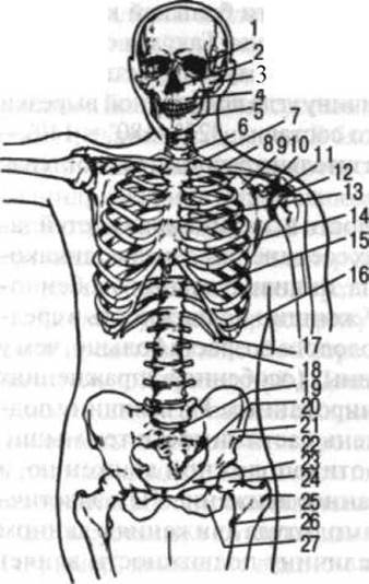
Fig. 17. Skeleton of the trunk, neck, and HEAD (anterior view):
1 — Orbit; 2 — Nasal cavity; 3 — Zygomatic bone; 4 — Maxilla; 5 — Mandible; 6 — 7th cervical vertebra; 7 — 1st rib; 8 — clavicle; 9 — scapula; 10 — coracoid process; 11 — acromion; 12 — head of humerus; 13 — body of Sternum; 14 — xiphisternum; 15 — costal Cartilage; 16 — bony part of rib; 17 — 12th rib; 18 — body of 1st lumbar vertebra; 19 — sacrum; 20 — coccyx; 21 — iliac fossa (ala of ilium); 22 — anterior superior iliac spine; 21 — pubic bone; 24 — head of Femur; 25 — greater trochanter; 26 — lesser trochanter; 27 — ischium
Vertebral column
The vertebral column serves as the primary rigid support of the trunk. It consists of 33–34 vertebrae and their interconnections.
The vertebral column is divided into 5 regions: the cervical region, consisting of 7 cervical vertebrae; the thoracic region, consisting of 12 thoracic vertebrae; the lumbar region, consisting of 5 lumbar vertebrae; the sacral region, consisting of 5 sacral vertebrae; and the coccygeal region, consisting of 4 or 5 coccygeal vertebrae. The sacral and coccygeal vertebrae fuse together to form distinct bones — the sacrum and the coccyx.
Vertebrae are classified as short spongy bones. Each vertebra features a body directed anteriorly and an arch located posteriorly to the body, which together enclose the vertebral foramen (Fig. 18).
The external surface of the vertebral body is covered with a layer of dense cortical bone, while its interior contains spongy bone composed of vertical and horizontal trabeculae arranged almost at right angles to one another. Via their superior and inferior surfaces, the vertebral bodies articulate with adjacent superior and inferior vertebrae through intervertebral discs. The anterior surface of the vertebral body, like its lateral and posterior surfaces, has a somewhat concave shape.

Fig. 18. Thoracic vertebra:
A - lateral view; B - anterior view; C - superior view; D - posterior view. 1 - vertebral body; 2 - articular facets for articulation with the head of the rib; 3 - superior articular process; 4 - articular facet on the transverse process for articulation with the tubercle of the rib; 5 - inferior articular process; 6 - spinous process; 7 - intervertebral notch; 8 - vertebral foramen
The vertebral foramina, stacked one above another, collectively form the vertebral canal, which houses the Spinal Cord and associated structures. At the point of attachment to the body, the vertebral arch features superior and inferior notches. The notches of adjacent superior and inferior vertebrae together form the intervertebral foramen, through which nerves, Blood Vessels, and Lymphatic vessels pass. Seven processes extend from the vertebral arch. The unpaired, posteriorly directed process is called the spinous process. It serves for the attachment of Ligaments and Muscles. The remaining processes are paired. These include the transverse processes, which project laterally from the vertebrae and lie approximately in the frontal plane, as well as the superior and inferior articular processes. The transverse processes serve for Muscle and ligament attachment, while the articular processes articulate with the corresponding processes of adjacent superior and inferior vertebrae.
The dimensions of vertebrae vary across different Regions of the vertebral column, depending on the magnitude of the mechanical load applied to a given region and the degree of muscular development. The greater the load, the larger the vertebrae. The lumbar vertebrae and the first sacral vertebra possess the maximum dimensions, as they bear the weight of the head, neck, trunk, and upper limbs, transmitting it to the lower limbs. The smallest vertebrae are the coccygeal vertebrae, which is attributed to the reduced load acting upon them and the regression of tail musculature.
The vertebrae of different regions of the vertebral column possess specific characteristics that distinguish them from one another.
Cervical vertebrae, starting from the 2nd, feature a small body, a large vertebral foramen, a bifid spinous process (with the exception of the seventh), and foramina in the transverse processes through which blood vessels supplying the Brain AND SPINAL cord pass. The superior surface of the cervical vertebral body is transversely concave, while the inferior surface is anteroposteriorly concave.
The 1st cervical vertebra, the atlas, lacks a body and a spinous process, consisting instead of a ring formed by anterior and posterior arches. On the posterior surface of the anterior arch lies a small articular facet for articulation with the dens of the axis — an unpaired process directed vertically upward.
On its sides, the atlas features massive thickenings known as lateral masses, which bear articular surfaces on their superior and inferior aspects for articulation with the Occipital bone and the 2nd cervical vertebra.
The 2nd cervical vertebra, the axis, features a process called the dens (odontoid process). Flanking the dens — which bears an anterior articular facet for articulation with the anterior arch of the atlas — are the superior articular facets for articulation with the inferior articular surfaces of the atlas.
The transverse processes of the cervical vertebrae bear tubercles; on the 6th vertebra, these are known as the carotid tubercles, named after the common carotid artery that runs anterior to each of them, where it can be compressed to palpate the pulse or stop bleeding. The 7th cervical vertebra, or vertebra prominens, features a well-developed spinous process that projects sharply beneath the Skin. It is easily palpable and serves as a landmark for counting vertebrae. The spinous process of the 2nd cervical vertebra can be palpated within the depression beneath the occipital bone.
Thoracic vertebrae feature articular facets on the lateral surfaces of their bodies for articulation with the heads of the Ribs. The 1st thoracic vertebra possesses a full facet for the 1st rib and a demifacet for the 2nd. The lower thoracic vertebrae, the 11th and 12th, each bear a single complete facet, whereas all other thoracic vertebrae possess two demifacets — one superior and one inferior. Two such demifacets, complementing one another between the bodies of two adjacent vertebrae, form a single depression that articulates with the head of a rib. Furthermore, the transverse processes of the thoracic vertebrae (except for the 11th and 12th) bear articular surfaces for articulation with the ribs. The spinous processes of the thoracic vertebrae are inclined and overlap one another like roof tiles, particularly in the middle section of the thoracic region.
Lumbar vertebrae are distinguished by massive bodies and feature a short, thick spinous process directed horizontally backward. Their articular processes lie approximately in the sagittal plane.
The sacrum is formed by the fusion of 5 sacral vertebrae toward the end of the second decade of life. It is a massive, triangular bone with its base directed upward and its apex downward. Its anterior, or pelvic, surface is concave and faces the pelvic cavity; the posterior, dorsal surface is rough and features crests. The sacrum exhibits lateral parts formed by the fusion of transverse processes, bearing auricular surfaces that articulate with the right and left hip bones. Pelvic and dorsal sacral foramina are located on the pelvic and dorsal surfaces of the sacrum, respectively, allowing the passage of nerves and blood vessels. Between these foramina on the pelvic surface lie transverse lines, which represent the fusion sites of the vertebral bodies. The dorsal surface of the sacrum features the median sacral crest along the midline, formed by the fusion of the spinous processes of the sacral vertebrae, and two lateral crests resulting from the fusion of the transverse processes. Near the Base of the sacrum anteriorly lies the promontory, while posteriorly are the articular processes that articulate with the articular processes of the 5th lumbar vertebra. Passing through the sacrum is the sacral canal, formed by the fusion of the bodies and arches of the sacral vertebrae as a continuation of the vertebral canal.
The coccyx most commonly consists of 4 vertebrae. The 1st is the most distinct, whereas the remaining ones typically present as small, spherical bones corresponding to vertebral bodies. Coccygeal vertebrae possess no other elements.
Palpating the spinous processes of the thoracic and lumbar vertebrae is best done with the spinal column in a flexed position, as this causes them to move slightly apart. When the back muscles and spinal ligaments are well-developed, these processes serve as reliable anatomical landmarks. For instance, the spinous process of the IV lumbar vertebra lies on the line connecting the iliac crests, while the spinous process of the I sacral vertebra is located just above the line passing between the posterior superior iliac spines.
JOINTS OF THE vertebrae. The spinal column features all Types of bone connections: continuous (syndesmoses, synchondroses, and synostoses) and discontinuous (intervertebral joints and joints between the spinal column and the Skull). Connections are categorized into those between vertebral bodies, vertebral arches, and articular processes. Intervertebral discs are situated between the vertebral bodies. Inside each disc lies a Nucleus pulposus, which is a remnant of the notochord; the periphery is enclosed by the anulus fibrosus, consisting of fibrocartilage whose fibers run in horizontal and oblique directions. Due to their elasticity, the nuclei pulposi tend to expand vertically, thereby helping to slightly separate the vertebral bodies. Thanks to the elasticity of the intervertebral discs, the spinal column can effectively cushion the shocks and impacts experienced during various physical activities (such as jumping and running). The intervertebral discs account for 1/4 of the height of the entire movable portion of the spinal column. It is well established that the range of motion between two adjacent vertebrae depends on the height of the intervertebral discs, as well as the transverse and anteroposterior dimensions of the vertebral bodies. Mobility is greater in regions where the discs are thicker, and vice versa. In the lumbar region, the height of each intervertebral disc is approximately 1/3 of the adjacent vertebral body; in the cervical region, it is 1/4; in the upper and lower PARTS OF THE thoracic region, 1/5; and in the middle part of the same region, 1/6. Under pressure, the elastic intervertebral discs expand transversely and anteroposteriorly while slightly decreasing in vertical height; however, once the axial load is removed, they return to their original shape. The anterior and posterior longitudinal ligaments run along the anterior and posterior surfaces of the vertebral bodies and intervertebral discs, respectively.
Very strong ligaments composed of yellow elastic fibers, appropriately called the ligamenta flava, are located between the vertebral arches. During Movements of the spinal column, especially flexion, these ligaments stretch and tense; upon returning to the starting position, they assist the muscles in extending the spine.
The interspinous ligaments are located between the spinous processes of the vertebrae, while the intertransverse ligaments lie between the transverse processes. The supraspinous ligament runs along the entire length of the spinal column over the tips of the spinous processes. As it approaches the skull, it expands in the sagittal direction and is referred to as the ligamentum nuchae.
The articular processes of the vertebrae are connected by joints that are flat in the upper segments of the spinal column and cylindrical in the lower segments, particularly in the lumbar region.
The connection between the atlas and the occipital bone—the atlanto-occipital joint—has distinctive features. It is a combined joint consisting of two anatomically distinct articulations. The articular surfaces of the atlanto-occipital joint are ellipsoid in shape, allowing for movement around two axes: transverse and sagittal.
Three joints between the atlas and the axis (epistropheus) also form a combined atlantoaxial joint with a single vertical axis of rotation. One of these is an unpaired cylindrical joint situated between the dens of the axis and the anterior arch of the atlas, while the other is a paired flat joint located between the inferior articular surface of the atlas and the superior articular surface of the axis.
The two joints (atlanto-occipital and atlantoaxial) located above and below the atlas Complement each other to form a connection that provides head mobility around three mutually perpendicular axes of rotation. The cruciform ligament of the atlas and the alar ligaments participate in reinforcing these joints. Between the atlas and the occipital bone lie two membranes, anterior and posterior, which close the gaps between these bones.
In youth, the sacrococcygeal symphysis features a joint cavity that transforms into a synchondrosis over the years. Movement in this joint occurs primarily in the anteroposterior direction. The range of motion of the tip of the coccyx in women is approximately 2 cm.
The spinal column as a whole. Viewed as a whole, the spinal column resembles two pyramids joined base-to-base.
The upper pyramid is formed by the vertebrae, which increase in size from the upper cervical vertebrae down to the V lumbar vertebra, whereas the lower pyramid consists of the sacral and coccygeal vertebrae, which decrease in size inferiorly.
The spinal column is not straight; it features natural curves: anterior curves known as lordoses, posterior curves known as kyphoses, and lateral curves to the right or left known as scolioses. The thoracic Kyphosis develops the earliest. In newborns, other anteroposterior curves of the spine are barely noticeable. Cervical lordosis appears as the baby begins to hold its head upright, while lumbar lordosis develops when the child starts standing. The spinal curves become clearly defined by the age of 5–6 years and are fully formed by 18–20 years. If an imaginary vertical line is drawn through the body's general center of gravity while a person stands completely upright (in an anthropometric stance), the lumbar lordosis lies approximately 5 cm anterior to this vertical line, the thoracic kyphosis lies about 2.5 cm posterior to it, and the cervical lordosis is situated 1.5 cm anteriorly—factors that accentuate these curves under METABOLISM/18.html">The Influence of gravity. Lordoses, kyphoses, and scolioses are best studied by observing the alignment of the line of spinous processes from posterior and lateral views.
Scolioses typically develop during the early years of a child's schooling due to asymmetrical body positioning, uneven muscle strain, and fatigue from prolonged motionless sitting. However, these curvatures can also manifest in preschool age. Physical Exercise and sports exert a highly beneficial influence on The Development of the spinal column, preventing the onset of poor posture and pathological lateral curvatures, while also serving as a powerful corrective tool for existing defects.
The curves of the spinal column enhance its Shock-absorbing properties as well as the capacity of the thoracic and pelvic cavities. Under external influences, these curves may fluctuate slightly throughout the day. Consequently, the total height of the human spinal column—and by extension, body length—is not constant. Daily fluctuations in body height typically range within 1 cm, though variations of 2–2.5 cm are not uncommon, and cases of daily height fluctuations reaching 4 or even 6 cm have been documented.
In the recumbent position, a person's body is 2–3 cm longer than in the standing position because in the former state, the intervertebral discs undergo slight decompression, whereas in the latter, gravity compresses them; when external compressive forces are absent, the discs expand and spinal curvatures become less pronounced.
The length of the spinal column accounts for approximately 40% of total body length. In men, it is 70–73 cm, and in women, 66–69 cm. Proportionate to total body height, the spinal column is relatively longer in women and children than in men. This is attributable not only to the height of the vertebral bodies but also to the greater relative height of the intervertebral discs. The relative height of the intervertebral discs decreases with age. In newborns, they make up more than 50% of the entire spinal column's height, whereas in adults, this figure is about 25%. Assuming a total spinal column height of 73 cm, the cervical region accounts for 13 cm, the thoracic region for 30 cm, the lumbar region for 18 cm, and the sacrococcygeal region for 12 cm. The spinal column reaches its definitive height by the age of 25.
Most body positions involve a downward axial load on the spinal column directed toward the sacrum, exerting a compressive effect on the intervertebral discs. During a handstand, this pressure is directed oppositely. Conversely, various types of suspension (such as hanging by the hands or knees) as well as support positions (e.g., on parallel bars) subject
the intervertebral discs not to compression, but rather to traction. One might intuitively expect this to increase the length of the thoracic and lumbar regions of the spinal column. However, measurements show that the direct distance between the spinous process of the VII cervical vertebra and the I sacral vertebra during suspension is not only no greater than, but actually less than, that in a normal standing posture. This reduction occurs because suspension and support positions accentuate the lumbar lordosis, which brings the spinous processes—particularly the lumbar ones—closer together. Furthermore, muscle tone plays a significant role by actively pulling the inferior vertebrae toward the superior ones.

Fig. 19. Lateral flexion of the trunk to the left. Graphic reconstruction based on several radiographs, demonstrating the curvature of various sections of the spinal column
Movements of the spinal column can occur around three axes of rotation: transverse (flexion and extension), anteroposterior (lateral bending to the right and left), and vertical (axial rotation or twisting to either side). Additionally, circumduction is possible as a combination of movements around multiple rotational axes.
The most mobile segments of the spinal column are the cervical and lumbar regions (Fig. 19), whereas the upper and lower sections of the thoracic region are less mobile; the middle thoracic section, spanning roughly from the III to the VII vertebra, exhibits very limited mobility. This is because the thoracic vertebrae articulate with the ribs and contribute to the bony Thorax. Furthermore, the spinous processes in this region are more tightly interlocked than in other parts of the spine.
The head possesses three axes of rotation relative to the spinal column: a transverse axis, around which forward nodding (flexion) and backward tilting (extension) occur; a sagittal axis, governing lateral tilts; and a vertical axis, allowing for right and left rotation. Quantified in degrees, this mobility is distributed as follows:
Table 1.
|
Axes of rotation |
Atlanto-occipital articulation |
Atlantoaxial articulation |
Cervical articulations (II–VII cervical vertebrae) |
Total |
|
Transverse: |
||||
|
flexion |
20 |
10 |
90 |
164 |
|
extension |
30 |
14 |
||
|
Anteroposterior: |
||||
|
lateral flexion |
15 |
0 |
30 |
45 |
|
Vertical: |
||||
|
rotation |
0 |
35 |
40 |
75 |
In addition to the ligamentous apparatus and muscles, auxiliary structures that help strengthen the vertebral column include the thoracic and abdominal cavities with their walls and Internal Organs. In individuals with high mobility of the spinal column, stabilization is achieved primarily through the action of muscles.
The Thorax
The thorax is formed by the thoracic region of the Vertebral Column and 12 pairs of ribs, connected anteriorly by the sternum (see Fig. 17).
The ribs consist of a bony part posteriorly and costal cartilage anteriorly. The upper 7 pairs of ribs attach their cartilages directly to the sternum and are called true ribs, while the lower 5 pairs do not reach the sternum and are referred to as false ribs. Among these, the VIII, IX, and often the X ribs attach their cartilages to the cartilage of the rib directly above them, forming the so-called costal arch (see Fig. 17). The lowest ribs, the XI and XII, do not connect to the superior ribs; they are embedded among muscles and are known as floating ribs. In 48% of cases, the X rib is also classified as a floating rib.
The bony part of a rib is a narrow, curved plate representing a typical long spongy bone. It features a central section—the body—and two extremities: posterior and anterior. At the posterior end, the rib has a head for articulation with the vertebral bodies. Anterior and lateral to the head lies a slightly narrowed segment, the neck. This is followed by the tubercle, which articulates with the transverse process of the vertebra. On the XI and XII ribs, these tubercles are absent. The body of each rib presents internal and external surfaces (which correspond to superior and inferior surfaces in the case of the I rib). Along the lower margin of the internal surface runs a groove that houses blood Vessels and nerves. The length of the ribs increases progressively from the I to the VII rib and decreases from the VIII to the XII.
The sternum (see Fig. 17) is an elongated, flat bone located in the anterior chest wall and classified among the long spongy bones. It comprises three parts: the manubrium, the body, and the xiphoid process. The manubrium features several notches: the superior jugular notch is unpaired, whereas the clavicular and costal notches are paired. The body of the sternum widens inferiorly and bears articular surfaces along its lateral margins for articulation with the cartilages of ribs III–VII. The xiphoid process, like the rest of the sternum, is flat. It connects to the sternal body via a synchondrosis, which typically undergoes ossification (synostosis) after the age of 30.
Both the ribs and the sternum are readily palpable. When counting the ribs, one should maintain continuous contact with the skin surface and place the fingertips into the intercostal spaces as each rib is palpated. Because the I rib is largely covered by the clavicle, the latter is palpated in its place, with the thumb positioned in the first intercostal space. Palpable landmarks on the sternum include its lateral borders, the superior border (where the jugular notch is easily identified), and the entire anterior surface of the sternum, including the xiphoid process.
Costosternal joints (Fig. 20). The true ribs (I–VII) articulate with the sternum via costal cartilages. The cartilage of the I rib fuses directly with the sternum, whereas the cartilages of the remaining six ribs (II–VII) articulate with it through small, flat sternocostal joints reinforced by anterior and posterior ligaments. These are known as radiate sternocostal ligaments. Their fibers extend onto the anterior and posterior surfaces of the sternum, blending with its periosteum to help form the Cytology/practical/45.html">Dense Connective Tissue membranes that cover the sternum both anteriorly and posteriorly.
Costovertebral joints. The heads of the ribs articulate with the bodies of the thoracic vertebrae to form the joints of the heads of the ribs, while the costal tubercles articulate with the transverse processes of the vertebrae to form the costotransverse joints.
The joints of the heads of the ribs are reinforced by radiate ligaments, whereas the costotransverse joints are secured by the costotransverse ligament, which spans from the transverse process of the vertebra to the tubercle and neck of the corresponding rib at each joint level.
In addition to this ligament, superior and lateral costotransverse ligaments are present. The former runs from the upper border of the rib neck to the lower border of the adjacent transverse process above, while the latter extends from the posterior surface of the rib neck to the bases of the transverse and spinous processes of the overlying vertebra.

Fig. 20. Articulations of the clavicle and ribs with the sternum:
1 — clavicle; 2 — costoclavicular ligament; 3 — radiate sternocostal ligament; 4 — costal arch; 5 — costoxiphoid ligament; 6 — sternocostal joint; 7 — bony part of the rib; 8 — costal cartilage; 9 — opened sternoclavicular joint containing the articular disc; 10 — interclavicular ligament
The joint of the head of the rib (plane joint) and the costotransverse joint (cylindrical joint) together constitute a combined articulation. The axis of rotation for this joint system passes approximately along the axis of the respective rib's neck. The backward extensions of the rotation axes for the right and left combined joints intersect at an angle that is obtuse in the upper ribs (especially the I rib joint) and acute in the lower ribs. Consequently, upon elevation, the upper ribs move predominantly forward, whereas the lower ribs move laterally.
The thoracic cavity presents superior and inferior thoracic apertures and 11 pairs of intercostal spaces (intercostalia). The superior aperture is bounded by the anterior surface of the body of the first thoracic vertebra, the first ribs, and the jugular notch of the sternum, while the inferior aperture is bounded by the body of the twelfth vertebra, the twelfth ribs, costal arches, and the xiphoid process. The shapes of both superior and inferior apertures may be rounded or anteroposteriorly flattened.
The Thorax as a Whole. The primary shapes of the thorax are cylindrical, conical, and flat. In addition, wide and short, as well as long and narrow variants are distinguished. A conical thorax is typically short, whereas a flat thorax is usually long. Transitional forms also occur.
The angle formed between the right and left costal arches is termed the infrasternal angle. This angle is relatively wide in individuals with a broad and short thorax and narrow in those with a slender and long thorax. In women, the thorax is generally somewhat shorter and narrower than in men.
The shape of The thoracic cage is closely related to the Morphology and topographical positioning of the internal organs. In a narrow and long thorax, The Heart is frequently vertically oriented ("drop-like heart"), and the aortic arch sits low; conversely, in a wide and short thorax, the heart lies more horizontally ("recumbent heart"), and the aortic arch is less curved and relatively high, occasionally reaching the level of the upper margin of the manubrium sterni.
The conformation of the thorax can be evaluated using three dimensions: vertical, transverse, and anteroposterior. The vertical dimension is measured with an anthropometer as the shortest distance between two reference levels—the superior border of the sternum and the anterior margin of the tenth ribs. Transverse and anteroposterior diameters are determined using a spreading caliper. The most precise assessment of thoracic dimensions in living subjects is achieved through radiological imaging.
Physical exercise exerts a profound influence on the shape and, even more so, the mobility of the thorax. According to A.A. Gladysheva, training in swimming, cross-country skiing, and wrestling leads to an increase in both thoracic dimensions and mobility in all directions. Gymnastic training primarily enhances the mobility of the lower thorax while producing little change in its overall size, whereas in cyclists and speed skaters, both the dimensions and the mobility of this region are reduced.
Last update: 08/08/2026
Editorial and Educational Adaptation: This material has been compiled based on the primary/original source text. The project team performed an editorial review, corrected technical inaccuracies, structured sections, and adapted the content for an educational format.
What was processed:
- elimination of formatting defects (OCR errors, structural breaks, corrupted characters);
- editorial organization of content;
- standardization of terminology in accordance with academic sources;
- verification of factual statements against the original source text.
All mentions of the author, publication year, and origin of the primary text have been preserved in accordance with the source.