Human Anatomy (with the Foundations of Dynamic and Sports Morphology) - Ivanitsky M. F. 2008
The Doctrine of Bones and Their Articulations
General Part
The solid support of The Human Body is the Skeleton, which consists of bones and their articulations. The skeleton protects deeper anatomical structures from injury (such as the Bone Marrow, the Central Nervous system, The Heart, and certain visceral Organs including the Lungs and pelvic organs). Bone movement is made possible by the action of skeletal Muscles Attached to them. In addition to their supportive, protective, and locomotor Functions, skeletal bones play a vital role in Mineral METABOLISM and hematopoiesis. Specifically, bones contain the body's primary mineral reserves (such as Calcium and phosphorus), storing them when in excess and mobilizing them as needed. The bone marrow located within the bones participates in The formation of Blood cellular elements.
Each bone comprising the skeleton is an individual organ constructed primarily of osseous tissue, while also containing other tissue types: dense Fibrous Connective Tissue forming the periosteum, Cartilage covering the articular surfaces of the bones, and Nervous Tissue.
The human skeleton consists of 206 bones—85 paired (170 in total) and 36 unpaired. In adult males, they account for 18% of total body weight, in females 16%, and in newborns 14%. Because Bone tissue undergoes a degree of dehydration with age, the specific gravity of the bones themselves increases over time.
Bone Structure. Cross-sections of bones reveal that they are fundamentally composed of compact and spongy bone tissue, which are built from individual bone lamellae. Within each lamella, the fibers run parallel to one another and are oriented in a single defined direction. In adjacent lamellae, the fiber orientation is nearly perpendicular, which imparts high structural strength to lamellar bone tissue.
In most bones, these lamellae form osteons. Each osteon consists of 5 to 20 concentrically arranged bone lamellae, resembling cylinders nested within one another. At the center of the osteon lies a cavity—the osteon canal—which contains blood Vessels and nerves. The caliber of osteon canals varies from 1/5 to 1/50 of a millimeter, depending on the size of the bone and the canal's position relative to the outer surface. Larger bones possess
wider bone canals. Superficially located canals are generally wider than those situated more deeply. Encased within special cavities in the walls of these cylindrical lamellae are bone Cells known as osteocytes. The spaces between adjacent osteons are filled by irregularly shaped interstitial lamellae.
Spongy bone substance is constructed from bone trabeculae that are arranged in a specific orientation and interconnect to form intricate networks. Each bone exhibits mechanically determined structural features: the course of its lamellae and trabeculae corresponds to the lines of compressive stress within that particular region of the skeleton (Fig. 13). The directional alignment of bone lamellae in two adjacent bones acts as a continuous trajectory interrupted only by joints. Notably, in skeletal complexes such as the FOOT, where each arch is composed of several independent bones, the overall orientation of the bone lamellae forms an arch-like configuration. A similar phenomenon can be observed in other areas of the skeleton. The spaces between the trabeculae of the spongy substance are filled with bone marrow.
Externally, bones are covered by the periosteum. This is a connective tissue membrane consisting of two layers: an outer fibrous layer and an inner osteogenic (bone-forming) layer. The periosteum is rich in Blood Vessels and nerves that send branches deep into the bone. Due to the presence of sensory nerve endings in the periosteum, bones are sensitive and painful when bruised. The periosteum performs protective, trophic (nutritive), neuroregulatory, and osteogenic functions. The cells of its inner layer are responsible for bone growth in thickness and the healing of fractures.
Class="center">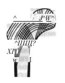
Fig. 13. Diagram showing the arrangement of bone lamellae and trabeculae of the spongy substance in the proximal Femur.
Inside the bone lies the bone marrow, which exists in two forms: red and yellow. Cytology/practical/86.html">Red bone marrow is a hematopoietic organ. It produces red Blood Cells and all granular forms of white blood cells. In newborns, only red bone marrow is present. In adults, it is located within the spongy substance of bones (in the epiphyses of long bones, vertebrae, Ribs, Sternum, pelvic bones, and cranial bones). Yellow bone marrow is found in the medullary cavities of the diaphyses of long bones and is rich in adipose cells.
Bone Shape. Bone Morphology is diverse and determined not only by inherited genetic traits but also by functional demands, external influences (such as the pull of attached muscles, the force of gravity acting on the bones, nutritional conditions, etc.). In areas where muscles attach to bones, roughness, tuberosities, and prominences develop. The stronger the muscles and ligaments attached to a given process or tuberosity, the more pronounced these features generally are. Consequently, the skeleton provides a reliable indicator of ligamentous robustness and Muscle strength in a given individual. In adults, these rough surfaces and bony prominences are better developed than in children, and more so in men than in women. Based on their structure, function, and development, bones are classified into tubular, spongy, and mixed (Fig. 14). Tubular bones are further subdivided into long and short, while spongy bones are classified as long, short, flat, and sesamoid.
Tubular bones are predominantly found in the Appendicular Skeleton (limbs), facilitating wide-amplitude movements.
A characteristic feature of tubular bones is that each possesses a central shaft, or diaphysis, containing a medullary cavity, and two expanded extremities, or epiphyses: a proximal epiphysis located closer to the trunk, and a distal epiphysis located farther from it. The region of the bone between the diaphysis and epiphysis is called the metaphysis. The ends of the bone feature articular surfaces, typically covered in unprocessed bone by hyaline cartilage (more rarely by fibrocartilage), which serve for articulation with neighboring bones. The compact bone layer in tubular bones is particularly well-developed in the diaphyseal region. Spongy substance is located internally, mainly within the proximal and distal epiphyses, where it is covered by only a thin shell of compact bone. The walls of the medullary cavity are formed by compact substance. In the Superficial layer of compact bone just beneath the periosteum lie the outer circumferential lamellae, while the inner layer facing the medullary cavity contains the inner circumferential lamellae, which encircle the marrow cavity like a sleeve; a layer of osteons lies between these circumferential systems (see Fig. 7). Spongy bones consist primarily of spongy substance enclosed by a thin layer of compact bone.
In flat bones, the spongy substance forms merely a thin layer (diplöe) sandwiched between the inner and outer plates of compact bone. Numerous Veins course through the spongy substance of the flat BONES OF THE Skull, where it is specifically referred to as diploë.
In the protective bones of the cranium, the inner plate (facing the cranial cavity) is thin, dense, and brittle. During cranial trauma, it may fracture and produce sharp bone fragments that can easily lacerate blood vessels and cause intracranial Hemorrhage, even when the outer surface of the bone appears undamaged.
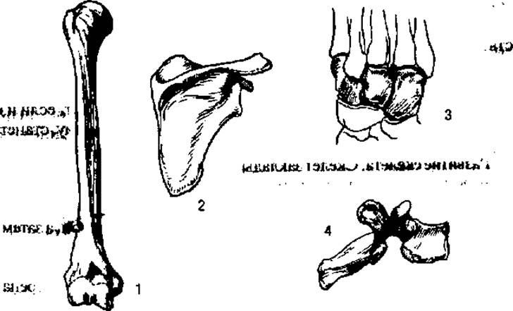
Fig. 14. Bones of various shapes:
1 - long bone (humerus); 2 - flat bone (scapula); 3 - short bones of the tarsus/metatarsus; 4 - irregular bone (vertebra)
Mixed bones consist of fused components that vary in shape and structure.
Chemical composition of Bones. Fresh bone from a living human or cadaver consists of 50% Water, with the remaining fraction comprising organic (12.4%) and inorganic (21.85%) substances. Bones used in anatomy practicals have a different composition due to preliminary preparation (maceration, degreasing, drying, and bleaching). In these prepared bones, organic matter accounts for about 1/3, and inorganic matter for roughly 2/3 of the total mass.
The organic component of bone is ossein, while the Inorganic Components are lime salts (calcium phosphate and calcium carbonate), along with sodium chloride.
The relative proportions of bone constituents vary among individuals and can change even within the same person depending on age, nutritional status, physical activity, and other factors. In childhood, the relative proportion of organic matter in bones is higher, endowing them with greater flexibility and lower rigidity; with advanced age, the relative amount of ossein decreases, leading to increased bone brittleness.
The organic and inorganic constituents of bone form a chemical compound with one another, making it impossible to distinguish ossein particles from lime salts under a Microscope, even at high magnification. However, Treatment with an acid (such as hydrochloric or nitric acid) can dissolve the lime salts—a process known as decalcification, which frees the bone from calcium salts. After soaking in an acid solution, the bone retains its shape while losing its rigidity, becoming so soft and flexible that it can be tied into a knot. Conversely, if ossein is removed from a bone by calcination (burning), the bone retains its form but becomes extremely brittle, crumbling into powder under pressure.
Development of the skeleton. The skeleton is initially laid down as condensed embryonic connective tissue—mesenchyme. At this early stage of development, the skeleton is not yet divided into separate regions. Later, the greater part of the skeleton becomes cartilaginous, and subsequently cartilage is replaced by bone tissue. This is how the Skeleton of the Trunk, limbs, and cranial base develops. These are referred to as secondary bones. At the same time, the bones of the cranial vault, certain facial bones, and partly the clavicle, which are primary bones, develop directly from the mesenchymal primordium; that is, they bypass the cartilaginous stage entirely, with the membranous stage transitioning directly into bone.
Bone Development and growth involve processes of both bone matrix formation and resorption. The deposition of bone tissue is carried out by specialized cells called osteoblasts, which rapidly proliferate, become surrounded by the bone matrix, and transform into mature bone cells, or osteocytes. Bone resorption is performed by large, multinucleated cells known as osteoclasts.
Osteogenesis on a connective tissue basis occurs through desmal ossification: the appearance of ossification centers within the central PARTS OF THE future bone. Most bones develop not just from one, but from several such ossification centers.
The development and growth of bones from a cartilaginous model take place via so-called perichondral and endochondral ossification. The former begins externally from the periosteum, where osteoblasts—Cells of the periosteum—synthesize bone tissue; the latter occurs inside the cartilaginous model of the future bone, where an ossification center emerges, leading to cartilage resorption and its replacement by trabeculae formed of bone tissue. The growth of a bone in thickness is accompanied not only by the deposition of bone tissue on the outside, but also by its resorption along the inner bone cavity, performed by osteoclasts.
The longitudinal growth of long tubular bones occurs due to Changes in the epiphyseal cartilages—those cartilaginous layers or growth plates located between the diaphysis and epiphyses. These bones grow in length from both ends, making them diepiphyseal. Short tubular bones (Phalanges of the fingers, metacarpals, metatarsals) grow in length through the epiphyseal cartilage at one end only, meaning they are monoepiphyseal. Although of lesser significance, the articular cartilage covering the epiphyses within the joint also serves as a zone of longitudinal growth. Following the Ossification of the epiphyseal cartilage at the end of the second decade of life, changes in the articular cartilage can only insignificantly increase bone length.
In humans, many bones are initially laid down and develop in several parts. Subsequently, these fuse to form a single monolithic bone. For instance, the hip bone develops from three major parts, each of which is initially referred to as a separate bone. By age 14–16, they fuse into a single continuous bone. During the growth process, tubular bones consist of three main parts, excluding individual ossification centers located at bony prominences. The diaphyses of bones begin to ossify before birth, whereas the epiphyses ossify afterward. Exceptions are the distal epiphysis of the femur and the proximal epiphysis of the Tibia, where ossification centers appear by the time of birth (their presence serves as an indicator of fetal full-term maturity). The fusion of all these parts occurs at various times, concluding by 16–18 years of age.
Short bones ossify in a manner similar to that observed in the epiphyses of tubular bones.
Flat bones grow either desmally (the skull) or endochondrally (the pelvis). After growth has ceased, bone fusion may occur. For example, the sutures between individual bones of the cranial vault obliterate with age, and in elderly individuals, this region typically appears as a single continuous monolithic bone. Growth in thickness occurs through apposition (deposition) of bone tissue On the surface of the bone.
Bone is a highly plastic tissue characterized by continuous processes of growth, metabolism, and remodeling regulated by the nervous and endocrine systems. In areas where a bone receives a greater and richer blood supply, it develops more rapidly. Observations of Postnatal bone development demonstrate that ossification proceeds more quickly in areas subjected to higher mechanical pressure compared to areas with lower pressure—for instance, in the lower limb faster than in the upper limb, and in the lower vertebrae compared to the upper ones. The growth intensity, dimensions, and specific surface relief of bones depend on mechanical loads. In the 1970s of the 19th century, P.F. Lesgaft formulated the rule stating that bone growth is determined by The activity of the surrounding muscles. Taking modern data into account, the functional laws of bone growth can be refined as follows:
a) mechanical loads that stimulate bone growth must be rhythmic in nature;
b) the activation of bone growth occurs at an optimal level of loading; insufficient or excessive loading inhibits their growth;
c) the response of a growing bone to mechanical loads is determined (among other factors) by the individual, unique CHARACTERISTICS OF THE physiological reaction norm to the load.
Engaging in Physical Exercise helps improve such mechanical properties of bone as resistance to fracture, bending, compression, tension, and torsion. In areas subject to maximum loading, the compact layer thickens, and the direction and architecture of the bone trabeculae change. The cancellous (spongy) bone tissue becomes coarser-meshed.
The Study of Bone Joints
Connections between bones are subdivided into continuous and discontinuous joints; continuous joints are further categorized into fibrous, cartilaginous, and bony joints. Discontinuous joints are referred to as synovial joints or articulations.
Fibrous joints (syndesmoses) include interosseous ligaments, interosseous membranes, and sutures.
Interosseous ligaments serve to reinforce various Types of bone connections (particularly stabilizing joints). They are connective tissue bands whose tensile strength is due to the fact that their constituent fibers do not run parallel, but rather intersect in a crossed and oblique course. Some of these ligaments can withstand very high tensile loads—exceeding 100 kg. They are composed primarily of Collagen fibers; however, some ligaments contain a significant amount of elastic fibers, rendering them less strong but more extensible.
Interosseous membranes are broad connective tissue sheets that connect adjacent bones over a considerable distance, such as the bones of the forearm (Fig. 15) or leg, and close certain bony apertures, such as the obturator foramen of the hip bone. They increase the surface area available for muscle attachment.
Sutures represent a narrow layer of connective tissue situated between two adjacent bones. They are characteristic of the skull bones. It is customary to distinguish serrate, squamous, and plane sutures (see Fig. 15). A serrate suture is formed by interlocking projections and indentations along the edges of one articulating cranial bone that fit into corresponding indentations and projections on the edge of the other bone (for example, the sagittal suture between the two parietal bones). A squamous suture features the overlapping of the beveled edge of one bone over the beveled edge of another, resembling fish scales or roof tiles (for example, the articulation between the temporal and parietal bones). A plane suture is characterized by the direct apposition of flat bone margins without the formation of interlocking processes (for example, the frontonasal suture).
2. Cartilaginous joints are subdivided into true cartilaginous joints, or synchondroses, and symphyses (symphyseal junctions).
Synchondroses are interposing layers of cartilage between bones. They possess significant strength and elasticity, enabling them to also function as Shock absorbers. The mobility of this type of joint is relatively limited and depends on the thickness of the cartilaginous layer. Examples of synchondroses include the intervertebral discs (see Fig. 15) between the vertebral bodies. These discs are constructed of fibrocartilage. Their elasticity is further enhanced by the gelatinous nuclei located within the central portion of the intervertebral discs.
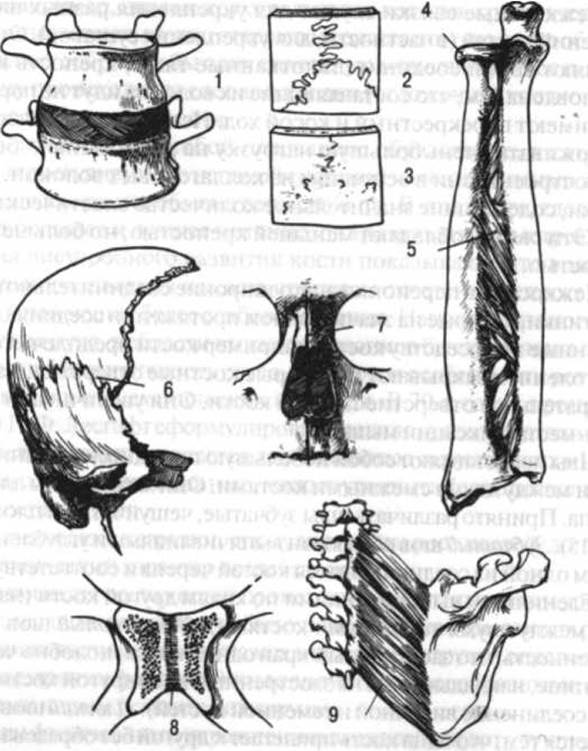
Fig. 15. Continuous bone connections:
1 - intervertebral disc (cartilaginous joint); 2 - serrate suture (a type of fibrous joint); 3 - the same suture undergoing obliteration (synostosis); 4 - annular ligament (fibrous joint); 5 - interosseous membrane (fibrous joint); 6 - squamous suture; 7 - plane suture; 8 - symphysis; 9 - connection of bones via muscle—syssarcosis
In regions where the cartilage experiences not only compressive forces but also intermittent tensile forces, a narrow slit-like cavity is present. In humans, an example of such a joint is the Pubic Symphysis (see Fig. 15).
Fibrous and cartilaginous joints are continuous in the sense that there is no gap or space between the connecting bones. Likewise, there is no gap between bones joined together by means of bone or Muscle tissue. These are also classified as continuous joints (see Fig. 15). The former are referred to as synarthroses (or synostoses), and the latter as synsarcosses.
Bony joints (synostoses) develop through the ossification of cranial sutures, the cartilaginous tissue between sacral vertebrae, epiphyseal cartilage, within the hip bone, and in other skeletal structures.
4. Synovial joints, or articulationes, are discontinuous joints characterized by A number of distinct features. Every joint comprises primary elements and accessory structures, also known as the auxiliary apparatus.
The primary elements of a joint include: the articular surfaces of the connecting bones, the articular capsule, the joint cavity, and the synovial fluid.
The articulating bone surfaces are covered with a layer of hyaline (or, more rarely, fibrous) cartilage. Its smooth surface facing the joint cavity facilitates the movement of one bone relative to another. The elasticity of the cartilage helps cushion the impacts and shocks that articulating bones experience during walking, jumping, and other movements. Furthermore, due to its elastic properties and ability to deform, the cartilage increases joint mobility and ensures the lubrication of articular surfaces under pressure. Microscopic Structural Features of the articular cartilage play a specific role in this process. Its surface facing the joint cavity features microscopic irregularities: 1st-order bends approximately 1000 µm in length, and 2nd-order bends around 50 µm. Under mechanical load, these irregularities flatten out, smoothing the articular surface (1st-order bends flatten at a specific pressure of 3.5 kg/cm2, and 2nd-order bends at 20 kg/cm2). Initially, only the marginal projections of the wavy surface are compressed, resulting in a relatively lower pressure deep within the cartilage. Part of the synovial fluid is thus displaced into these deeper zones. A highly viscous fluid containing hyaluronic acid remains between the contacting surfaces of the cartilage-covered bone ends, enabling the joint to function even under heavy compression of the articulating surfaces, despite increased friction. As the pressure on the cartilage decreases, the fluid from its deeper layers flows back into the joint cavity, and the coefficient of friction between the articular surfaces drops.
Cartilage surfaces are typically congruent, meaning their shapes correspond to one another: if one bone features a convexity, the apposing bone features a concavity. The heads of long bones are covered with thicker hyaline cartilage in their central, most prominent part, and thinner cartilage toward the periphery. Conversely, in the corresponding articular fossae, the cartilage is thinner in the center and thicker at the margins.
The articular capsule, or Joint Capsule, consists of two layers: an outer fibrous layer and an inner synovial layer, from which synovial joints derive their name.
The fibrous layer of the articular capsule represents the continuation of the periosteum of one articulating bone into the periosteum of the other. The bundles of fibers within this layer run in various directions: the more superficial ones longitudinally, and the deeper ones transversely.
The synovial layer is formed of loose connective tissue and extends as far as the articular cartilages. Its inner surface, facing the joint cavity, is smooth, shiny, and lined with a layer of endothelial cells.
The thickness of the articular capsule varies. Generally, it is thicker in areas not covered by muscles and thinner elsewhere. The joint cavity is a slit-like space bounded by the articulating bone surfaces and the capsule, and filled with synovial fluid produced by the endothelial (synovial) layer of the capsule.
Accessory joint structures include synovial folds and villi, intra-articular discs, menisci and labra, as well as ligaments. Synovial folds are outgrowths of the capsule's synovial layer filled with adipose tissue; they occupy free spaces within the joint when articulating surfaces are incongruent and act as shock absorbers. Villi are abundant on the inner surface of the synovial layer and serve as the site of production and resorption of synovial fluid.
Intra-articular discs are plate-like cartilaginous structures located inside the joint cavity that divide it into two compartments, thereby enhancing joint mobility. Unlike discs, menisci are incomplete structures featuring a central opening; their outer margin is thickened and fused with the joint capsule, whereas the inner, sharp edge remains free. Menisci improve bone congruence, cushion shocks and impacts, and facilitate a wider range of movements. Articular labra (rims) are composed of fibrous cartilage and attach along the margins of articular fossae, increasing the contact area between articulating bones and promoting a more even distribution of pressure.
Joint stability is maintained by the following factors.
1. Tension of accessory ligaments. The ligamentous apparatus varies significantly among joints. In some cases, ligaments appear as thickened Regions of the joint capsule (e.g., the iliofemoral ligament); in others, they lie at some distance from the capsule (e.g., the sacrospinous and sacrotuberous ligaments); and in still others, they are located intra-articularly (e.g., the cruciate ligaments of the knee). While stabilizing joints, ligaments simultaneously act as brakes limiting excessive bone mobility. Systematic exercise can increase the elasticity of the ligamentous apparatus and the range of motion within the joint.
2. Muscle tension from muscles crossing the joint. This is particularly true for highly mobile joints (such as the shoulder joint), where a loose capsule cannot play a significant stabilizing role.
3. Atmospheric pressure. This also plays a vital role in keeping articulating surfaces in contact. For instance, if the soft tissues around the hip joint of a suspended cadaver are severed without damaging the capsule, atmospheric pressure alone is sufficient to keep the articular surfaces in apposition, even though the weight of the lower limb tends to pull them apart. However, if the joint capsule is also damaged, air enters the cavity, resulting in immediate Separation of the surfaces.
4. Molecular adhesion between articular surfaces. In joints where closely apposed bone surfaces correspond perfectly and share identical radii of curvature (congruent joints, such as the hip joint), molecular attraction forces keep the surfaces together. The synovial fluid also exerts a cohesive sealing effect.
Joint shape and Classification. The degree of mobility in a given joint depends on its structural features, primarily the shape of the articulating bone surfaces. Joints are conventionally classified according to their shape (Fig. 16).
Fig. 16. Types of synovial joints categorized by shape and the number of mutually perpendicular axes of rotation:
1 - ball-and-socket (shoulder), triaxial; 2 - cotyloid / nut-shaped (hip), triaxial; 3 - ellipsoid (wrist), biaxial; 4 - saddle (carpometacarpal joint of the thumb), biaxial; 5 - hinge (interphalangeal), uniaxial; 5a - compound (elbow), comprising three joints (a - humeroradial, ball-and-socket; b - humeroulnar, hinge, uniaxial; c - proximal radioulnar, pivot, uniaxial); 6 - combined (proximal and distal radioulnar), uniaxial pivot joints; 7 - plane (intertarsal joints: a - navicular bone, b - medial cuneiform bone, c - intermediate cuneiform bone, d - lateral cuneiform bone, e - cuboid bone, f - Metatarsal Bones); 8 - trochoid / pivot-like hinge (talocrural / ankle), uniaxial (functionally acts as a hinge joint)
Ball-and-socket joints are the most mobile. They possess an infinite number of axes of rotation passing through the center of the bone HEAD, among which three mutually perpendicular axes are classically distinguished: 1) transverse, or frontal; 2) anteroposterior, or sagittal; and 3) vertical, or longitudinal.
Movement around the transverse axis permits flexion and extension in the limbs, and forward and backward bending in the trunk and head; around the anteroposterior axis—abduction and adduction in the limbs, and lateral bending in the trunk and head; and around the vertical axis—internal and external rotation (pronation and supination) in the limbs, and rotational movements in the trunk and head (collectively termed rotation). Additionally, ball-and-socket joints permit a specialized movement known as circumduction.
A classic example of a ball-and-socket joint is the shoulder joint.
Not all ball-and-socket joints allow movement around all three axes. For example, the metacarpophalangeal joints permit movement only around the transverse and anteroposterior axes, whereas active rotation around the vertical axis is precluded due to the absence of the requisite musculature and the restrictive tension of stabilizing ligaments.
Multi-axial joints include the ball-and-socket (or cotyloid) joint, in which the head of a bone fits deeply into the articular cavity. Movements in this joint are similar to those in a spheroidal joint, although their range of motion is considerably smaller. A prime example of a ball-and-socket joint is the hip joint.
Ellipsoid joints feature two axes of rotation—transverse and anteroposterior. They allow for flexion and extension, abduction and adduction, as well as circumduction. Rotation inward or outward is not possible. In certain joints, such as the radiocarpal joint, a slight passive rotation can occur due to the Elastic properties of the articular cartilage.
Saddle joints are also classified as biaxial. The articular surfaces of the bones meeting in these joints somewhat resemble the shape of a saddle. Besides adduction, abduction, flexion, and extension, these joints also permit circumduction. A classic example of a saddle joint is the carpometacarpal joint of the thumb. When discussing this joint, the terms "opposition" and "reposition" are used instead of "flexion" and "extension". Another biaxial type is the condyloid joint, which serves as an intermediate form between ellipsoid and ginglymus joints. The knee joint is a typical example.
Hinge (ginglymus) and pivot joints are classified as Uniaxial joints. Pure hinge joints are found, for example, between the phalanges of the fingers. Hinge joints operate around a single frontal axis of rotation, which permits flexion and extension. Pivot joints have articular surfaces shaped like a segment of a cylinder. These joints allow for inward and outward rotation around a vertical axis (as in the radioulnar joint) or side-to-side rotation (as in the atlantoaxial joint).
Plane joints are characterized by articular surfaces that form segments of a sphere with a large radius and minimal curvature. Movements in these joints are restricted to a slight gliding of one articular surface against another, partly facilitated by the deformation of the articular cartilage. Examples of plane joints include the interconnections between many of the carpal or Tarsal Bones.
Some joints feature movements that are functionally interdependent. For instance, movement in one temporomandibular joint cannot occur without simultaneous movement in the contralateral joint. Such paired joints are collectively referred to as a combined joint.
Joints containing intra-articular discs essentially consist of two joint cavities and are termed bicompartmental joints (e.g., the sternoclavicular and temporomandibular joints). Joints formed by the interaction of only two bones are called simple joints, whereas those involving three or more bones are classified as compound joints. An example of the former is the interphalangeal joint, while the elbow and radiocarpal joints serve as examples of the latter.
The degree of mobility in a joint depends on the congruence of its articulating surfaces (in terms of surface area). The greater the congruence, the lower the mobility, and vice versa. For example, the articular surface of the humeral head is significantly larger than the glenoid cavity of the scapula, making the shoulder joint one of the most mobile in the body. In plane joints (such as the intercuneiform JOINTS OF THE tarsus), the articulating surfaces are completely congruent, resulting in virtually negligible mobility.
The maximum angle of flexion, extension, abduction, and adduction in a given joint can be roughly estimated by subtracting the angle of the articular surface with the lesser curvature from the angle of the articular surface with the greater curvature. For instance, to determine the mobility of the humeroulnar joint, one subtracts the angular magnitude of the trochlear notch of the ulna from the angular magnitude of the humeral trochlea. In this case, 320° − 180° = 140°, representing the approximate range of motion available to the ulna relative to the humerus.
Thus, the degree of mobility in Bone Articulations depends on their structural characteristics. It varies among individuals depending on age, sex, personal traits, and level of physical conditioning. On average, mobility is greater in women than in men, in younger individuals than in older adults, and in trained athletes (especially those engaging in flexibility training) compared to untrained persons. The range of motion is also influenced by the extensibility of antagonistic muscles located opposite the direction of movement, as well as the strength of the prime movers. The more elastic the former and the stronger the latter, the greater the range of motion in the joint, and vice versa. Ambient Temperature also affects mobility: movements typically have a smaller range in a cold environment than in a warm one. Even the time of day impacts joint mobility, being lower in the morning than in the evening.
Questions regarding the potential for increasing joint mobility ("flexibility") through specialized training and the factors determining this mobility have been extensively explored in the works of researchers such as N.G. Ozolin, G.G. Topolyan, L.E. Lebedyanskaya, and E.D. Gevlich. According to E.D. Gevlich, the amplitude of joint movement is inversely proportional to the magnitude of applied strength loads. However, a decrease in joint range of motion caused by strength training is not absolutely inevitable; it can be prevented by properly combining strength exercises with stretching exercises for the same muscle groups.
Last update: 08/08/2026
Editorial and Educational Adaptation: This material has been compiled based on the primary/original source text. The project team performed an editorial review, corrected technical inaccuracies, structured sections, and adapted the content for an educational format.
What was processed:
- elimination of formatting defects (OCR errors, structural breaks, corrupted characters);
- editorial organization of content;
- standardization of terminology in accordance with academic sources;
- verification of factual statements against the original source text.
All mentions of the author, publication year, and origin of the primary text have been preserved in accordance with the source.