MEDICAL BIOLOGY, HUMAN ANATOMY, PHYSIOLOGY AND PATHOLOGY - Ya.I. Fedoniuk 2010
ANATOMY, PHYSIOLOGY, PATHOLOGY
CHAPTER 2. MUSCULOSKELETAL SYSTEM (THE MECHANISM OF MOVEMENT)
THE DOCTRINE OF BONES AND THEIR JOINTS (OSTEOLOGY AND ARTHROSYNDESMOLOGY). GENERAL OSTEOLOGY AND ARTHROSYNDESMOLOGY DATA
Osteology is The Study of bones.
The Skeleton (sceleton) is a complex of dense structures that develop from mesenchyme. The entire human skeleton consists of the BONES OF THE HEAD, trunk, upper and lower extremities (Fig. 2.2).
Class="center">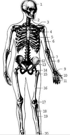
Fig. 2.2. Human skeleton; anterior view.
The skeleton performs the following Functions:
1) mechanical support (due to the attachment of soft Tissues to various PARTS OF THE skeleton);
2) movement (owing to the lever-like Structure OF THE bones);
3) protection (by forming bony canals and cavities such as the Skull, rib cage, and pelvis);
4) metabolic (especially Mineral METABOLISM, as bone serves as a reservoir for mineral salts, including phosphorus, potassium, calcium, iron, etc.);
5) hemopoietic (Bone Marrow located within the bones);
6) diagnostic (utilizing radioactive isotopes and X-rays).
Classification of Bones (Fig. 2.3):
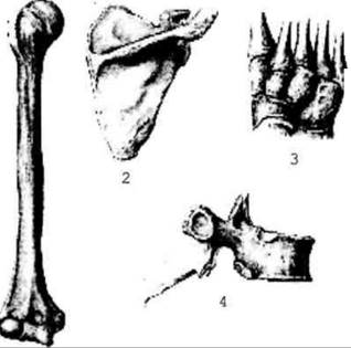
Fig. 2.3. Shapes of bones
1. Long bones feature a shaft (diaphysis) and extremities (proximal and distal epiphyses). Between the diaphysis and epiphysis lies the "growth zone" or metaphysis, which drives longitudinal bone growth.
2. Short bones, for example, the Phalanges of the fingers.
3. Flat bones are broad and serve a protective function (for instance, the scapula).
4. Irregular (mixed) bones combine elements of flat and spongy bones (e.g., vertebrae).
5. Pneumatic bones contain air-filled cavities (such as the Maxilla).
6. Sesamoid bones (for example, the Patella).
Bone as an organ. A bone (os) as an organ consists of various
tissues: the main one being proper Bone tissue, as well as Cartilage, hematopoietic, and adipose tissue, which are penetrated by Blood Vessels and nerves. Bone tissue consists of cellular elements and intercellular substance (see Chapter 2). Among all the Tissues of the body, the intercellular
substance of bone is distinguished by a high content of inorganic substances, which provides the mechanical strength of the skeleton. Degreased, bleached, and dried bone (macerated) consists of 1/3 organic substances, known as ossein, and 2/3 inorganic substances (macro- and microelements). In a living Organism, bone contains up to 50% Water, 28.15% organic substances, including 15.75% fat, and 21.85% inorganic substances, which are represented by compounds of calcium, phosphorus, magnesium, and other elements.
If a bone is placed in acid, the salts dissolve while the organic matter remains. Such a bone can be tied into a knot without breaking. If a bone is burned, the organic matter Burns away while the salts remain, leaving the bone hard but brittle. It is precisely the simultaneous presence of both organic and inorganic substances that imparts strength and elasticity to bones.
Thus, bone strength (mechanical properties) is ensured by the physicochemical unity of organic and inorganic substances, as well as by the architecture of bone tissue.
Each bone consists of compact and spongy bone tissue, The ratio of which varies in different bones and even within the same bone. The universal structural unit of mature bone tissue is the lamella, which forms trabeculae in spongy bone and osteons in compact bone.
An osteon is formed by concentric lamellae that enclose a central canal containing Blood Vessels and nerves (Fig. 2.4). Osteons in compact bone are arranged in an orderly fashion and oriented in accordance with the greatest load applied to the bone.
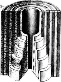
Fig. 2.4. Cross-section of an osteon
Compact bone tissue is always located On the surface of bones, forming a thick layer in the diaphyses of long bones and a thin outer layer in their epiphyses, as well as in spongy and flat bones. Spongy bone tissue is usually located inside the bone. In this tissue, bone lamellae form trabeculae of varying directions that enclose spaces filled with Cytology/practical/86.html">Red bone marrow. Inside long bones, There is a medullary cavity, which is filled with red bone marrow during the prenatal period and in newborns, and is subsequently replaced by yellow bone marrow.
Externally, the bone, with the exception of articular surfaces, is covered by the periosteum. The periosteum consists of a superficial fibrous layer formed by bundles of Collagen fibers and a deep osteogenic layer (containing osteoblasts and osteoclasts). The periosteum, permeated with blood vessels, provides Nutrition to the bone tissue; the cellular elements of the osteogenic layer ensure bone growth in width and its regeneration. The articular surfaces of the bone are covered with articular cartilage. Between the epiphysis and diaphysis of tubular bones lies the epiphyseal cartilage plate (growth plate), through which the bone grows in length.
Arthrosyndesmology is the science of bone Joints and Connections.
Bone connections unite the bones of the skeleton into a single whole. These joints possess a diverse structure and such physical properties as strength, elasticity, and mobility. These properties depend on the specific function of the given connection.
The following Types of bone connections are distinguished (Fig. 2.5).
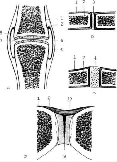
Fig. 2.5. Types of bone connections
1. Continuous joints (synarthroses). There is a layer of Connective Tissue, cartilage, or Muscle between the bones. They are immovable or slightly movable.
2. Discontinuous joints (diarthroses, synovial joints) (articulatio). They are characterized by the presence of a cavity between the bones. Functionally, they are more mobile.
3. Semi-joints (symphyses). This is a transitional form between continuous and discontinuous connections. In this case, there is a cleft within the connective tissue (cartilage) located between the bones.
There are four types of continuous connections:
1. Syndesmosis - when connective tissue remains in the gap between the bones after birth, such as the interosseous membrane in the forearm.
2. Synchondrosis - when the connective tissue in the gap between the bones is replaced by cartilage, such as between the 1st rib and the Sternum.
3. Synostosis - when the connective tissue in the gap between the bones is replaced by bone tissue, such as in the hip bone.
4. Synsarcosis - a connection between bones mediated by striated Muscles, such as the connection of the scapula with the spine and Ribs, or the skull with the Hyoid bone.
Discontinuous connections (synovial joints) are formed only when four necessary components are present, namely: two articular surfaces, a Joint Capsule, synovial fluid, and a joint cavity. Synovial fluid lubricates the articular surfaces to reduce friction during movement, and the capsule tightly encloses the joint cavity.
In addition to these four mandatory components of a joint, some articulations contain additional elements, namely: synovial villi, synovial folds, bursae; fat pads, discs, menisci, intra-articular ligaments, and sesamoid bones.
All these additional elements appear in joints that bear a high functional load.
All joints are classified by the number of articular surfaces, by shape, and by function.
According to the number of articular surfaces, joints are distinguished into:
1. Simple (if two bones participate in The formation of the joint).
For example, the interphalangeal joints.
2. Complex (if more than two bones participate in the Formation of the joint). For example, the elbow joint.
3. Complex joints with intra-articular structures (if there are discs or menisci inside the joint).
For example, the knee joint.
4. Combined (if the joint consists of two anatomically separated joints that function simultaneously). For example, the temporomandibular joint.
According to their function, we distinguish joints as follows:
1. Uniaxial - when movements in the joint are possible around only one axis.
2. Biaxial - when movements in the joint are possible around two axes.
3. Polyaxial - when movements in the joint are possible around three axes.
Depending on their shape, Uniaxial joints can be ginglymus (hinge) and pivot; Biaxial joints include ellipsoidal, condylar, and saddle joints; Polyaxial joints include ball-and-socket, plane, and cotyloid (cup-shaped) joints (Fig. 2.6).
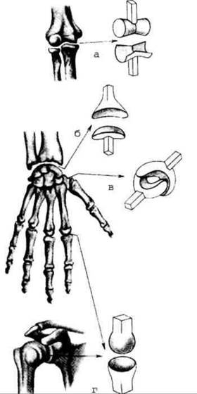
Fig. 2.6. Shapes of articular surfaces
a - hinge; b - ellipsoidal; c - saddle; d - ball-and-socket.
Depending on The structure of the articulating surfaces (shape, size, etc.), movements around various axes are possible in joints. In joint biomechanics, the following axes are distinguished: frontal, sagittal, and vertical.
Movements such as flexion and extension occur around the frontal axis; abduction and adduction occur around the sagittal axis; rotation occurs around the vertical axis, meaning the bone moves around its longitudinal axis.
Circumduction is a sequential movement around all three axes, in which the free end of a bone or limb (e.g., the hand) describes a circle.
The range of motion in joints depends on the shape of the joint, the number and arrangement of ligaments reinforcing the joint, and the position and degree of stretch of the Muscles surrounding the joint.
Last update: 08/08/2026
Editorial and Educational Adaptation: This material has been compiled based on the primary/original source text. The project team performed an editorial review, corrected technical inaccuracies, structured sections, and adapted the content for an educational format.
What was processed:
- elimination of formatting defects (OCR errors, structural breaks, corrupted characters);
- editorial organization of content;
- standardization of terminology in accordance with academic sources;
- verification of factual statements against the original source text.
All mentions of the author, publication year, and origin of the primary text have been preserved in accordance with the source.