HUMAN ANATOMY ATLAS - G.L. Bilich - 2014
Articular System
The Skeleton, together with the Muscles, performs supporting and locomotory Functions due to the fact that all bones are interconnected, forming rigid fixed structures or movable bone levers. Bones and Their Joints constitute the passive part of The Musculoskeletal System, whereas muscles represent its active part. The Nature of these connections depends on the Structure and function of a particular skeletal link.
During human ontogenesis, most bone joints undergo two Selection/3.html">Stages of development: initially, they form continuous connections, some of which subsequently transform into discontinuous ones (synovial joints or diarthroses). During the sixth week of embryonic development, clefts form within the mesenchyme connecting the bone rudiments, followed by The Development of articular cartilages, capsules, and ligaments.
All connections between bones in The Human Body are divided into three major groups: continuous connections; hemiarthroses, or symphyses; and discontinuous, or synovial joints (diarthroses) (Table 21).
Continuous connections (synarthroses) are bone junctions mediated by various types of Connective Tissue. There is no cleft or cavity between the articulating bones. Continuous connections are extremely robust, but mobility within them is limited or entirely absent. Depending on the type of tissue uniting the bones, fibrous, cartilaginous, and bony joints are distinguished (Fig. 118).
In strong fibrous joints (juncturae fibrosae), bones are united by dense Fibrous connective tissue. These include syndesmoses, sutures, and gomphoses.
A syndesmosis (syndesmosis) is a bone junction mediated by ligaments and membranes whose Collagen fibers merge with the periosteum, transitioning into it without a sharp boundary. Ligaments (ligamenta) are thick bundles or bands formed by Dense Fibrous Connective tissue that extend from one bone to another, reinforcing the skeletal connections and limiting their range of motion. Most ligaments are formed by bundles of collagen fibers. However, some ligaments consist of elastic fibers, such as the ligamenta flava spanning between the vertebral arches. They stretch during spinal flexion and, thanks to their elasticity, shorten again, facilitating spinal extension.
Interosseous membranes (membranae interosseae) are connective tissue sheets stretched between the shafts (diaphyses) of the long tubular BONES OF THE forearm and leg. They firmly hold one bone relative to the other and serve as an attachment site for many muscles. Interosseous membranes are formed by parallel bundles of collagen fibers organized into layers running from one bone to the other.
Sutures (suturae) are junctions between the bone margins of the cranial vault mediated by thin layers of fibrous connective tissue. With age, the collagen fibers undergo calcification, and the fibrous connective tissue transforms into coarse-fibered Bone tissue. The periosteum continues uninterrupted across the suture line. Depending on the configuration of the articulating bone margins, serrated, plane, and squamous sutures are distinguished. In a serrated suture (sutura serrata), the serrated edge of one bone fits into the complementary recesses of the other bone (for example, between the parietal bones, the frontal and parietal bones, and the parietal and occipital bones). A plane, or harmonious, suture (sutura plana) is formed by even (flat) edges of two articulating bones (found in the facial bones of the Skull). In a squamous suture (sutura squamosa), the bevelled edges of flat bones overlap one another like scales (for example, the junction between the squamous part of the Temporal bone and the Parietal bone).
Class="center">Table 21. Types of bone Connections
Types of Connections |
Type |
Characteristics |
Mobility |
Joints and Articulations (Examples) |
1. Continuous Connections. Junctions mediated by dense fibrous connective tissue, Cartilage, or bone. Joint cavity is absent |
||||
Ligaments, membranes |
Junctions mediated by connective tissue whose fibers fuse with the periosteum |
Movable |
Ligaments (interspinous, ligamenta flava, intertransverse, supraspinous, etc.), interosseous membranes of the forearm and leg, etc. |
|
Sutures: |
Junctions mediated by a thin connective tissue layer between bones |
Immovable |
Connections of skull bones — cranial sutures |
|
Fibrous (syndesmoses) |
serrated |
The serrated edge of one bone interlocks with the gaps between the Teeth of the other |
Immovable |
Coronal, sagittal, lambdoid sutures |
plane |
The even edge of one bone articulates with the even edge of another |
Immovable |
Frontonasal, frontolacrimal, lacrimomaxillary, median and transverse palatine sutures, etc. |
|
squamous |
The bevelled edge overlaps a similar edge of the other bone like a scale |
Immovable |
Squamosal suture |
|
gomphosis |
Articulating ROOT of a tooth with the walls of the bony dental alveolus via the periodontium |
Immovable |
Dentoalveolar joints |
|
Cartilaginous (synchondroses) |
Junctions mediated by cartilage. Strong and resilient |
Slightly movable |
Connections between diaphyses and epiphyses of long tubular bones (in childhood), cranial synchondroses, synchondrosis of the xiphoid process of the Sternum, etc. |
|
Bony (synostoses) |
Junctions mediated by bone tissue resulting from the ossification of fibrous or cartilaginous joints |
Immovable |
Bone formed at the site of former synchondroses |
|
2. Hemiarthroses |
||||
Symphyses |
Junctions mediated by cartilage or connective tissue containing a small cleft |
Slightly movable |
Pubic Symphysis, intervertebral symphyses |
|
3. Discontinuous — Synovial Connections (Joints) |
||||
Presence of an articular cavity containing synovial fluid, an articular capsule, and cartilage-covered articular surfaces |
Movements around one, two, or three axes |
|||
Simple |
Two articular surfaces participate in the articulation |
Shoulder, hip, interphalangeal, sacroiliac joints, etc. |
||
Joints |
Complex |
Three or more articular surfaces participate in the articulation |
Elbow, wrist, ankle, talocalcaneonavicular, cuneonavicular joints, etc. |
|
Combined |
Two anatomically isolated joints that function together |
Right and left temporomandibular, atlanto-occipital, lateral atlantoaxial, costovertebral, proximal and distal radioulnar joints |
||
Composite (Complex structure with meniscus/disc) |
Presence of a disc or meniscus located between the articulating surfaces, dividing the joint cavity into two parts |
Knee, sternoclavicular, temporomandibular joints |
||

Fig. 118. Continuous connections:
A — syndesmosis; B — synchondrosis; C — symphysis; D, E, F — gomphosis (dentoalveolar junction); G — serrated suture; H — squamous suture; I — plane (harmonious) suture; J — interosseous membrane; K — ligaments
A variant of fibrous joints is the gomphosis, or dentoalveolar syndesmosis (gomphosis), which is the connection of a tooth root with the bone tissue of the dental alveolus via the periodontium, a thin layer of connective tissue. Sutures and gomphoses represent strong, elastic, slightly movable, or practically immobile connections between the bones of the skull.
Cartilaginous joints, or synchondroses (synchondrosis), are connections between bones mediated by fibrocartilage or hyaline cartilage which, like other cartilage types, consists of a small number of chondrocytes located in narrow lacunae and an intercellular matrix. The Extracellular matrix is formed by bundles of collagen fibers consisting of type I collagen (about 90%) and type II collagen (about 10%), possessing a highly ordered structure determined by the direction of tension and compression forces. Connective tissue fibers and the bundles they form are oriented parallel to each other. The largest bundles, 40–70 nm thick, are interconnected by fine fibrils, while The amount of amorphous ground substance in fibrous cartilage is small.
Synchondroses are characterized by strength, elasticity, and limited mobility, the volume and amplitude of which depend on the thickness and STRUCTURE OF THE cartilage layer between the bones. It is extremely rare for the cartilage between articulating bones to persist throughout life. Such synchondroses are permanent (e.g., intervertebral discs and the junction of the 1st costal cartilage with the sternum). In most synchondroses, the cartilaginous layer between bones persists only up to a certain age (e.g., spheno-occipital synchondrosis), after which the cartilage is replaced by bone tissue.
Bony connections, or synostoses (synostosis), appear as synchondroses ossify between individual bones of the cranial base, the bones making up the hip bone, etc.
Symphyses (from Greek *sphysis* — growing together) are also cartilaginous joints lacking an articular capsule. However, the core of the cartilage contains a small cleft-like cavity filled with synovial fluid. Such joints possess: 1) articular surfaces of bones covered with articular cartilage; 2) an articular capsule; 3) an articular cavity; 4) synovial fluid. Articular surfaces are typically covered with hyaline cartilage. Only in the temporomandibular and sternoclavicular joints is the cartilage fibrous. The thickness of the cartilage ranges from 0.2 to 6.0 mm and is directly dependent on the functional load experienced by the joint. The greater the load, the thicker the cartilage. Articular cartilage lacks Blood Vessels and perichondrium. It contains 75–80% Water and 20–25% dry matter, of which about half is collagen bound to Proteoglycans. Water, nutrients, and other substances freely diffuse into the cartilage from the synovial fluid through the extracellular matrix.
These include the intervertebral symphyses and the pubic symphysis. The articulating bones are connected by fibrocartilage formed by interweaving S-shaped bundles of collagen fibers that blend into the periosteum and the hyaline cartilage covering the opposing symphyseal surfaces. Cartilage Cells are embedded among the fibers.
Synovial joints, or diarthroses (articulationes synoviales), are discontinuous bone connections in which an articular cavity is always present between the articulating bones (Fig. 119). Every joint features: 1) bone articular surfaces covered with articular cartilage; 2) an articular capsule; 3) an articular cavity; 4) synovial fluid. Articular surfaces are usually covered with hyaline cartilage. Only in the temporomandibular and sternoclavicular joints is the cartilage fibrous. Cartilage thickness ranges from 0.2 to 6.0 mm and is directly proportional to the functional load on the joint. The greater the load, the thicker the cartilage. Articular cartilage lacks blood vessels and perichondrium. It consists of 75–80% water and 20–25% dry matter, approximately half of which is collagen bound to proteoglycans. Water, nutrients, and other molecules freely penetrate the cartilage from the synovial fluid via diffusion through the extracellular matrix.

Fig. 119. Structure of a synovial joint (diagram)
Synovial fluid (synovia) plays the primary role in nourishing the cartilage. Additionally, cartilage Nutrition is supported by diffusion from the capillaries of the underlying subchondral bone plate.
The articular cartilage is separated from the bone by a convoluted electron-dense line that forms numerous projections directed toward the cartilage, into which sinusoidal blood capillaries penetrate. Normally, plates of osteoid tissue are always present between the cartilage and the bone capillaries.
Articular cartilage protects the articular ends of bones from mechanical stress, reduces pressure, and distributes it evenly across the bone surface. Joint deformations occurring during movement are reversible. For instance, pressure alters the mutual arrangement of collagen fibers and proteoglycan aggregates, causing the cartilage to flatten. The higher the proteoglycan content in the cartilage, which retains water, the greater its resistance to compression. The ability of articular cartilage to return to its original state after compression is most pronounced in its superficial zone. With Aging, the Elastic properties of articular cartilage decline.
The articular capsule (capsula articularis), which attaches near the margins of the articular surfaces of the articulating bones or at some distance from them, fuses firmly with the periosteum, forming a closed joint cavity. The capsule consists of two layers. The outer layer is represented by a thick fibrous membrane (membrana fibrosa) formed of fibrous connective tissue, whose collagen fibers run predominantly longitudinally from one bone to the other. In places, the fibrous membrane forms thickenings known as ligaments (ligamenta), which reinforce the Joint Capsule. Ligaments may lie within the thickness of the capsule (capsular ligaments) or outside it (extracapsular ligaments). In addition, there are intracapsular ligaments located deep within the joint and covered externally by the synovial membrane. The thickness and shape of ligaments depend on the Structural Features of the joint and the gravitational forces acting upon it. Ligaments not only reinforce the joint but also guide and restrict movements. They are exceptionally strong; for example, the tensile strength of the iliofemoral ligament reaches 350 kg, and that of the long plantar ligament, 200 kg. Ligaments act as passive brakes, limiting joint movement. At their attachment sites to the bone, ligaments transition into fibrocartilage.
Similar to the periosteum, the articular capsule is rich in blood vessels and nerve endings that penetrate into the synovial layer. The inner layer of the joint capsule is formed by a thin, smooth, glistening synovial membrane — membrana synovialis (stratum synoviale) — which lines the fibrous membrane from the inside and extends onto the bone surface not covered by articular cartilage. The synovial membrane comprises flat and villous parts. The villous part features numerous small projections extending into the joint cavity, known as synovial villi, which are exceptionally rich in blood vessels. These branching villi significantly increase the surface area of the synovial membrane. The number of villi and folds in the synovial lining is proportional to the mobility of the joint. Through these villi, ultrafiltration of synovial fluid from the bloodstream into the joint cavity occurs, as well as the reabsorption of substances from it. Lymphatic vessels do not penetrate the villi; instead, they are located in the flat portion of the synovial membrane, forming plexuses that terminate in dilated lacunae. The proper (flat) lamina of the synovial membrane resembles a basement membrane and is composed of fine reticular and collagen fibers along with a discontinuous layer of ground substance.
If the articulating surfaces do not match each other adequately (are incongruent), the synovial membrane typically forms various synovial folds (plicae synoviales). In larger folds, such as those in the knee joint, accumulations of adipose tissue are present.
The synovial membrane lines joint outpouchings (diverticula) and bursae, and surrounds intra-articular ligaments and tendons. Along with collagen fibers, the synovial membrane contains an Abundance of elastic fibers.
Synovial fluid, present in small amounts within the joint cavities, contains 95% water, with the remainder consisting of Proteins, mucopolysaccharides, salts, glucose, and other substances. The protein content in synovial fluid varies considerably across different joints. Synovial fluid forms a thin protective film over the superficial zone of the articular cartilage, lubricates the cartilage-covered joint surfaces, eliminates friction between them, and simultaneously provides trophic support to the articular cartilage. The quantity and composition of synovia, as well as the thickness of the protective film, depend on the functional load on the joint. Even in large joints such as the knee or hip, its volume does not exceed 2–4 ml. The pressure within the joint cavity is subatmospheric.
Under normal conditions in a living human, the joint cavity is a narrow cleft situated between the cartilage-covered articular surfaces and bounded by the synovial membrane. The shape of the joint cavity depends on the geometry of the articulating surfaces, the presence or absence of auxiliary structures (synovial folds, articular discs, or menisci), and intra-articular ligaments.
Articular surfaces rarely match each other completely in shape. To achieve congruence (from Lat. *congruens* — agreeing, corresponding), joints contain a series of auxiliary structures, such as cartilaginous discs, menisci, and labra. For instance, the temporomandibular joint features an articular disc fused with the capsule along its outer margin. Typically, an articular disc divides the joint cavity into two compartments. Articular menisci are crescent-shaped plates of cartilage or connective tissue situated between the articular surfaces. The knee joint contains half-ring-shaped medial and lateral menisci located between the articular surfaces of the Femur and Tibia.
Discs and menisci consist of fibrocartilage (collagenous cartilage) containing orderly bundles of collagen fibers oriented parallel to the lines of tension and pressure. The fibers in discs and menisci are composed predominantly of type I collagen (90%), with type II collagen also present (up to 10%). The amount of amorphous ground substance is small and rich in sulfated glycosaminoglycans. Elongated chondrocytes enclosed in basophilic, birefringent capsules lie within the superficial layers of the cartilage. The central portion of a meniscus or disc consists of thick parallel bundles of collagen fibers separated by tendon cells with numerous flat, wing-like processes. Discs and menisci are capable of shifting during movement. They smooth out irregularities in the articulating surfaces, render them congruent, and cushion shocks and impacts during motion.
The articular labrum (labrum articulare), located along the margin of a concave articular surface, complements and deepens it. Its base is attached to the edge of the articular surface, while its inner concave surface faces the joint cavity. For example, along the margin of the lunate articular surface of the hip bone's acetabulum, there is the acetabular labrum, which deepens the articular surface of the hip joint and improves its fit with the spherical femoral HEAD. Articular labra are structurally similar to menisci, but dense regular collagenous (fibrous) tissue, resembling tendon structure, predominates within them.
Classification of Joints. Depending on the number of articular surfaces and their interrelationships, joints are classified into simple joints (*articulationes simplices*), formed by two articulating bones, and compound joints (*articulationes compositae*), involving more than two bones; combined and complex joints (anatomical classification of joints). If two or more anatomically independent joints function together, they are termed combined joints (e.g., both temporomandibular joints). In complex joints, discs or menisci are present between the articulating surfaces, dividing the joint cavity into two compartments.
Joints are also categorized According to the shape of their articular surfaces and the number of axes of rotation around which movements occur within these joints (biomechanical classification of joints) (Table 22, Figs. 120, 121).
The shape of the articulating surfaces determines the number of axes around which movement can take place. Accordingly, joints are classified into uniaxial, biaxial, and multiaxial. For convenience, the shape of an articular surface is compared to a segment of a surface of revolution, with each joint shape corresponding to a specific number of axes of rotation. Thus, Uniaxial joints include pivot (cylindrical) and hinge joints. When a straight line rotates around a parallel axis, a cylindrical surface of revolution is generated. A pivot joint (*articulatio cylindrica*) has one convex articular surface shaped as a cylinder segment and a corresponding concave articular surface that fits the cylinder. Pivot joints include the median atlantoaxial joint and the proximal and distal radioulnar joints. In a hinge joint (*ginglymus*), the trochlea is a cylinder with a groove or ridge oriented perpendicular to the cylinder's axis. The opposing articular surface features a matching depression or projection. Examples of hinge joints include the interphalangeal JOINTS OF THE hand. A variation of the hinge joint is the screw joint, or trochoid joint (*articulatio trochoidea*). The difference between a screw and a hinge is that the groove is not perpendicular to the axis of rotation but follows a spiral orientation. An example of a screw joint is the humeroulnar joint.
Table 22. Classification of diarthroses (synovial joints) based on the shape of their articular surfaces
Number of axes |
Joint shape |
Characteristics of articular surfaces |
Direction of movement |
Examples |
Pivot (Cylindrical) |
Convex articular surface forms a cylinder segment. Concave surface matches the cylinder's convexity |
Rotation around the vertical longitudinal axis of the joint; rotational Movements of the head, Rotation of the radius (pronation and supination) |
Proximal and distal radioulnar, median atlantoaxial |
|
Uniaxial |
Hinge |
Articular surface is a cylinder segment with a ridge; the articular socket features a corresponding groove |
Rotation around the transverse frontal axis (flexion, extension) |
Interphalangeal joints of hand and FOOT, ankle joint |
Screw (Trochoid) |
A variation of the hinge joint. The ridge and groove are angled relative to the axis of rotation |
Around the transverse frontal axis (flexion, extension) |
Humeroulnar |
|
Ellipsoid |
Articular surfaces are ellipse segments: one slightly convex, the other slightly concave |
Around the transverse frontal axis (flexion, extension) and the sagittal anteroposterior axis (adduction and abduction) |
Radiocarpal, metacarpophalangeal, metatarsophalangeal |
|
Biaxial |
Saddle |
Saddle-shaped articular surfaces reciprocally embrace one another |
Around the transverse frontal axis (flexion, extension) and the sagittal anteroposterior axis (adduction, abduction) |
Carpometacarpal joint of the thumb, calcaneocuboid, sternoclavicular |
Condyloid |
A transitional form between hinge and ellipsoid joints |
Around the transverse frontal axis (flexion and extension) and the longitudinal axis (rotation) |
Knee, atlanto-occipital, temporomandibular |
|
Ball-and-socket (Spheroid) |
Articular surfaces comprise a sphere segment and a corresponding socket (depression) |
Around the frontal axis (flexion and extension), sagittal axis (adduction and abduction), and longitudinal axis (rotation) |
Shoulder, humeroradial |
|
Multiaxial |
Cotyloid (Cup-shaped) |
A variation of the ball-and-socket joint with a deeper articular socket |
Around the frontal axis (flexion and extension), sagittal axis (adduction and abduction), and longitudinal axis (rotation) |
Hip |
Plane |
Articular surfaces are flat |
Around the frontal axis (flexion and extension), sagittal axis (adduction and abduction), and longitudinal axis (rotation). Range of motion is limited |
Zygapophysial, lateral atlantoaxial, acromioclavicular, carpometacarpal II–V, sacroiliac, cuneonavicular, tarsometatarsal |

Fig. 120. Synovial joints. TYPES OF JOINTS by shape and number of axes of rotation:
1 — uniaxial joints: 1, 2 — hinge joints; 3 — pivot joint; B — Biaxial joints: 4 — ellipsoid joint; 5 — condyloid joint; 6 — saddle joint; C — triaxial joints: 7 — ball-and-socket joint; 8 — cup-shaped joint; 9 — plane joint
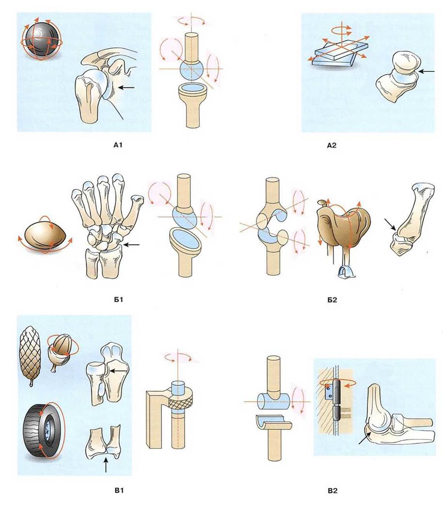
Fig. 121. Movements in joints (diagram):
A — triaxial (multiaxial) joints: A1 — ball-and-socket joint; A2 — plane joint; B — biaxial joints: B1 — ellipsoid joint; B2 — saddle joint; C — uniaxial joints: C1 — pivot joint; C2 — hinge joint
Biaxial joints include ellipsoid, condyloid, and saddle joints. The articular surfaces of an ellipsoid joint (*articulatio ellipsoidea*) are shaped as an ellipse, forming a convexity (articular head) and a concavity (articular socket). Rotation in an ellipsoid joint occurs around two mutually perpendicular axes. An example of an ellipsoid joint is the radiocarpal joint. The condyloid joint (*articulatio bicondylaris*) shares structural similarities with both hinge and ellipsoid joints. Its articular head is ellipse-shaped, but unlike a hinge joint, its articular surface is located on a condyle. For instance, the knee and atlanto-occipital joints are condyloid (the former is also complex, and the latter is combined).
In a saddle joint (*articulatio sellaris*), the articular surfaces resemble two saddles nested together with axes intersecting at right angles. The saddle joint of the thumb's carpometacarpal joint is unique to humans and enables the opposition of the thumb to the other fingers. This characteristic saddle joint structure underlies human manual labor capabilities.
Multiaxial joints include ball-and-socket, cup-shaped, and plane joints. In a ball-and-socket joint (*articulatio spheroidea*), the convex articular surface, shaped as a sphere segment, forms the articular head. The concave articular surface (articular socket) on the opposing bone matches this convexity. However, The surface of the articular head is typically larger than the socket, allowing for a wide range of motion in ball-and-socket joints. A prime example is the glenohumeral (shoulder) joint.
Movements in ball-and-socket joints occur around the frontal, sagittal, and longitudinal axes. Around the frontal axis, flexion takes place—whereby the angle between the articulating bones decreases—along with extension, where the joint angle increases up to 180°, straightening the limb. Around the sagittal axis, adduction occurs—bringing one of the articulating bones closer to the median plane (towards the torso)—as well as abduction, which moves the bone away from it. During rotation, a bone revolves around its longitudinal axis in either direction.
Circumduction is a sequential movement around all axes, in which the free end of the moving bone or limb (such as the hand) traces a circle. The greater the difference in angular dimensions (in degrees) between the articulating surfaces, the greater the range (amplitude) of motion. Conversely, when the articular surfaces are nearly equal in extent, the range of motion in the joints is minimal. Joint mobility is also influenced by the number and arrangement of stabilizing ligaments, as well as the position and distensibility of the surrounding muscles.
A specialized variant of the ball-and-socket joint is the cotylica or cup-shaped joint (articulatio cotylica), which features a very deep articular fossa enclosing more than half of the spherical head. Consequently, the disparity in angular dimensions between the spherical head and the socket is small, making movements in this type of joint restricted. The hip joint serves as a prime example of a cup-shaped joint, which is also referred to as a nut-shaped joint.
Plane joints (articulatio plana) are also classified as a variation of ball-and-socket joints. Their articular surfaces resemble segments of a sphere with a very large diameter. Movements in plane joints occur around three mutually perpendicular axes. However, the range of motion is restricted because the surfaces are nearly flat and the difference in their angular dimensions is slight. Examples of plane joints include the intercarpal and tarsometatarsal joints. Thus, the overall range of motion in any joint is determined by its Anatomical Structure and the disparity in angular dimensions of its articular surfaces.
Last update: 08/08/2026
Editorial and Educational Adaptation: This material has been compiled based on the primary/original source text. The project team performed an editorial review, corrected technical inaccuracies, structured sections, and adapted the content for an educational format.
What was processed:
- elimination of formatting defects (OCR errors, structural breaks, corrupted characters);
- editorial organization of content;
- standardization of terminology in accordance with academic sources;
- verification of factual statements against the original source text.
All mentions of the author, publication year, and origin of the primary text have been preserved in accordance with the source.