ATLAS OF HUMAN ANATOMY - G.L. Bilich - 2014
THE LOCOMOTOR APPARATUS
SKELETAL SYSTEM
BONES OF THE LOWER LIMB
Bones of the Free Lower Limb
The Femur (thigh bone) is the largest long bone in The Human Body, consisting of a shaft and two epiphyses (Fig. 103). The proximal epiphysis features the femoral HEAD (caput femoris) for articulation with the hip bone within the hip joint. At the center of the head lies the fovea of the head of the femur (fovea capitis ossis femoris), which serves as the attachment site for the ligament of the head of the femur (Fig. 104). A topographical and anatomical indicator reflecting the degree of coverage of the femoral head by the acetabulum is Wiberg's angle. In adults, this angle is 20—25° and is formed by lines drawn from the center of the femoral head through the edge of the acetabulum and vertically upward from the center of the head. The long femoral neck (collum femoris) connects the head to the shaft, forming an angle of approximately 130° with it. In the region of the femoral neck, an inconstant bony projection (spur)—Bigelow's septum—may occasionally be present (Bigelow, Henry Jacob, 1818—1890, American surgeon and anatomist).
Class="center">
Fig. 103. Right femur (A — anterior view, B — posterior view, C — orientation of the bony trabeculae of the Head and Neck relative to the applied load):
1 — Patellar surface; 2 — Lateral condyle; 3 — Lateral epicondyle; 4 — Shaft of femur; Body of femur; 5 — Lesser trochanter; 6 — lnter- trochanteric line; 7 - Greatcr trochanter; 8 — Head of femur; 9 — Neck of femur; 10 — Trochanteric fossa; 11 — Intertrochanteric crest; 12— Pectineal line; Spiral line; 13 — Gluteal tuberosity; 14 — Lateral lip; 15 — Medial lip; 16 —Linea aspera; 17 — Medial supracondylar line; 18 — Lateral supracondylar line; 19 — Poplileal surface; 20 — Intercondylar line; 21 — Intercondylar fossa; 22 — Medial condyle; 23 — Medial epicondyle; 24 — Adductor tubercle
At the junction of the neck and shaft, There are two bony prominences for Muscle attachment: the greater trochanter (trochanter major), located superiorly and laterally, and the lesser trochanter (trochanter minor), situated at the inferior margin of the neck, medially and posteriorly. On the Medial surface of the greater trochanter, facing the femoral neck, is the trochanteric fossa (fossa trochanterica)—known as Allen's fossa (Allen, Harrison, 1841—1897, American anatomist). Anteriorly, between the two trochanters, runs the intertrochanteric line (linea intertrochanterica); posteriorly, they are connected by the intertrochanteric crest (crista intertrochanterica). An important topographical and anatomical landmark in orthopedic surgery is Langenbeck's triangle, an area bounded by the anterior superior iliac spine, the greater trochanter, and the femoral neck (Langenbeck, Bernhard Rudolf Konrad, 1810—1887, German surgeon). Another key landmark used to determine the Displacement of the greater trochanter during a fracture is Schoemaker's line, drawn from the greater trochanter to the anterior superior iliac spine. In a normal anatomical position, these lines intersect above the umbilicus on both sides, whereas in a femoral neck fracture, they intersect below the umbilicus (Schoemaker, Jan, 1871—1940, Danish surgeon).
The shaft of the femur (corpus femoris) is cylindrical in shape, convex anteriorly, and slightly twisted along its longitudinal axis. Its anterior and lateral surfaces are smooth, while the posterior surface features the linea aspera, which divides into medial and lateral Lips (labium mediale et labium laterale) diverging superiorly and inferiorly to provide attachment for Muscles. Superiorly, these lips extend toward the greater and lesser trochanters. The lateral lip is significantly thicker than the medial one and forms the gluteal tuberosity (tuberositas glutea), the insertion site for the gluteus maximus muscle. The medial lip continues into the rough pectineal line (linea pectinea) for the attachment of the pectineus muscle. At the inferior epiphysis of the femur, both lips diverge to enclose the triangular popliteal surface (facies poplitea). These tubercles, lines, and lips serve as sites of origin and insertion for muscles acting on the Hip and knee joints.
The distal epiphysis of the femur is wide and forms two rounded condyles (Fig. 105). The medial condyle (condylus medialis) is larger than the lateral condyle (condylus lateralis). Posteriorly, the two condyles are separated by the deep intercondylar fossa (fossa intercondylaris). Anteriorly, the articular surfaces of the condyles merge to form the concave patellar surface (facies patellaris), against which the posterior surface of the Patella rests. Superior to the articular surface of the medial condyle lies the medial epicondyle (epicondylus medialis), while a smaller lateral epicondyle (epicondylus lateralis) is located on the lateral side. These epicondyles serve as the origin sites for muscles extending down to the leg. Occasionally, in the center of the radiographic shadow of the distal epiphysis in a lateral projection, Ludloff's spot is observed—an irregular area of radiolucency caused by normal anatomical variations in the Bone Structure, specifically a sparser trabecular pattern (Ludloff, Karl, 1864—1954, German surgeon).
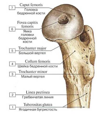
Fig. 104. Proximal epiphysis of the right femur, medial view:
1 — Gluteal tuberosity; 2 — Pectineal line; Spiral line; 3 — Lesser trochanter; 4 — Neck of femur; 5 — Greater trochanter; 6 — Fovea forligament ofhead; 7— Headoffemur
The patella is a large sesamoid bone embedded in the tendon of the quadriceps femoris muscle (Fig. 106). The Base of the patella (basis patellae) points superiorly, while its apex (apex patellae) points inferiorly. The posterior flattened articular surface (facies articularis)—known as Lenoir's surface of the patella—articulates with the patellar surface of the femur (Lenoir, Camille Alexandre Henri, born 1867, French surgeon and anatomist). The rough, convex anterior surface (facies anterior) is easily palpable through the Skin.
The leg consists of two bones: the Tibia is located medially, and the Fibula is located laterally (see Figs. 84, 85). Each of these bones has a shaft and two thickened epiphyses. The proximal epiphysis of the tibia forms the knee joint with the femur. Distally, both BONES OF THE leg articulate with the Bones of the FOOT to form the ankle joint. The interosseous space of the leg (spatium interosseum cruris) lies between these two bones.
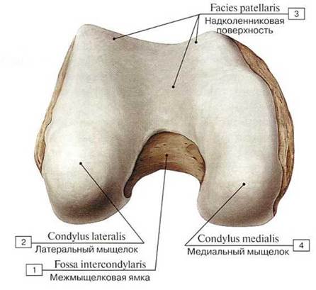
Fig. 105. Distal epiphysis of the right femur, anterior view:
1 — lntcrcondylar fossa; 2 — Lateral condyle; 3 — Patellar suiface; 4 — Mcdial condyle

Fig. 106. Right patella (A — anterior surface, B — articular surface, C — lateral view)
1 — Base of patella; 2 — Anterior surface; 3 — Apex of patella; 4 — Articular surface
The tibia [tibia] is the thicker bone of the leg. Its proximal thickened epiphysis bears the medial and lateral condyles [condylus medialis et condylus lateralis], which feature the superior articular surface [facies articularis superior] for articulation with the femoral condyles. Between the articular surfaces of the tibial condyles lies the intercondylar eminence [eminentia intercondylaris], which includes the medial intercondylar tubercle [tuberculum intercondylare mediale] and the lateral intercondylar tubercle [tuberculum intercondylare laterale]. Anterior to these tubercles is the anterior intercondylar area [area intercondylaris anterior], and posteriorly lies the posterior intercondylar area [area intercondylaris posterior]; both serve as attachment sites for the cruciate ligaments. On the lateral aspect of the lateral condyle, slightly posteriorly, is the fibular articular surface [facies articularis fibularis] for connection with the fibula, thereby forming the tibiofibular joint (Fig. 107).
On the lateral condyle of the tibia is Gerdy's tubercle, a prominence to which the iliotibial tract attaches [Gerdy, Pierre Nicolas, 1797—1856, French physician, anatomist, and pathologist].
The three-sided body of the tibia (corpus tibiae) features a sharp anterior border (margo anterior) that is easily palpable beneath the skin. This border thickens superiorly to form the tibial tuberosity (tuberositas tibiae), which serves as the attachment site for the quadriceps femoris. The sharp lateral border faces the fibula and is known as the interosseous border (margo interosseus), while the medial border (margo medialis) is somewhat rounded. The tibial body presents three surfaces: the medial surface (facies medialis), which is smooth and lies directly subcutaneous, and the lateral (facies lateralis) and posterior (facies posterior) surfaces, both of which are covered by muscles. The posterior surface displays the oblique, inferomedially directed soleal line (linea musculi solei), running from the posterior margin of the lateral condyle.
The distal end of the tibia is expanded, bearing a fibular notch (incisura fibularis) on its lateral margin for articulation with the fibula, thereby forming the tibiofibular syndesmosis. The medial malleolus (malleolus medialis) projects downward from the medial side of the tibia. Posterior to the malleolus lies a shallow malleolar groove (sulcus malleolaris), which transmits the tendon of the tibialis posterior muscle. On the lateral aspect of the medial malleolus is an articular facet (facies articularis malleoli) that is continuous with the inferior articular surface (facies articularis inferior) of the tibia. Together with the articular surface of the fibula and the talus of the tarsus, these surfaces form the ankle joint.
The fibula is a slender bone whose proximal, expanded end features the head of the fibula (caput fibulae). This terminates in an apex (apex capitis fibulae), and on its medial side bears the articular facet of the head of the fibula (facies articularis capitis fibulae) for articulation with the tibia. The body of the fibula (corpus fibulae) is triangular in cross-section and presents three borders: the anterior border (margo anterior), posterior border (margo posterior), and the sharp medial interosseous border (margo interosseus). These borders demarcate three surfaces: the lateral surface (facies lateralis), posterior surface (facies posterior), and medial surface (facies medialis).
The thickened distal epiphysis of the fibula forms the lateral malleolus (malleolus lateralis), on the medial surface of which is a smooth articular facet (facies articularis malleoli) for articulation with the talus. The posterior margin of the malleolus features the malleolar fossa (fossa malleoli lateralis), which gives attachment to the tendons of the fibular muscles (Fig. 108).
At the very Water/144.html">Origin of the lateral head of the gastrocnemius muscle, a sesamoid bone known as the Vesalian bone is frequently found.
Figures 109 and 110 show radiographs of the bones of the leg.
The bones of the foot (ossa pedis), much like those of the hand, are divided into three groups: the Tarsal Bones (ossa tarsi), Metatarsal Bones (ossa metatarsalia), and the bones of the toes (ossa digitorum pedis) (Figs. 111, 112, 113).
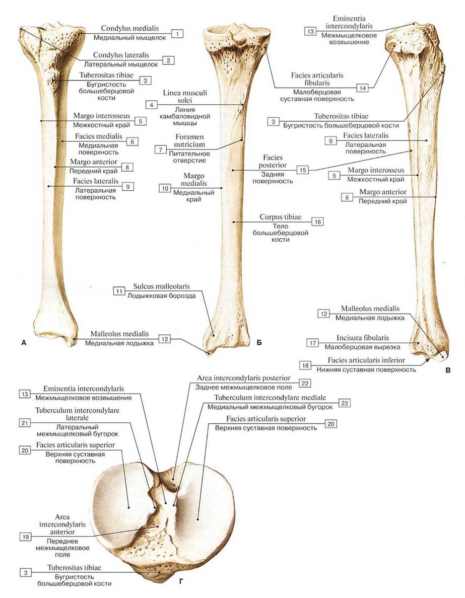
Fig. 107. Right tibia (A — anterior view, B — posterior view, C — lateral view, D — proximal epiphysis, superior view):
1 — Medial condyle; 2 — Lateral condyle; 3 — Tibial tuberosity; 4 — Soleal line; 5 — Interosseous bordcr; 6 — Medial surfacc: 7 — Nutrient foramen; 8 — Anterior border; 9 — Lateral surface; 10 — Medial border; 11— Malleolar groove; 12 — Medial malleolus; 13— Intercondylareminence; 14 — Fibular articular facet; 15 — Posterior surface; 16 — Shaft of tibia; Body of tibia; 17— Fibular notch; 18 — Inferior articular surface; 19 — Anterior intercondylar area; 20 — Superior articular surface; 21 — Lateral intercondylar tubercle; 22 — Lateral intercondylar area; 23 — Medial intercondylar tubercle

Fig. 108. Right fibula (A — anterior view, B — posterior view, C — medial view,
D — articular surfaces of the inferior epiphyses of the bones of the leg):
1 — Apex ofhead; 2 — Head of fibula; 3 — Neck of fibula; 4 — Lateral surface; 5 — Medial surface; 6 — Interosseous bordcr; 7— Medial crcst; 8 — Anterior border; 9 — Lateral malleolus; 10— Malleolar fossa; 11 — Articular facet; 12 — Shaft of fibula; Body of fibula; 13 — Posterior border; 14 — Posterior surface; 15 — Nutrient foramen; 16 — Articular facet; 17 — Articular facet; 18 — Inferior articular surface; 19 — Fibula; 20 — Tibia; 21 — Medial malleolus; 22— Malleolar groove

Fig. 109. Anteroposterior radiograph of the bones of the leg:
1 — medial surface and medial border of the tibia; 2 — interosseous border of the fibula; 3 — lateral surface and interosseous border of the tibia; 4 — lateral surface of the fibula; 5 — tibial tuberosity; 6 — medullary cavity of the tibia; 7 — interosseous space (after A. Yu. Vasiliev)

Fig. 110. Lateral radiograph of the bones of the leg:
1 - anterior border of the fibula; 2 — anterior border of the tibia; 3 — tibial tuberosity; 4 — posterior surface of the tibia; 5 — posterior surface of the fibula (after A. Yu. Vasiliev)
The tarsus consists of seven short bones arranged in two rows. The proximal (posterior) row contains two large bones: the talus and the calcaneus. The remaining five tarsal bones (the navicular, cuboid, and three cuneiforms) form the distal (anterior) row of the tarsus (see Figs. 111, 112, 113).
The talus comprises a body (corpus tali), a head (caput tali), and a constricted neck (collum tali). The body of the talus is large, and its superior surface features the trochlea of the talus (trochlea tali), which bears three articular surfaces. The superior surface (facies superior) articulates with the inferior articular surface of the tibia. The other two articular surfaces are located on either side of the trochlea. The medial malleolar surface (facies malleolaris medialis) and the larger lateral malleolar surface (facies malleolaris lateralis) articulate with the articular surfaces of the tibial and fibular malleoli to form the ankle joint (Fig. 114).

Fig. 111. Bones of the right foot, superior view:
1 — Calcaneal tuber; 2 — Body оf talus; 3 — Neck of talus; 4 — Head of talus; 5 — Talus; 6 — Navicular; 7 — Intermediate cuneiform; Middle cuneiform; 8 — Medial cuneiform; 9 — Base of metatarsal; 10 — Shaft of metatarsal; Body of metatarsal; 11 — Head of metatarsal; 12 — Metatarsal [1]; 13 — Base of phalanx; 14 — Shaft of phalanx; Body of phalanx; 15 — Head of phalanx; 16 — Proximal phalanx[1]; 17 — Distal phalanx [l]; 18 — Distal phalanx [V]; 19 — Middle phalanx[V]; 20— Proximal phalanx [V]; 21 — Lateral cuneiform; 22— Tubero-sity of fifth metatarsal bone [V]; 23 — Cuboid; 24 — Calcaneus; 25— Posterior process

Fig. 112. Bones of the right foot, inferior view:
1 — Calcaneus; 2 — Cuboid: 3 — Tuberosily of cuboid: 4 — Groovc for tendon of flbularis longus; Groove for tendon of peroneus longus; 5 — Tuberosity of fifth metatarsal bone [V]; 6 — Metatarsal [V]; 7 — Proximal phalanx [V]; 8 — Middle phalanx [V]; 9 — Distal phalanx [V]; 10 — Distal phalanx [I]; 11 — Proximal phalanx [I]; 12 — Sesamoid bones; 13 — Metatarsal [I]; 14— Medial cuneiform; 15 - Intermediate cuneiform; Middle cuneiform; 16 — Lateral cuneiform; 17— Navicular; 18 — Headoftalus; 19— Neckoftalus; 20— Bodyoftalus; 21 — Sustentaculum tali; Talar shclf; 22 —Talus, posterior process

Fig. 113. Bones of the right foot (A — medial view, B — lateral view):
1 — Distal phalanx [I]; 2 - Proximal phalanx [I]; 3 — Head of phalanx; 4 — Shaft of phalanx; Body of phalanx; 5 — Base of phalanx; 6 — Head of metatarsal; 7 — Shaft of metatarsal; Body of metatarsal; 8 — Base of metatarsal; 9 — Metatarsal [I]; 10 — Medial cunciform; 11 — Navicular; 12 — Head of talus; 13 — Neck of talus; 14 — Body of talus; 15 — Sustentaculum tali; Talar shelf; 16 — Calcaneal tuber; 17— Calcaneus, medial process; 18 — Medial tubercle; 19— Lateral tubercle; 20 — Talus, posterior process; 21 — Cuboid; 22— Calcaneus, lateral process; 23— Calcaneus; 24— Intermediate cuneiform; Middle cuneiform; 25 — Lateral cuneiform; 26 — Distal phalaax [V]; 27 — Middle phalanx [V]; 28 — Proximal phalanx [V]; 29— Metatarsal [V]; 30— Tuberosity offifth metatarsal bone [V]

Fig. 114. Right talus (A — superior view, B — inferior view):
1 — Posterior process, lateral tubercle; 2 — Groove for tendon of flexor hallucis longus; 3 — Posterior process, medial tubercle; 4 — Body of talus; 5 — Medial malleolar facet; 6 — Head of talus; 7 — Navicular articular surface; 8 — Anterior lacet for calcaneus; 9 — Neck of talus; 10 — Lateral process; 11 — Lateral malleolar facet; 12 — Trochlea of talus, superior facet; 13 — Middle facet for calcaneus; 14 — Sulcus tali; 15 — Posterior calcaneal articular facet
Posterior to the body of the talus projects the posterior process of the talus (processus posterior tali — Stieda's process, which features a groove for the tendon of the flexor hallucis longus muscle; Ludwig Stieda, 1837–1918, German anatomist). This groove divides the process into a medial tubercle (tuberculum mediale) and a lateral tubercle (tuberculum laterale). The posterior talofibular ligament attaches to this process. On the inferior aspect of the talus, there are three articular surfaces for articulation with the calcaneus: the anterior calcaneal articular facet (facies articularis calcanea anterior), the middle calcaneal articular facet (facies articularis calcanea media), and the posterior calcaneal articular facet (facies articularis calcanea posterior). Between the middle and posterior calcaneal facets lies the sulcus of the talus (sulcus tali), to which the talocalcaneal ligaments of the subtalar joint attach. The head of the talus is directed anteriorly and medially. It bears a rounded navicular articular surface (facies articularis navicularis) for articulation with the navicular bone. According to Henle, the head of the talus is not a segment of a sphere, but rather flattened from above and medially, resembling an orange — Henle's "orange" (Friedrich Gustav Jacob Henle, 1809–1885, German anatomist and pathologist).
The calcaneus is the largest bone of the foot, located beneath the talus (Fig. 115). Posteriorly, the body of the calcaneus forms a prominent projection, the calcaneal tuberosity (tuber calcanei). On the superior surface of the calcaneus, there are three articular facets corresponding to the calcaneal articular surfaces of the talus: the anterior talar articular facet (facies articularis talaris anterior), the middle talar articular facet (facies articularis talaris media), and the posterior talar articular facet (facies articularis talaris posterior). Between the middle and posterior articular facets runs the calcaneal sulcus (sulcus calcanei), which, together with the corresponding sulcus on the talus, forms the tarsal sinus (sinus tarsi) occupied by the ligament connecting these two bones. The entrance to the sinus is located on the lateral side of the foot. Extending medially from the calcaneus is a short, thick process known as the sustentaculum tali (talar shelf). On the lateral surface of the calcaneus lies the groove for the tendon of the fibularis (peroneus) longus muscle (sulcus tendinis musculi peronei longi). At the anterior end of the calcaneus is the cuboid articular surface (facies articularis cuboidea) for articulation with the cuboid bone (see Fig. 115).

Fig. 115. Right calcaneus (A — medial view, B — lateral view):
1— Sustentaculum tali; Talar shelf; 2 — Articular surface for cuboid; 3 — Middle talar articular surfacc; 4 — Anterior talar articular surface; 5— Posterior talar articular surface; 6— Groove fortendon of flexor hallucis longus; 7— Medial process; 8 — Calcaneal tuber; 9 — Groove for tendon of fibularis longus; Groove fortendon of peroneus longus; 10 — Fibular trochlea; Peroneal trochlea; Peroneal tubercle; 11 — Calcaneal sulcus; 12 — Lateral process
The navicular bone (os naviculare) is situated in the medial part of the foot between the talus posteriorly and the three cuneiform bones anteriorly. The proximal (posterior) concave surface of the bone serves for articulation with the head of the talus. The distal (anterior) surface of the navicular bone features three articular facets for connection with the three cuneiform bones. On its medial margin is the tuberosity of the navicular bone (tuberositas ossis navicularis) for the attachment of the tibialis posterior muscle. On the lateral side of the navicular bone, an inconstant articular surface may be present for articulation with the cuboid bone (Fig. 116).
The cuneiform bones (ossa cuneiformia) — medial, intermediate, and lateral — lie anterior to the navicular bone. The medial cuneiform bone (os cuneiforme mediale), the largest of the three, articulates with the base of the first metatarsal bone. The intermediate cuneiform bone (os cuneiforme intermedium) forms a joint with the second metatarsal bone. The lateral cuneiform bone (os cuneiforme laterale) articulates with the third metatarsal bone (see Fig. 116).
The cuboid bone (os cuboideum) is located on the lateral side of the foot between the calcaneus posteriorly and the two lateral metatarsal bones anteriorly. Articular surfaces are present where these bones make contact with one another. On the medial side of the cuboid bone, an articular facet for the lateral cuneiform bone is visible, and slightly posterior to it lies the surface for articulation with the navicular bone. On the inferior (plantar) surface, the tuberosity of the cuboid bone (tuberositas ossis cuboidea) is found, anterior to which runs the groove for the tendon of the fibularis (peroneus) longus muscle (sulcus tendinis musculi peronei longi) (see Fig. 116).
The metatarsal bones (ossa metatarsalia) consist of five short tubular bones. The shortest and thickest of these is the first metatarsal bone, while the longest is the second. Each metatarsal bone comprises a body (corpus), a head (caput), and a base (basis). The bodies of the metatarsals are three-sided in cross-section, with their dorsal (superior) surface convex. The bases of the metatarsals are expanded, and each bears an articular surface for articulation with the tarsal bones. The base of the first metatarsal forms a joint with the medial cuneiform bone. The bases of the second and third metatarsals articulate with the intermediate and lateral cuneiforms, respectively, whereas the bases of the fourth and fifth metatarsals articulate with the cuboid bone. The metatarsal heads are laterally compressed. The anterior articular surface of each metatarsal head is rounded and serves to articulate with the base of the proximal phalanx of the corresponding digit. The head of the first metatarsal is divided on its plantar surface into two facets, which articulate with two sesamoid bones. On the lateral side of the fifth metatarsal bone is the tuberosity of the fifth metatarsal bone (tuberositas ossis metatarsalis V) for the attachment of the fibularis (peroneus) brevis muscle (see Figs. 112–117). Between the bases of the First and Second metatarsals, an accessory metatarsal bone often develops — Gruber's bone (syn.: intermetatarsal bone, os intermetatarseum) (Wenzel Leopold Gruber, 1814–1890, Austrian anatomist who worked in Russia). When the tuberosity of the fifth metatarsal bone remains separate, an accessory tarsal bone known as Vesalius's bone is formed. Another accessory bone of the foot is the os trigonum (or accessory triangular bone / prehallux) — Bardeleben's bone (Karl von Bardeleben, 1849–1918, German anatomist).
The Phalanges of the foot (ossa pedis) are considerably shorter than the phalanges of the hand. Like the fingers, the toes possess three phalanges: a proximal phalanx (phalanx proximalis), a middle phalanx (phalanx media), and a distal phalanx (phalanx distalis). Only the great toe (hallux) has two phalanges: proximal and distal (see Figs. 111–116). The phalanges are short tubular bones, each comprising a body (corpus phalangis), a head (caput phalangis), and a base (basis phalangis). The bodies of the proximal and middle phalanges are slightly curved dorsally. The base of each proximal phalanx features a flattened socket for articulation with the head of the corresponding metatarsal bone. The shallow sockets on the bases of the middle and distal phalanges are divided by a crest and serve for articulation with the head of the adjacent proximal phalanx. Each distal (terminal) phalanx terminates in a tuberosity (tuberositas phalangis distalis).

Fig. 116. Bones of the right foot, superior view (A — bases of the proximal phalanges, B — bases of the metatarsals,
C — cuneiform and cuboid bones, D — navicular and cuboid bones):
1 — Navicular; 2 — Medial cuneiform; 3 — Intermediate cuneiform; Middlc cuneiform; 4 — Lateral cuneiform; 5— Base of metatarsal [1]; 6 — Base of proximal phalanx [I]; 7 — Mctatarsals [I—V]; 8 — Base of metatarsal [V]; 9 — Tubcrosity of fifth metatarsal bone [V]; 10— Cuboid
Figure 117 shows a radiograph of the foot.

Fig. 117. Radiograph of the foot, direct plantar projection:
1 — calcaneus; 2 — talus; 3 — navicular bone; 4 — cuboid bone; 5 — medial cuneiform bone; 6 — intermediate cuneiform bone; 7 — lateral cuneiform bone; 8 — head of metatarsal bone; 9 — sesamoid bone; 10 — proximal phalanges; 11 — middle phalanges; 12 — distal phalanges; 13 — base of metatarsal bone (according to A.Yu. Vasilyev)
Last update: 08/08/2026
Editorial and Educational Adaptation: This material has been compiled based on the primary/original source text. The project team performed an editorial review, corrected technical inaccuracies, structured sections, and adapted the content for an educational format.
What was processed:
- elimination of formatting defects (OCR errors, structural breaks, corrupted characters);
- editorial organization of content;
- standardization of terminology in accordance with academic sources;
- verification of factual statements against the original source text.
All mentions of the author, publication year, and origin of the primary text have been preserved in accordance with the source.