Human Anatomy (with Fundamentals of Dynamic and Sports Morphology) - Ivanitsky M. F. 2008
The Doctrine of Bones and Their Articulations
Skeleton of the Upper Limb
BONES OF THE upper limb
The Skeleton of the Upper Limb is divided into two parts: the Pectoral Girdle and the free upper limb. The pectoral girdle connects the free upper limb to the trunk, serves as an origin and insertion point for Muscles, and increases the range of motion of the upper limb. The skeleton of the pectoral girdle includes the scapula, clavicle, sternoclavicular joint, and acromioclavicular joint.
The free upper limb consists of three segments: the arm, forearm, and hand. It includes the humerus, ulna, and radius; the shoulder, elbow, radioulnar (proximal and distal), and radiocarpal joints; as well as the Bones and joints of the hand.
The scapula (Fig. 27) is a flat, triangular bone located on the posterior surface of the trunk. It has three borders—superior, medial, and lateral; three angles—lateral, inferior, and superior; and two surfaces—costal and dorsal. The superior border features a notch. The medial border faces THE Vertebral Column, while the lateral border faces the axillary fossa.
The lateral angle is thickened and features a glenoid cavity for articulation with the HEAD of the humerus. Above and below the glenoid cavity are two tubercles: the supraglenoid tubercle, which serves as the origin for the tendon of the long head of the biceps brachii, and the infraglenoid tubercle, the Water/144.html">Origin of the tendon of the long head of the triceps brachii. The inferior angle of the scapula lies at the level of the 7th–8th Ribs and is easily palpable beneath the Skin, while the superior angle is located between the superior and medial borders of the scapula.
The costal surface of the scapula faces The thoracic cage, is concave, and forms the subscapular fossa. The dorsal surface is convex and features a spine that extends from the medial border to the lateral angle. Above the spine is the supraspinous fossa, and below it is the infraspinous fossa, which house the Muscles of the same names. The spine of the scapula is easily palpable beneath the skin. Laterally, it transitions into the acromion, the most laterally protruding point of which is called the shoulder or acromial point, used in measuring shoulder width. Below the acromion lies the coracoid process, which serves for the attachment of muscles and ligaments.
The clavicle (see Fig. 27) is a bone curved in an S-shape along its long axis. It is positioned horizontally at the anterior and superior aspect of the Thorax at the border of the neck and is easily palpable throughout its entire length. The clavicle has two ends—sternal and acromial. The former is thickened and has an articular surface for articulation with the Sternum; the latter is flattened and articulates with the acromion. The superior surface of the clavicle is smooth and even, whereas the inferior surface is rough, as it attaches to the thorax and scapula via Ligaments and Muscles.
The function of the clavicle is to help stabilize THE POSITION OF the scapula, keeping the shoulder joint at a certain distance from the thoracic cage.
The humerus (see Fig. 21) is a typical long tubular bone comprising a shaft, a proximal end, and a distal end (epiphysis). At its proximal end, the head can be distinguished. It faces the scapula and features an articular surface separated from the rest of the bone by the anatomical neck, along the margin of which the capsule of the shoulder joint attaches. Below the anatomical neck on the lateral side are two tubercles for Muscle attachment: the greater tubercle, facing laterally, and the lesser tubercle, facing anteriorly. A crest extends downward from each tubercle. Between the tubercles and crests lies a groove through which the tendon of the long head of the biceps brachii runs. The narrowest part of the humerus just below the tubercles is called the surgical neck, as fractures frequently occur here. On the lateral surface of the bone shaft is the deltoid tuberosity for the attachment of the deltoid muscle, while the radial groove spirals downward and outward across the posterior surface.
Class="center">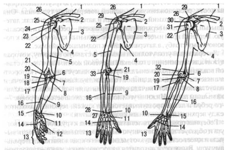
Fig. 2.7. Bones of the upper limb. Anterior view in three arm positions (A — arm in neutral position, forearm semi-pronated; B — arm pronated; C — arm in neutral position, forearm supinated): 1 — clavicle; 2 — superior angle of the scapula; 3 — medial border of the scapula; 4 — inferior angle of the scapula; 5 — humeral bone; 6 — medial epicondyle of the humerus; 7 — trochlea of the humerus; 8 — coronoid process of the ulna; 9 — radius; 10 — styloid process of the radius; 11 — trapezium; 12 — proximal phalanx of the thumb; 13 — proximal, middle, and distal Phalanges of the fingers; 14 — Metacarpal bones; 15 — head of the ulna; 16 — shaft of the ulna; 17 — radial tuberosity; 18 — neck of the radius; 19 — head of the radius; 20 — capitulum of the humerus; 21 — lateral epicondyle of the humerus; 22 — surgical neck; 23 and 30 — anatomical neck; 24 — head of the humerus; 25 — greater tubercle; 26 — acromion; 27 — Carpal Bones; 28 — styloid process of the ulna; 29 — coracoid process of the scapula; 31 — lesser tubercle of the humerus; 32 — coronoid fossa; 33 — olecranon of the ulna
When the deltoid muscle is developed through athletic training, one observes not only an enlargement of the deltoid tuberosity but also an increase in the thickness of the compact layer of the humerus.
The distal end of the humerus forms the condyle, the articular surface of which articulates with the forearm bones. The medial part of its articular surface, which articulates with the ulna, is called the trochlea of the humerus, while the lateral part, which articulates with the radius, is called the capitulum of the humerus. Anteriorly, above the trochlea, lies the coronoid fossa, and posteriorly lies the olecranon fossa; during flexion and extension of the forearm, the processes of the ulna enter these fossae. On both sides of the distal end of the humerus are the medial and lateral epicondyles, which are easily palpable beneath the skin—especially the medial one, on the posterior side of which runs the ulnar nerve groove.
The ulna (see Fig. 27). This is a typical tubular, three-sided bone. The anterior surface of the bone is separated from the posterior by a sharp interosseous border. Its proximal end is thickened and features a trochlear notch for articulation with the humerus. The trochlear notch is bounded posteriorly by the olecranon and anteriorly by the coronoid process. The olecranon is easily palpable, especially when the elbow is flexed. At the Base of the coronoid process lies the ulnar tuberosity, to which the brachialis muscle attaches, and on the lateral side is the radial notch for articulation with the head of the radius. The distal end of the ulna features a thickening—the head of the ulna. On the lateral side, the head forms an articular surface for articulation with the radius, and inferiorly with the articular disc (triangular fibrocartilage). The styloid process of the ulna extends from the head. The ulna is palpable beneath the skin posteriorly along its entire length.
The radius (see Fig. 27), like the ulna, is a long tubular bone. Unlike the ulna, the radius has a thickened distal end. The proximal end features a head, on the superior surface of which is a articular facet for articulation with the capitulum of the humerus, while its margin bears an articular circumference for articulation with the ulna. Below the head are the neck and the radial tuberosity. The latter serves for the attachment of the tendon of the biceps brachii.
At the distal end of the radius is an articular surface for articulation with the carpal bones. On the lateral side of this end is the styloid process, palpable beneath the skin, and on the medial side is the ulnar notch for articulation with the head of the ulna.
The sharp border of the radius facing the ulna is called the interosseous border. The interosseous membrane of the forearm attaches here.
Most of the radius lies deep among the muscles; however, just below and posterior to the lateral margin of the humeral condyle (in the "fossa of beauty"), its head can be palpated, and distally—the entire lower end of the bone, including the styloid process.
The hand has three segments: the carpus, metacarpus, and digits (fingers), which in turn consist of individual phalanges.
The carpal bones (see Fig. 27). The eight small carpal bones are irregular in shape and arranged in two rows (Fig. 30). They are classified as short spongy bones.
The proximal row comprises (from the thumb side): the scaphoid, lunate, triquetrum, and pisiform bones.
The distal row also comprises four bones: the trapezium, trapezoid, capitate, and hamate, the latter of which features a hook pointing toward the palmar side of the hand.
The proximal row of carpal bones forms a convex articular surface directed toward the radius, while the distal row forms an irregular surface for articulation with the proximal row.
The carpal bones do not lie in a single plane; instead, they form a sulcus on the palmar surface and a convexity on the dorsal surface. This sulcus serves as a passageway for the tendons of the finger flexor muscles. Its medial margin is bounded by the pisiform bone and the hook of the hamate, which are easily palpable, while the lateral margin is formed by two bones—the scaphoid and the trapezium.
The metacarpal bones. The metacarpus consists of five short tubular bones. The metacarpal bone of the first digit is shorter than the others but more massive. The second metacarpal bone is the longest. Each metacarpal bone consists of a base, a shaft, and a head.
The bases of the metacarpal bones articulate with the carpal bones. The bases of the first and fifth metacarpal bones feature saddle-shaped articular surfaces, while the bases of the remaining metacarpals have flat articular surfaces. The shafts of the metacarpal bones are convex dorsally and concave on the palmar side. The metacarpal heads present spheroidal articular surfaces and articulate with the proximal phalanges of the digits.
The metacarpal bones are easily palpable on the dorsal aspect of the hand, and their heads become visible when the hand is clenched into a fist.
The bones of the digits—the phalanges—are short tubular bones (see Fig. 27). Each digit consists of three phalanges: proximal, middle, and distal. The sole exception is the first digit (thumb), which has only two phalanges: proximal and distal. The proximal phalanges are the longest, whereas the distal ones are the shortest. Each phalanx comprises a central section, the shaft, and two extremities. The proximal extremity forms the base of the phalanx, and the distal extremity forms its head. The articular surfaces of the distal and middle phalanges, as well as the articular surface of the heads of the proximal phalanges, are trochlear (pulley-shaped) in form; articulating with one another, they form ginglymoid (hinge) joints. The articular surface at the base of the proximal phalanges articulates with the metacarpal head via a spheroidal joint.
In addition to the aforementioned bones, the hand also contains sesamoid bones. These are embedded within the substance of muscle tendons (most frequently those passing toward the thumb and index finger from the palmar side of the hand) and serve to increase the moment arm of the muscles that attach to them.
Articulations of the Bones of the Upper Limb
The bones of the pectoral girdle form two joints: the sternoclavicular and acromioclavicular joints.
The sternoclavicular joint (Fig. 28) is formed by the clavicular notch of the sternum and the sternal end of the clavicle. It is a simple, bicompartmental joint owing to the presence of an articular disc within the joint cavity, which divides it into two chambers.
The shape of the joint approaches that of a saddle. However, by virtue of the articular disc, it Functions as a ball-and-socket (spheroidal) joint. Movements within it are possible around three mutually perpendicular axes: the sagittal axis (elevation and depression), the vertical axis (protraction and retraction), and the transverse axis (slight rotation, as well as circumduction). During circumduction, the lateral end of the clavicle describes an ellipse with a height of 10 cm and an anteroposterior diameter of approximately 12 cm.
The Joint Capsule is thin. It is reinforced by the interclavicular ligament, which connects the sternal ends of both clavicles; the costoclavicular ligament, extending from the Cartilage of the 1st rib to the Inferior surface of the clavicle; and the anterior and posterior sternoclavicular ligaments. The anterior ligament checks posterior Displacement of the clavicle, while the posterior ligament checks anterior displacement. The fan-shaped bundles of the sternoclavicular ligament, spreading from top to bottom, prevent the clavicle from being displaced downward under the action of forces tending to depress its acromial end. This helps maintain the clavicle in a horizontal position even when carrying loads or when subjected to external forces (such as a barbell in weightlifting, a partner in acrobatics, etc.). Furthermore, this joint is reinforced by the subclavius muscle.
The position of the sternoclavicular joint cleft can be determined by Palpation. If, while studying movements in this joint, one finger is placed on the jugular notch of the sternum and another on the clavicle, it can be verified that during clavicular movements, the finger resting on the clavicle shifts slightly, whereas the finger positioned on the sternum remains stationary.
Occasionally, between the 1st rib and the clavicle at the site of the costoclavicular ligament, a costoclavicular joint develops (most frequently in individuals engaged in manual labor, associated with enhanced mobility of the clavicle).
The acromioclavicular joint (see Fig. 28) is formed by the acromial end of the clavicle and the acromion of the scapula. It is a simple, plane joint that may occasionally undergo synchondrosis. The joint is reinforced by two ligaments: the coracoclavicular ligament, extending from the coracoid process to the inferior surface of the clavicle, and the acromioclavicular ligament. Movements in the joint are limited. The greatest range of scapular motion is observed around the sagittal axis.

Fig. 28. Joints and ligaments of the pectoral girdle and the shoulder joint:
1 - trapezoid ligament (part of the coracoclavicular ligament); 2 - conoid ligament; 3 - costoclavicular ligament; 4 - anterior sternoclavicular ligament; 5 - interclavicular ligament; 6 - articular disc; 7 - tendon of the long head of the biceps brachii muscle; 8 - synovial Sheath of the tendon; 9 - coracoacromial ligament; 10 - acromioclavicular ligament (after G.F. Ivanov)
As a rule, the scapula and clavicle move simultaneously.
In addition to the aforementioned ligaments, the scapula possesses two intrinsic ligaments: the coracoacromial ligament and the superior transverse scapular ligament. The former resembles a triangular lamina extending from the acromion of the scapula to the coracoid process. It participates in protecting the shoulder joint from above by forming the so-called coracoacromial arch (fornix humeri). The second ligament bridges across the scapular notch, converting it into a foramen.
The glenohumeral (shoulder) joint (see Fig. 28) is formed by the head of the humerus and the glenoid cavity of the scapula. It is a simple, incongruent, ball-and-socket joint. The articular surface of the humeral head is significantly larger than the glenoid cavity of the scapula. The depth of the cavity is increased by means of the glenoid labrum, which runs along its margin.
The articular capsule originates near the glenoid labrum and attaches to the anatomical neck of the humerus. The capsule is thin (0.1–0.5 cm) and loose. In males, it is thicker than in females. Its thickness is reduced in the superior part of the anterior wall of the joint and in the central part of the posterior wall; consequently, during a fall onto the hand with supination of the humerus, rupture of the anterior section of the capsule frequently occurs, causing the humeral head to be displaced anteriorly and medially beneath the coracoid process of the scapula. When the arm is abducted and subjected to a displacing force, rupture of the inferior portion of the capsule may occur, resulting in downward displacement of the humeral head.
The Ligamentous apparatus of the joint is represented solely by the coracohumeral ligament, which extends from the coracoid process to the joint capsule. The tensile strength of this ligament ranges from 0.4 to 1.9 kg/mm2 in males, and from 0.2 to 1.5 kg/mm2 in females. The peak tensile strength of the ligament is observed between the ages of 22 and 35 years. Its elongation under load is minimal. Fibres of the muscles passing adjacent to the shoulder joint also blend into the capsule.
The joint possesses three mutually perpendicular axes of rotation: transverse, anteroposterior, and vertical (Fig. 29). Movements possible around the transverse axis are forward movement (flexion) and backward movement (extension); around the anteroposterior axis—abduction and adduction; and around the vertical axis—medial rotation (pronation) and lateral rotation (supination). In addition, circumduction is possible in the shoulder joint. Movements in the shoulder joint are typically combined with Movements of the pectoral girdle, As a result of which the outstretched upper limb can trace approximately a hemisphere. Movement occurring solely within the shoulder joint has a considerably smaller amplitude. The upper limb can be abducted up to the horizontal plane, i.e., by approximately 90°. Further movement, by which the arm is raised upward, occurs primarily through the motion of the scapula and clavicle, assisted by coupled movements of the vertebral column. From the natural resting position of the arm, these independent movements are small. They can be increased by elevating or abducting the arm, which is of great importance in sports such as basketball, volleyball, tennis, etc. Extension in the shoulder joint of a raised arm (the wind-up in throwing a ball or striking a ball) increases by 10–15° compared to the initial position at a 30° angle, and by 15–20° at a 45° angle. If the arm is elevated to a vertical position, rotational movements are possible through approximately 90°; with the arm lowered, through 135°. The range of motion in the shoulder joint is as follows: flexion — 90°, extension — 45°, abduction — 90°, adduction — 30°, supination — 85°, pronation — 85°.

Fig. 29. Axes of Rotation of the upper limb joints (shoulder, elbow, wrist, and interphalangeal joints):
a - frontal (transverse); b - sagittal (anteroposterior); c - vertical
One of the distinctive Features of the shoulder joint is that the tendon of the long head of the biceps brachii muscle passes directly through its cavity; the tension of this tendon reinforces the joint.
The elbow joint consists of three joints: the humeroulnar, humeroradial, and proximal radioulnar joints. All of them share a single joint capsule and joint cavity, thus forming a compound joint.
The humeroulnar joint is formed by the trochlea of the humerus and the trochlear notch of the ulna. It is a hinge joint with a single transverse axis of rotation that allows for flexion and extension (see Fig. 29).
The humeroradial joint is formed by the capitulum of the humerus and the head of the radius. It has a ball-and-socket shape and three axes of rotation. Flexion and extension of the forearm occur around the transverse axis, while pronation and supination take place around the vertical axis. The sagittal axis is not utilized because the interosseous membrane is stretched between the forearm bones. The articular cleft of the humeroradial joint is easily palpable in the depression located on the posterior surface of the forearm, at its proximal end on the radial side (the "beauty pit"). The proximal radioulnar joint is formed by the articular surfaces of the radial head and the ulnar notch. This is a cylindrical joint with a single vertical axis that allows for pronation and supination of the forearm.
Flexion and extension around the transverse axis, as well as pronation and supination around the vertical axis, are possible in the elbow joint (see Fig. 29).
The trochlea of the humerus spans approximately 320°, whereas the trochlear notch of the ulna spans 180°; thus, the range of motion around the transverse axis—namely, flexion and extension of the forearm—is 140° (320° - 180° = 140°). The range of motion for pronation and supination of the forearm is also approximately 140°. As a result of systematic athletic training, the pronation-supination mobility of the radius relative to the ulna can reach 180°, and even more with the application of an external force.
Flexion and extension involve simultaneous movements in the humeroradial and humeroulnar joints, whereas pronation and supination involve simultaneous movements in the humeroradial, proximal radioulnar, and distal radioulnar joints.
The elbow joint is reinforced by the ulnar collateral ligament, extending from the medial epicondyle of the humerus to the ulna; the radial collateral ligament, which runs from the lateral epicondyle, loops around the head of the radius anteriorly and posteriorly, and attaches to the ulna; and the annular ligament of the radius, which encircles the radial head and attaches to the ulna. The purpose of the collateral ligaments is to prevent movements around the sagittal axis. Slight abduction of the ulna is possible during extension and pronation of the forearm, while adduction occurs during flexion and supination. Lateral swaying of the ulna is facilitated by a fat pad located in the medial part of its semilunar notch at the base of the olecranon.
The strength of the elbow joint ligaments is quite high; however, only the annular ligament of the radius ruptures along its length, whereas the collateral ligaments typically tear away from their bone attachment sites. The ulnar collateral ligament tears at a load of 77.2 kg in individuals aged 20–35, and at 29 kg in individuals aged 61–90. The relative elongation of this ligament is 123%, and its tensile strength is 0.07 kg/mm2. The relative elongation of the radial collateral ligament is 160%, and its tensile strength is 0.26 kg/mm2.
In individuals with highly developed musculature, full extension of the elbow joint is frequently impossible, which can be attributed not only to the robust Development of the olecranon of the ulna, but also to the increased tone of the forearm flexor muscles. Conversely, in individuals with poorly developed musculature, hyperextension of this joint may be observed (more commonly in women than in men).
The forearm bones—the ulna and the radius—are connected at their ends by the proximal and distal radioulnar joints, while along the rest of their length, the space between them is bridged by the interosseous membrane of the forearm, which firmly binds them together (see Fig. 29) without impeding the movement of the radius relative to the ulna.
The proximal and distal radioulnar joints are cylindrical in shape. They form a single combined joint that permits pronation and supination around a vertical axis of rotation (see Fig. 29). During these movements, the ulna remains stationary while the radius rotates around it; the axis of rotation passes through the center of the radial head and through the head of the ulna. In the supinated position, the forearm bones lie parallel to each other, whereas in the pronated position, the radius crosses the ulna.
The radiocarpal joint is formed by the radius and the proximal row of carpal bones: the scaphoid, lunate, and triquetrum. The ulna is separated from the radiocarpal joint by a cartilage known as the articular disc.
The radiocarpal joint is ellipsoidal in shape. It allows for flexion and extension around the transverse axis, and adduction and abduction around the sagittal axis (Fig. 29). Pronation and supination of the hand around the vertical axis occur alongside the corresponding movements of the forearm. A slight passive rotational movement (by 10–12°) is possible due to the elasticity of the articular cartilage. The articular cleft of the radiocarpal joint can be palpated from the dorsal surface of the hand during flexion and extension.
The midcarpal joint is situated between the two rows of carpal bones (Fig. 30). It features a complex, irregularly shaped surface. The range of mobility in this joint is approximately 85° for both flexion and extension of the hand, 40° for adduction, and 20° for abduction. In addition, circumduction is also possible.
The ligamentous apparatus of the hand is highly complex. Ligaments are located on the palmar, dorsal, medial, and lateral surfaces of the wrist, as well as between individual carpal bones. The primary ligaments are the radial and ulnar collateral ligaments of the wrist. The former extends from the styloid process of the radius to the scaphoid, and the latter from the styloid process of the ulna to the triquetrum.
The ligaments located on the palmar surface of the hand constitute a numerous group that forms the robust palmar ligament apparatus of the wrist. It connects the carpal bones to one another, as well as to the radius, ulna, and metacarpal bones. The flexor retinaculum is stretched between the bony prominences on the radial and ulnar sides of the palmar surface of the hand. It converts the carpal sulcus into the carpal tunnel, through which the finger flexor tendons and the median nerve pass.
The dorsal ligaments of the hand are less developed than the palmar ones. They interconnect the carpal bones, forming thickenings of the capsules that envelop the joints between these bones. In addition to PALMAR AND DORSAL ligaments, the second row of carpal bones also possesses interosseous ligaments.
The carpometacarpal joints are the Articulations between the distal row of carpal bones and the bases of the metacarpal bones. These joints have limited mobility, permitting a gliding motion of 5–10° in either direction.
An exception is the carpometacarpal joint of the thumb, which is formed by the trapezium and the base of the first metacarpal bone. This joint does not communicate with the cavities of other joints. It has a saddle shape and two mutually perpendicular axes of rotation. These axes allow for adduction and abduction, opposition and reposition, as well as circumduction. Thanks to the opposition of the thumb to all other fingers, the grasping capabilities of the hand are significantly enhanced.
The range of mobility in the carpometacarpal joint of the thumb is 45–60° for abduction and adduction, and 35–40° for opposition and reposition. Despite the strength of this joint capsule, which can withstand a load of 65–100 kg, dislocations can occur and frequently become habitual.
All other carpometacarpal joints are plane joints.

Fig. 30. JOINTS OF THE forearm and hand in a frontal section:
1 - ulna; 2 - articular disc; 3 - pisiform bone; 4 - triquetral bone; 5 - hamate bone; 6 and 12 - capitate bone; 7 - carpometacarpal joint; 8 - metacarpal bones (at the plane of section through their shafts and bases); 9 - carpometacarpal joint of the thumb; 10 - trapezoid bone; 11 - trapezium bone; 13 - scaphoid bone; 14 - midcarpal joint; 15 - radiocarpal joint; 16 - radius
Because the bones of the distal carpal row and the four metacarpal bones (II–V) have limited mobility relative to one another and feature a very strong capsular-ligamentous apparatus, they are grouped into a single functional unit—the rigid foundation of the hand.
The metacarpophalangeal joints are formed by the heads of the metacarpal bones and the bases of the proximal phalanges. These joints are spheroidal in shape and, accordingly, possess three mutually perpendicular axes of rotation. Flexion and extension, abduction and adduction, as well as circumduction, take place around these axes. Pronation and supination are strictly passive and can only be performed by grasping one of the fingers of one hand with the other. Active movements are impossible due to the absence of dedicated working muscles and the presence of ligaments that restrict such motions. Flexion and extension in the metacarpophalangeal joints range from 90—100°, while abduction and adduction range from 45—50°. The metacarpophalangeal joints are reinforced by collateral ligaments located on each side of the joint. In addition, deep transverse metacarpal ligaments run between the heads of the metacarpal bones (excluding the 1st and 2nd), binding the metacarpal heads together and preventing excessive lateral displacement. The absence of a ligament between the heads of the 1st and 2nd metacarpal bones allows for greater mobility of the thumb.
The interphalangeal joints of the hand are ginglymus (hinge) joints. They feature a single transverse axis of rotation (see Fig. 29), around which flexion and extension occur. The range of motion is 110—120° for the proximal interphalangeal joints and 80—90° for the distal ones. All interphalangeal joints are reinforced by well-defined ligaments situated on their medial, lateral, and palmar aspects. While not impeding flexion and extension of the phalanges, these ligaments effectively restrict lateral movements.
The hand as a whole. As Friedrich Engels noted, the hand is not only an organ of labor, but also its product. This applies most of all to the hand, which is involved in a vast array of occupational and everyday activities. Because the hand does not perform a weight-bearing function, it lacks the arched Structure seen in the FOOT (see p. 103) and exhibits exceptional mobility across all its segments.
The fingers of the hand are elongated in shape. The proportion of their longitudinal dimensions is expressed by the "digital formula": III>IV>II>I (in order of decreasing finger length). There are also the so-called ulnar (IV>II) and radial (II>IV) variants of this formula. The former is observed more frequently in adults than in children; on the right hand more often than on the left; in men more often than in women; and more commonly in manual laborers, who typically have a broader hand. The right hand is broader than the left (in right-handed individuals). Handedness has genetic roots: among right-handed parents, left-handed children account for 2.1%; if one parent is left-handed, the figure is 17.2%; and if both are left-handed, it reaches 46%. Asymmetry in hand dimensions is further amplified by the preferential use of one upper limb in labor activities and by unbalanced athletic training.
Last update: 08/08/2026
Editorial and Educational Adaptation: This material has been compiled based on the primary/original source text. The project team performed an editorial review, corrected technical inaccuracies, structured sections, and adapted the content for an educational format.
What was processed:
- elimination of formatting defects (OCR errors, structural breaks, corrupted characters);
- editorial organization of content;
- standardization of terminology in accordance with academic sources;
- verification of factual statements against the original source text.
All mentions of the author, publication year, and origin of the primary text have been preserved in accordance with the source.