Human Anatomy, Part 1 - K. A. Dyubenko, A. K. Kolomiysev, Yu. B. Chaykovsky 2002
Special Part
Articulations of the upper limb bones, juncturae membri superioris — Articulations of the forearm bones
The hand as a whole
The hand, or manus, is the distal segment of the upper limb. Its structural framework comprises 27 bones interconnected by joints and ligaments, along with Muscles, Skin, Blood Vessels, and nerves.
As the most vital part of the upper limb, the hand performs motor, sensory, and communicative Functions. It holds particular significance for individuals with visual or speech impairments.
The Skeletal Structure of the hand consists of the Carpal Bones (eight short spongy bones), Metacarpal bones (five short tubular bones), and Phalanges (fourteen bones). The carpal bones are arranged in two rows: distal and proximal. Counting from the thumb, the proximal row comprises the scaphoid, lunate, triquetrum, and pisiform bones. The proximal row of carpals articulates with the forearm, forming the Base of the hand.
The distal row consists of the trapezium, trapezoid, capitate, and hamate bones. By articulating with the bases of the three metacarpals (II–IV), the distal carpal bones form the rigid foundation of the hand (V. M. Tonkov).
The BONES OF THE hand articulate to form the intercarpal, carpometacarpal, metacarpophalangeal, and interphalangeal joints, all reinforced by ligaments. When muscles are relaxed, these ligaments can withstand an average load of about 7 kg. Heavy physical stress on the hand is contraindicated in children before Puberty, as it can impair The Development of the hand muscles and Connective Tissue sheaths.
Physiologically, all JOINTS OF THE hand function as components of a single joint complex. Consequently, the intercarpal, distal radioulnar, radiocarpal, carpometacarpal, and intercarpal joints are collectively referred to as the joint of the hand.
Driven by labor throughout evolutionary history, the hand acquired characteristic traits inherent to modern humans. According to L. P. Asatiani, K. I. Moshkara, and E. I. Danilova, the primary changes include the enlargement of metacarpal bones I–II compared to the others, the strengthening of the second and third carpometacarpal joints, and the saddle-shaped configuration of the first carpometacarpal joint. Additional features include The ability to oppose the thumb, enlarged distal phalanges and carpal bones (particularly the capitate), the complete fusion of the central bone with the scaphoid, and The formation of the carpal tunnel, or canalis carpi, beneath the retinaculum flexiorum.
Powered by Muscles Attached to its bones, joints, and ligaments, the hand performs a complex array of movements, functioning as a unified whole. Hand movements are precise, rapid, coordinated, and diverse, having reached a high degree of refinement through labor and skilled activities such as playing musical instruments or performing athletic exercises. This is achieved through the coordinated motion of the fingers: maximal flexion and extension, adduction and abduction relative to the middle finger, and the straightening of the hand and individual digits.
The thumb possesses specialized, distinct movements. Acted upon by eight muscles, it performs adduction, abduction, opposition, reposition, and circumduction. Nineteen short muscles attach to individual bones of the hand, positioning it as required.
The primary function of the hand is grasping, gripping, and holding objects, bringing them closer to the body or moving them away. In some cases, the hand also serves as a support organ (for instance, when an athlete performs a handstand). Thus, the hand operates as a lightweight, elegant system of levers that ensures rapid, coordinated movements through flexible articulations. It has played an exceptionally crucial role in Human Evolution and remains equally vital in modern life. Philosophers and scientists have long recognized its significance; as Lucretius (1st century BCE) noted: "The hands were given to us as servants on the right and on the left, so that with their aid we might accomplish what life requires."
Hand anomalies. There is no universally accepted Classification of Congenital hand malformations. They are broadly divided into congenital defects involving an increase or decrease in the size and number of bones, termed hyperplastic and hypoplastic anomalies, respectively. Hyperplastic anomalies include Syndactyly—the fusion of fingers or toes (Fig. 94); hyperdactyly and hyperphalangism—enlargement of a digit or phalanx (Fig. 95); and Polydactyly and polyphalangism—an increased number of digits and phalanges.
1 E. Apert described a clinical syndrome (1906) involving Skull deformity caused by early synostosis combined with syndactyly and other malformations. The cranial anomaly is characterized by anterior acrocephaly ("tower skull") and significant facial alterations (exophthalmos, prominent Nose, etc.). Syndactyly manifests as the complete fusion of all digits of the hand. It is an inherited anomaly transmitted via an autosomal dominant Gene.
Class="center">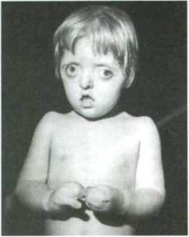
Fig. 94. Apert syndrome (acrocephalosyndactyly)
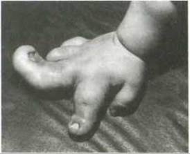
Fig. 95. Hyperdactyly and hyperphalangism
Hypoplastic anomalies include brachydactyly—reduced size of a digit; brachphalangia—reduced size of the phalanges; and hypophalangism—a reduced number of phalanges.
Last update: 08/08/2026
Editorial and Educational Adaptation: This material has been compiled based on the primary/original source text. The project team performed an editorial review, corrected technical inaccuracies, structured sections, and adapted the content for an educational format.
What was processed:
- elimination of formatting defects (OCR errors, structural breaks, corrupted characters);
- editorial organization of content;
- standardization of terminology in accordance with academic sources;
- verification of factual statements against the original source text.
All mentions of the author, publication year, and origin of the primary text have been preserved in accordance with the source.