Human Anatomy, Part 1 - K. A. Dyubenko, A. K. Kolomiytsev, Y. B. Chaykovsky 2002
Special Part
Articulations of the Bones of the Lower Limb, juncturae membri inferioris - Articulations of the Pelvic Girdle, juncturae cinguli pelvici
All Types of bone connections—both intermittent and continuous—are observed in the JOINTS OF THE pelvis. The pelvic bones feature a complex ligamentous apparatus. The sacrotuberous ligament, lig. sacrotuberale, extends from the lateral margin of the sacrum and coccyx to the ischial tuberosity (Fig. 96). The sacrospinous ligament, lig. sacrospinale, originates at the same site as the previous one, intersects with the sacrotuberous ligament, and attaches to the ischial spine. Both ligaments convert the greater and lesser sciatic notches into the corresponding greater and lesser sciatic foramina (foramen ischiadicum majus et minus), through which Muscles, Blood Vessels, and nerves pass. The obturator foramen is closed by the fibrous obturator membrane (membrana obturatoria), except for its upper-lateral margin, where a small opening remains, leading into the obturator canal (canalis obturatorius). The obturator Vessels and nerves pass through this canal.
The iliolumbar ligament, lig. iliolumbale, extends from the transverse process of the fifth lumbar vertebra to the posterior iliac crest.
The Pubic Symphysis, symphysis pubica, belongs to a special type of synchondrosis and is located in the median plane. Between the articular surfaces of the pubic bones, which are covered with hyaline Cartilage, lies an interpubic disc (discus interpubicus) containing a narrow cavity. This cavity forms during the second year of life. In women during Pregnancy, the pubic symphysis may exhibit some mobility due to the softening of the cartilage. The pubic symphysis is reinforced by the superior pubic ligament, lig. pubicum superius, and the arcuate pubic ligament, lig. arcuatum pubis, located inferior to the symphysis.
The sacroiliac joint, articulatio sacroiliaca, is formed by the auricular surfaces of the sacrum and ilium. According to the shape of its articular surfaces, it is classified as a plane, tightly fitting joint (amphiarthrosis). The articulating surfaces are covered with fibrocartilage.
The joint is reinforced by strong ligaments (see Fig. 96):
- interosseous sacroiliac ligaments, ligg. sacroiliaca interossea;
- anterior sacroiliac ligaments, ligg. sacroiliaca anteriora;
- posterior sacroiliac ligaments, ligg. sacroiliaca posteriora;
- the iliolumbar ligament, lig. iliolumbale, which extends from the transverse process of the fifth lumbar vertebra to the iliac crest (crista iliaca).
The Sacrococcygeal joint, articulatio sacrococcygea. The connection between the sacrum and the coccyx resembles the joints between the bodies of true vertebrae. This connection is reinforced by the following ligaments:
- ventral sacrococcygeal ligament, lig. sacrococcygeum anterius [ventrale];
- superficial posterior sacrococcygeal ligament, lig. sacrococcygeum posterius [dorsale] superficiale;
- deep posterior sacrococcygeal ligament, lig. sacrococcygeum profundum [dorsale];
- lateral sacrococcygeal ligament, lig. sacrococcygeum laterale.
Class="center">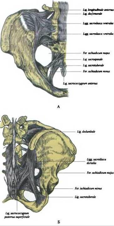
Fig. 96. Ligaments of the pelvis:
A — anterior view; B — posterior view
Last update: 08/08/2026
Editorial and Educational Adaptation: This material has been compiled based on the primary/original source text. The project team performed an editorial review, corrected technical inaccuracies, structured sections, and adapted the content for an educational format.
What was processed:
- elimination of formatting defects (OCR errors, structural breaks, corrupted characters);
- editorial organization of content;
- standardization of terminology in accordance with academic sources;
- verification of factual statements against the original source text.
All mentions of the author, publication year, and origin of the primary text have been preserved in accordance with the source.