Human Anatomy (with the Fundamentals of Dynamic and Sports Morphology) - Ivanitsky M. F. 2008
The Doctrine of Bones and Their Joints
Skeleton of the Lower Limb
BONES OF THE Lower Limb
The Skeleton of the Lower Limb is divided into the Pelvic Girdle (or pelvic girdle skeleton) and the free lower limb skeleton.
The pelvic girdle connects the free lower limb to the trunk. It includes the hip bone, the sacroiliac joint, and the pelvic joints. The free lower limb is divided into the thigh, leg (crus), and FOOT. Its skeleton comprises the Femur, Tibia, Fibula, Patella, hip joint, knee joint, tibiofibular joints, ankle joint, as well as the Bones and joints of the foot.
The hip bone is classified as a flat bone. Posteriorly, it articulates with the sacrum, and anteriorly, the two hip bones join each other at the Pubic Symphysis, forming the pelvis.
The three bones that make up the hip bone—the ilium, ischium, and pubis—contribute to The formation of the acetabulum, which serves to articulate the pelvis with the femoral HEAD. Each of these bones develops independently, but by the age of 14–16, or slightly earlier, they fuse together.
The ilium is located superior to the acetabulum (Fig. 31). It consists of a thickened portion, the body, which participates in forming the acetabulum, and the ala (wing), which is a broad, thin plate thickened at its margins. Its upper margin is called the iliac crest, to which the Abdominal Muscles attach. Anteriorly, the iliac crest terminates in the anterior superior iliac spine, and slightly below it lies the anterior inferior iliac spine. On the posterior border of the iliac ala are the posterior iliac spines—superior and inferior—which serve for the attachment of muscles and ligaments.
The internal surface of the ilium forms a depression known as the iliac fossa, which provides support for the abdominal viscera and serves as the origin for the iliacus Muscle. Posteriorly and medially on the ilium is the auricular surface, which articulates with the sacrum. Inferiorly, the iliac fossa is bounded by the arcuate line, which separates the greater pelvis from the lesser pelvis. Posterior to the auricular surface lies the iliac tuberosity, an attachment site for Ligaments and Muscles.
The ischium is located inferior to the acetabulum (see Fig. 31). It has a body that continues into the ramus of the ischium, which joins the pubic bone. At the bend of the ischium, a prominence is formed, known as the ischial tuberosity. Superior to it lies the ischial spine, which separates the greater sciatic notch from the lesser sciatic notch.
The pubic bone (see Fig. 31) consists of a body, as well as superior and inferior rami. The body of the bone contributes to the Formation of the acetabulum. The rami are angled toward each other and, at their junction, form the symphyseal surface for articulation with the pubic bone of the opposite side. Slightly lateral to this surface is the pubic tubercle, from which the pubic crest extends, continuing into the arcuate line of the ilium.
Together with the pubic bone, the ischium bounds the obturator foramen, which is covered by the obturator membrane.
Class="center">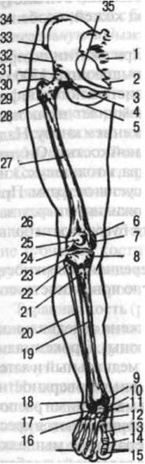
Fig. 31. Bones of the lower limb (anterior view):
1 — sacrum; 2 — coccyx; 3 — pubic bone; 4 — obturator foramen; 5 — ischium; 6 — medial epicondyle of the femur; 7 — medial condyle of the femur; 8 — medial condyle of the tibia; 9 — medial malleolus; 10 — head of the talus; 11 — navicular bone; 12 — medial cuneiform bone; 13 — 1st metatarsal bone; 14 — proximal phalanx of the 1st digit; 15 — distal phalanx of the 1st digit; 16 — cuboid bone; 17 — calcaneus; 18 — lateral malleolus; 19 — shaft of the tibia; 20 — shaft of the fibula; 21 — tibial tuberosity; 22 — head of the fibula; 23 — lateral condyle of the tibia; 24 — lateral condyle of the femur; 25 — patella; 26 — lateral epicondyle of the femur; 27 — shaft of the femur; 28 — intertrochanteric line; 29 — greater trochanter; 30 — neck of the femur; 31 — head of the femur; 32 — ilium; 33 — anterior superior iliac spine; 34 — iliac crest; 35 — ischial spine
The femur (see Fig. 31) is the largest long tubular bone. Its shaft is cylindrical in shape and slightly curved anteriorly. A linea aspera (rough line) runs along its posterior surface. On the proximal epiphysis of the femur is its head, which bears an articular surface for articulation with the acetabulum. In the center of the head is a pit (fovea capitis) to which the ligament of the head of the femur, located inside the hip joint, attaches. The head is connected to the bone shaft by the neck, the axis of which forms an angle of approximately 130° with the longitudinal axis of the femoral shaft. At the junction of the neck and shaft are two prominences: the greater and lesser trochanters. Anteriorly, they are connected by the intertrochanteric line, and posteriorly by the well-defined intertrochanteric crest, both of which serve for muscle attachment. The distal end of the femur expands into two condyles—the medial (larger) and lateral (smaller)—with an intercondylar fossa between them. The femoral condyles feature articular surfaces for articulation with the tibia and the patella. The radius of curvature of the condyle surfaces (viewed in profile) decreases posteriorly, giving their contour the shape of a spiral segment. On the lateral surfaces of the femur, slightly above the articular surfaces of the condyles, are prominences—the medial and lateral epicondyles—to which ligaments attach. These prominences, like the condyles, are easily palpable beneath the Skin, especially when the leg is flexed at the knee joint.
The mechanical strength of the femur is exceptionally high. When subjected to compression along its longitudinal axis, it can withstand loads exceeding 1,500 kg.
The patella (see Fig. 31) is located anterior to the distal epiphysis of the femur. In shape, it somewhat resembles a biconvex lens with a blunter upper edge and tapering downward. The patella is the largest sesamoid bone. It increases the leverage of the quadriceps femoris muscle, within the tendon of which it is embedded, and additionally protects the joint from trauma. By means of a ligament, which is a continuation of the quadriceps tendon, the patella attaches to the tibial tuberosity.
It is readily palpable beneath the skin. Its anterior surface is rough, while its posterior surface is smooth and features an articular surface that articulates with the femur.
The tibia (see Fig. 31) is situated on the medial side of the leg (on the side of the great toe). Its proximal end is expanded and forms two condyles: medial and lateral. On the superior aspect of the condyles are articular surfaces for articulation with the femoral condyles, and between them lies the intercondylar eminence, to which the cruciate ligaments of the knee joint are fixed. The lateral condyle features an articular surface for articulation with the head of the fibula.
The shaft of the tibia is triangular in cross-section. It has three surfaces: medial, lateral, and posterior. The medial surface is separated from the lateral surface by the anterior border, which is easily palpable under the skin, as is the entire Medial surface of the tibia. The posterior and lateral surfaces are covered by muscles. Superiorly, the anterior border transitions into the prominent tibial tuberosity, which serves for the attachment of the patellar ligament.
The posterior surface bears the rough soleal line (linea musculi solei), to which the soleus muscle attaches. The distal epiphysis has a downward-projecting prominence on the medial side—the medial malleolus—a fibular notch on the lateral side for articulation with the fibula, and an inferior articular surface for articulation with the foot.
The fibula (see Fig. 31), like the tibia, is a long tubular bone. It is located laterally in the leg. Its proximal end terminates in a head that articulates with the tibia, while its distal end forms the lateral malleolus. Both the head and the lateral malleolus of the fibula are easily palpable beneath the skin. On the medial side of the lateral malleolus is an articular surface that articulates with the talus. A groove runs along its posterior surface, lodging the tendons of the fibular (peroneal) muscles.
There is a space between the bones of the leg, which is spanned by the interosseous membrane of the leg. Distally, the leg transitions into the foot.
The foot consists of three parts: the tarsus, metatarsus, and digits (Phalanges). The Tarsal Bones include the talus, calcaneus, navicular, cuboid, and three cuneiform bones—medial, intermediate, and lateral (counted from the side of the great toe). All of these are short spongy bones. The metatarsus consists of five short tubular Metatarsal Bones. The digits consist of phalanges, which are also short tubular bones.
The talus (Fig. 32) is positioned between the distal ends of the leg bones and the calcaneus, acting as a sort of bony meniscus between the bones of the leg and the foot. It comprises a body and a head.
The superior surface of the body bears a pulley-shaped articular surface (trochlea) that serves for articulation with the bones of the leg; the anterior surface of the head features an articular surface for articulation with the navicular bone; the medial and lateral sides of the body bear articular surfaces for articulation with the malleoli; and the inferior surface articulates with the calcaneus.
The calcaneus (see Fig. 32) forms the posteroinferior part of the tarsus. It is the largest bone of the foot. It consists of a body and the calcaneal tuberosity, which projects posteriorly. Superiorly, the bone features articular surfaces for articulation with the talus, anteriorly with the cuboid, and medially with a shelf-like projection known as the sustentaculum tali.
The navicular bone (see Fig. 32) is located at the medial margin of the foot, anterior to the talus, posterior to the cuneiforms, and medial to the cuboid. Its medial border presents a downward-facing tuberosity that is easily palpable beneath the skin and serves as a landmark for determining the height of the medial longitudinal arch of the foot.
The cuboid bone (see Fig. 32) lies on the lateral border of the foot and articulates with the calcaneus, navicular, lateral cuneiform, and the 4th and 5th metatarsal bones. Its plantar surface features a groove that lodges the tendon of the fibularis (peroneus) longus muscle.
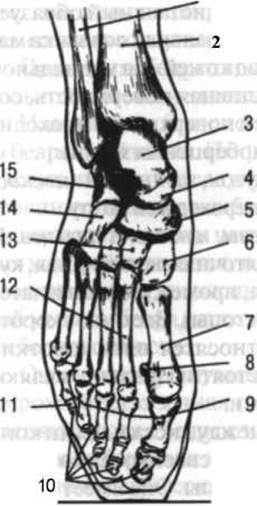
Fig. 32. Bones of the foot (dorsal aspect). Position of the foot within a shoe on the toes (drawn from a radiograph):
1 — fibula; 2 — tibia; 3 — trochlea of the talus; 4 — head of the talus; 5 — navicular bone; 6 — medial cuneiform bone; 7 — 1st metatarsal bone; 8 — sesamoid bone; 9 — proximal phalanges; 10 — distal phalanges; 11 — middle phalanx of the 5th digit; 12 — 2nd–5th metatarsal bones; 13 — intermediate and lateral cuneiform bones; 14 — cuboid bone; 15 — calcaneus
The cuneiform bones (see Fig. 32)—medial, intermediate, and lateral—lie anterior to the navicular bone, posterior to the first three metatarsals, and medial to the cuboid. The wider part of the medial cuneiform is directed inferiorly and the narrower part superiorly, whereas the reverse is true for the other two cuneiforms. Together with the cuboid and the bases of the metatarsals, these three bones contribute to the formation of the transverse arch of the foot, which is convex superiorly and concave inferiorly.
The metatarsal bones (see Fig. 32). Each of the five metatarsals is a long (tubular) bone. They are divided into a base, a body (shaft), and a head.
The shafts of the metatarsals are slightly convex on the dorsal aspect of the foot and concave on the plantar aspect. In shape, these bones resemble three-sided prisms. The 2nd metatarsal is the longest, while the 1st is the shortest and thickest. The bases of the metatarsals bear articular surfaces for articulation with the tarsal bones as well as with adjacent metatarsals, while their heads feature surfaces for articulation with the proximal phalanges of the toes. The 5th metatarsal has a prominence on its lateral margin—the tuberosity—which is easily palpable beneath the skin.
All metatarsal bones can be easily palpated from the dorsal side because they are covered by a relatively thin layer of soft Tissues; on the plantar side, they lie deep beneath a mass of muscles and subcutaneous adipose tissue. The metatarsals do not lie in a single plane, but rather form a transverse arch.
The bones of the toes (see Fig. 32). The toes consist of phalanges. Similar to the fingers, the great toe (hallux) has two phalanges, while the remaining toes have three each. Frequently, the two phalanges of the 5th toe fuse together, resulting in a two-phalangeal digit. Proximal, middle, and distal phalanges are distinguished. They differ from the phalanges of the hand in being shorter, particularly the distal ones. As in the hand, sesamoid bones are present in the foot. Here they are much better developed and are located in the region of the joints between the first and fifth metatarsals and their proximal phalanges. These sesamoid bones enhance the transverse arching of the metatarsus in its anterior region. Another sesamoid bone is found within the tendon of the fibularis (peroneus) longus muscle, positioned within the groove on the plantar surface of the cuboid bone. Additionally, small sesamoid bones are occasionally encountered between the proximal and distal phalanges of the great toe.
JOINTS OF THE lower limb
The bones of the pelvic girdle are connected to the sacrum and to each other by the sacroiliac joint and the pubic symphysis. The sacroiliac joint is formed by the auricular surfaces of the sacrum and the ilium. It is a simple, plane joint. Movements within it are restricted (only 3–5°). The joint is reinforced by numerous ligaments. Among these, the ventral and dorsal sacroiliac ligaments are located on the anterior and posterior surfaces of the joint, the interosseous ligaments lie within the joint, and the iliolumbar ligament extends from the 4th and 5th lumbar vertebrae to the ilium. Furthermore, the sacroligamentous structures—the sacrospinous and sacrotuberous ligaments—run from the sacrum to the ischial spine and ischial tuberosity respectively; by reinforcing the sacroiliac joints, they convert the greater and lesser sciatic notches into the greater and lesser sciatic foramina.
The pubic symphysis is formed by the opposed surfaces of the pubic bones, between which lies a cartilaginous disc. It belongs to the category of amphiarthroses (or symphyses), since a small cavity typically forms within the center of the Cartilage, or disc, around the second year of life, converting the primary cartilaginous joint (synchondrosis) into a secondary cartilaginous joint (symphysis). This cartilage is fibrocartilage, though it transitions to hyaline cartilage immediately adjacent to the bones. The symphysis is reinforced inferiorly by the arcuate pubic ligament. Depending on the site of pelvic weight-bearing—the femoral heads in the standing position or the ischial tuberosities in the sitting position—the constituent cartilage of the symphysis undergoes compression or tension. The pubic symphysis, together with the sacroiliac joints, ensures the structural integrity and rigidity of the pelvis.
The Pelvis as a whole. The pelvis forms a closed bony ring composed of the right and left hip bones, the sacrum, and the coccyx. It is divided into an upper, larger division—the greater (false) pelvis—and a lower, smaller division—the lesser (true) pelvis. The terminal line (pelvic brim) separating the greater and lesser pelvis begins posteriorly at the sacral promontory, follows the arcuate line, and reaches the pubic tubercle anteriorly.
The greater pelvis is formed mainly by the iliac wings and partly by the pubic bones, whereas the walls of the lesser pelvis are formed below the terminal line not only by the sacrum, coccyx, and hip bones, but also by the sacrospinous and sacrotuberous ligaments and the obturator membranes.
The greater pelvis serves as support for the abdominal viscera and as an attachment site for the abdominal wall muscles. The lesser pelvis resembles a canal with two apertures—an inlet and an outlet. It contains the Urinary Bladder and rectum, and, in females, additionally the Uterus and Vagina.
The pelvis exhibits marked Sexual Dimorphism: the female pelvis is broader and shorter than the male pelvis. The iliac wings of the female pelvis are more flared and positioned more vertically than in males. The pelvic inlet is larger in females, and the lesser pelvis narrows less inferiorly than in males, which is associated with a lesser curvature of the anterior surface of the sacrum and a wider divergence of the ischial tuberosities in women. The inferior pubic rami form an arch where they meet in females and intersect at a right angle relative to the ischial rami (in the male pelvis, the pubic arch forms an angle of 70–75°).
A comparison of the pelvic inlet and outlet reveals that the inlet has a greater transverse diameter, while the outlet possesses a greater anteroposterior diameter due to the mobility of the coccyx.
Relative to the horizontal plane of the body, the plane of the pelvic inlet has an inclination of 55–75°. This inclination is greater in the standing position than when seated. Viewed in profile, when standing, the anterior superior iliac spines and the anterior surface of the pubic symphysis lie approximately in the same coronal plane. Pelvic inclination can decrease significantly when sitting, depending on spinal mobility. Body weight is transmitted via THE Vertebral Column to the sacrum, and from the sacrum in two directions: toward the pubic symphysis and toward the ischial tuberosities.

Fig. 33. Hip joint (anterior view):
1 - sacrospinous ligament; 2 - sacrotuberous ligament; 3 - obturator membrane; 4 - pubofemoral ligament; 5 - acetabular labrum; 6 — iliofemoral ligament
The hip joint (Fig. 33) is formed by the acetabulum of the hip bone and the head of the femur. The depth of the acetabulum is increased by the acetabular labrum, which attaches to its rim. The capsule of the hip joint is exceptionally strong owing to the ligamentous apparatus woven into it. The strongest ligament is the iliofemoral ligament, which can withstand loads of up to 300 kg. It originates just below the anterior inferior iliac spine and attaches, radiating in a fan-like manner, to the intertrochanteric line. The ischiofemoral and pubofemoral ligaments are considerably weaker than the iliofemoral ligament. They extend to the femur from the ischium and pubis, respectively. In the standing position, these ligaments, like the iliofemoral ligament, are under tension. They act to restrict extension of the thigh; the iliofemoral ligament also limits abduction and adduction, the pubofemoral limits abduction, and the ischiofemoral limits medial rotation (pronation). Deep within the Joint Capsule lies the circular zone (orbicular zone), which encircles the femoral neck like a ring. Within the joint cavity is the ligament of the head of the femur (ligamentum teres), which runs from the acetabulum to the femoral head. This ligament is not under tension. If the joint capsule of a cadaveric hip is severed, the femoral head easily dislocates from the acetabulum.
Apparently, the ligament of the head of the femur does not serve to reinforce the hip joint, but rather fulfills another function: Blood Vessels run within the ligament, and it provides them with protection; furthermore, the ligament acts as an elastic cushion for the femoral head and helps absorb shocks experienced by the body during various movements. However, in asymmetric body postures where the pelvis is tilted obliquely, the ligament of the head of the femur on the side of the weight-bearing (typically straightened) leg becomes taut and contributes to the stabilization of the hip joint.
The hip joint is a simple, spherical (nut-shaped) joint. Much like the shoulder joint, it has three axes of rotation: a transverse axis, around which hip flexion (forward movement) and extension (backward movement) take place; an anteroposterior axis, which governs abduction and adduction; and a vertical axis, which allows for supination and pronation. In addition, the hip joint permits circumduction of the thigh.
Thigh mobility in the hip joint is more limited than shoulder mobility in the shoulder joint for several reasons: first, the articulating bone surfaces in the hip joint show a much greater degree of congruence; second, the Ligamentous apparatus of the hip joint is significantly stronger; and third, the hip joint is surrounded by substantially more powerful muscles.
The range of hip mobility during flexion and extension is 120°, with approximately 105° dedicated to flexion and 15° to extension. Passive flexion can reach 150–160°. The degree of flexion increases when this movement is combined with slight limb abduction (which is important, for example, when a jumper clears the bar) as well as with knee flexion. Thus, with the lower leg flexed at the knee, hip flexion reaches 118° in women and 112° in men, whereas with the lower leg extended, it is only 84° and 87°, respectively. This limitation in mobility is due to two main factors: first, when the lower leg is extended, the center of gravity of the thigh shifts distally, increasing the torque of gravity (making flexion harder to execute with the same muscular strength); and second, the passive insufficiency of the hamstring muscles.
Mobility in the hip joint around its transverse axis can be increased through systematic training; for instance, when performing the splits (anteroposterior), the iliofemoral ligament of the "rear" leg and the hamstring and knee flexors/extensors of the "front" leg are stretched. As the ligamentous apparatus undergoes intensive stretching, The Role of the muscles in stabilizing the hip joint increases. With good muscle tone, this reinforcement is entirely sufficient to keep the femoral head securely within the acetabulum.
Hip abduction is possible only within a range of 40–60°, and adduction within 15–30°; however, when the limb is flexed at the knee, both abduction and adduction increase to 74–80°. The degree of hip abduction depends on THE POSITION OF the thigh. In a supinated position ("turned-out position"), the degree of abduction is significantly greater than in the neutral position where the toes point forward. With a supinated thigh, the greater trochanter does not obstruct abduction because it does not jam against the upper rim of the acetabulum; consequently, during a side leg raise ("side kick"), the thigh is always kept in a more supinated position.
Supination and pronation range from 15° to 40°; when the hip is flexed, the range of these movements nearly doubles.
Under METABOLISM/18.html">The Influence of systematic training, the mobility of the thigh around the vertical axis of the hip joint increases. For instance, the turned-out foot position (at a 180° angle relative to one another), characteristic of classical ballet and dance, is primarily determined by the positioning of the femurs within the hip joints. This position is relatively easy to assume when both feet are supported by the floor. However, if one foot is slightly elevated, maintaining this posture becomes impossible. In children who do not practice choreography, "active turnout"—achieved solely through Muscle contraction—averages 130° in girls and 121° in boys; in children of the same age undergoing systematic dance training, "active turnout" is greater, averaging 146° in girls and 134° in boys, and by ages 16–18 it reaches an average of 152° in girls and 145° in boys. These figures represent the combined mobility of the right and left hip joints. The range of outward rotation in each individual joint constitutes half of this total value, although it should be noted that joint mobility—particularly in the hips—is not strictly symmetrical.
When the body is fully extended (standing or lying down), the thigh is in one of its extreme positions. The "neutral position," in which the entire ligamentous apparatus of the hip joint is maximally relaxed, occurs when the thigh is slightly flexed, abducted, and supinated. In this position, the mobility of the thigh around all three axes of rotation is approximately equal. It should also be emphasized that individual variations in hip joint mobility can be exceptionally wide.
The knee joint (Fig. 34) is formed by the femoral condyles, the superior articular surface of the tibia, and the patella. It is a complex, condylar-hinge joint. From an extended position, it Functions as a hinge joint. However, as flexion progresses, pronation and supination can occur due to the decreasing radius of curvature of the articular surface of the femoral condyles.
Expressed in degrees, this mobility breaks down as follows: active flexion is 130°, passive flexion provides an additional 30°, and hyperextension from the neutral position adds another 10–12°. Thus, total mobility during flexion reaches 170°. As the knee flexes, its collateral ligaments slacken, thereby permitting a degree of rotational and circumduction movement. Passive pronation and supination in the knee joint are possible within a range of 10° when the lower leg is extended.
The knee joint possesses low congruence: the femoral condyles are highly convex, while the concavity on the tibial condyles is shallow. This congruence is improved by two menisci—medial and lateral—located inside the joint (Fig. 34). In addition, the menisci cushion the shocks and impacts experienced by the body during movement (walking, running, jumping, etc.) and contribute to a more even distribution of weight from the thigh onto the tibia. The menisci are roughly semilunar in shape. Their outer margin is thickened, while the inner edge is sharp. The medial meniscus is larger than the lateral one, which correlates with the larger size of the medial femoral condyle. Both menisci are connected anteriorly by the transverse ligament of the knee, and their ends are attached to the intercondylar eminence of the tibia. The joint capsule of the knee is thin and loose.

Fig. 34. Knee joint (anterior view):
1 — posterior cruciate ligament; 2 — anterior cruciate ligament; 3 — tibial collateral ligament; 4 — medial meniscus; 5 — patellar ligament; 6 — patella; 7 — interosseous membrane of the leg; 8 — anterior ligament of the fibular head; 9 — fibular collateral ligament
The knee joint features A number of ligaments, including the tibial and fibular collateral ligaments running from the medial and lateral epicondyles of the femur to the tibia and fibula. Located intra-articularly are the cruciate ligaments (see Fig. 34): the anterior cruciate ligament originates from the inner surface of the lateral femoral condyle, runs downward, forward, and medially, and attaches to the anterior intercondylar area of the tibia; the posterior cruciate ligament originates from the inner side of the medial femoral condyle, runs downward, backward, and laterally, and attaches to the posterior intercondylar area of the tibia. Situated in the posterior wall of the knee joint capsule is the strong oblique popliteal ligament, which is partly a continuation of the tendon fibers of the semimembranosus muscle. Running anteriorly to the knee joint is the tendon of the quadriceps femoris, which attaches to the patella. The continuation of this tendon is the patellar ligament, which extends down to the tibial tuberosity. This ligament is separated from the joint cavity by a bursa and a significant accumulation of loose Connective Tissue.
The cruciate ligaments are crucial for stabilizing the knee joint: the anterior ligament prevents the femur from sliding backward, while the posterior prevents it from sliding forward. They can also exert a braking or limiting effect during the extension and flexion of the lower leg at the knee. During extension, the anterior fibers of the anterior ligament and the posterior fibers of the posterior ligament are stretched, and vice versa during flexion. Consequently, tears of the cruciate ligaments can occur during abrupt flexion-extension movements (for example, when kicking a ball in soccer).
The collateral ligaments prevent sideways Displacement of the articulating bones, while also limiting extension and rotation. Their braking effect on extension manifests at angles of 183–185°, meaning even during slight hyperextension. Supination movements are checked by the tibial collateral ligament, whereas pronation movements are checked by the fibular ligament. Possessing minimal elasticity, these ligaments do not stretch when movements exceed the specified range of mobility—instead, they tear.
Among the Components of the knee joint, the menisci are injured most frequently, with the medial meniscus suffering damage more often than the lateral one (the latter being more prone to cystic degeneration). One reason for this is the adhesion of the medial meniscus to the tibial collateral ligament and its greater mobility during supination of the lower leg in a semi-flexed knee position. The lower incidence of injury to the lateral meniscus is due to its ability to shift during the contraction of the popliteus muscle. Meniscus injuries occur more frequently in soccer players than in other athletes, affecting both the weight-bearing and the non-weight-bearing leg.
The synovial membrane of the knee joint has a complex Structure. It lines the inner surface of the joint capsule and, as it approaches the intra-articular cruciate ligaments, envelops them from the front and sides, forming numerous folds containing adipose tissue. The synovial membrane contains numerous synovial villi, which are particularly abundant around the patella. A large number of bursae are located in the vicinity of the knee joint.
The Location OF THE knee joint line, as well as the patellar ligament itself, is easily palpable from the front both when the lower leg is extended and, especially, when it is flexed. The femoral and tibial condyles can be felt on either side of the patella. Posteriorly, the joint line cannot be palpated because the popliteal fossa is covered by a thick layer of soft tissue.
Connections of the bones of the leg. An interosseous membrane, similar to the one found in the forearm, is located between the bones of the leg—the tibia and fibula. In addition, the head of the fibula articulates with the tibia via a planar joint reinforced anteriorly and posteriorly by ligaments, which represent capsular thickenings where the periosteum of one bone transitions into that of the other. The distal ends of the leg bones are joined by a syndesmosis, though a small joint cavity communicating with the ankle joint may also be present here.
The ankle joint is formed by the bones of the leg and the talus. The medial and lateral malleoli of the leg bones form a mortise that grasps the trochlea of the talus (Fig. 35).
The ankle joint is complex and hinge-shaped. Around the transverse axis passing through the trochlea, it allows for plantarflexion (movement toward the SOLE OF THE foot) and dorsiflexion (movement toward the dorsum of the foot). Because the trochlea narrows posteriorly, slight adduction and abduction around the vertical axis become possible as the foot is plantarflexed. The joint is reinforced by ligaments located on its medial and lateral sides (see Fig. 35). On the medial side lies the triangular medial (deltoid) ligament, running from the medial malleolus to the navicular, talus, and calcaneus. On the lateral side, the joint is stabilized by the calcaneofibular and talofibular ligaments. The ligaments of the ankle joint possess substantial strength. Among the ligaments situated on the lateral side of the joint, the calcaneofibular is the strongest.
In the standing position, dorsiflexion of the foot is possible within a range of 15–25°, plantarflexion is 45–50°, abduction and adduction are 12° each, and pronation and supination range up to 13°.
One of the characteristic age-related Features of the ankle joint is that in adults it exhibits greater mobility toward the plantar surface of the foot, whereas in children—particularly newborns—mobility is greater toward the dorsum of the foot, which is associated with the developmental CHARACTERISTICS OF THE foot.
The position of the ankle joint cleft is clearly visible from the front, especially when the muscles on the anterior aspect of the lower leg, whose tendons pass through this area, are relaxed.
The subtalar joint is formed by the talus and calcaneus. It is located in the posterior region of these bones. The articular surfaces of the connecting bones are congruent, forming a spiral-shaped joint. It is a simple joint enclosed in a thin capsule reinforced with small ligaments.
In the anterior region, situated between the talus and the calcaneus, lies another joint that is separate from the previous one and has a more complex structure.
The talocalcaneonavicular joint is formed by three bones: the talus (via its head), the calcaneus (via its anterosuperior articular surface), and the navicular bone (via its posterior surface). This is a complex, ball-and-socket joint. A common axis of rotation runs anteroposteriorly between the talus and the calcaneus, enabling foot pronation and supination. During pronation, the medial border of the foot moves downward while the lateral border rises; during supination, the reverse occurs. Among the ligaments supporting this joint, the interosseous talocalcaneal ligament is The most significant. Functioning simultaneously, the subtalar and talocalcaneonavicular joints form a combined articulation.

Fig. 35. Joints and ligaments of the right foot:
1 and 9 — medial (deltoid) ligament; 2 — plantar calcaneonavicular ligament; 3 — long plantar ligament; 4 — plantar cuneonavicular ligament; 5 — plantar tarsometatarsal ligament; 6 — dorsal tarsometatarsal ligaments; 7 — dorsal cuneonavicular ligaments; 8 — talonavicular (dorsal) ligament
Complementing one another, the ankle, subtalar, and talocalcaneonavicular joints allow the foot to perform flexion and extension, adduction and abduction, pronation and supination, and circumduction. The range of foot mobility around the frontal axis reaches 90°, and around the sagittal axis, 55°. One of the age-related characteristics of bone positioning and joint movement in the foot is that, with age, a certain degree of pronation occurs along with a lowering of the medial part of the longitudinal arch. The foot of a child, particularly During the first year of life, exhibits a distinct supinated posture; consequently, when starting to walk, children frequently place their weight not on the entire plantar surface, but solely on its lateral border.
The calcaneocuboid joint is formed by the calcaneus and the cuboid bone. It is simple in structure and flat in shape. Its joint capsule is reinforced by accessory ligaments, the strongest of which is the long plantar ligament running from the calcaneus to the bases of the 2nd–5th metatarsal bones. Together with the talonavicular joint, this articulation forms a single transverse tarsal joint characterized by limited mobility. On the dorsal side, it is reinforced by the bifurcated ligament, which connects the calcaneus to the navicular and cuboid bones.
The joints between the cuneiform, navicular, and cuboid bones are flat and possess minimal mobility. They are strongly reinforced by ligaments located predominantly on the dorsal and plantar aspects of the foot.
The tarsometatarsal joints are located between the tarsal and metatarsal bones and are generally flat in shape, except for the joint between the medial cuneiform and the 1st metatarsal bone, which may sometimes be classified as a saddle joint. These joints are well-supported by ligaments situated on both the dorsal and plantar sides of the foot.
The metatarsophalangeal joints are spheroidal in shape, although their mobility is relatively limited. A distinctive feature of the 1st metatarsophalangeal joint is the presence of two sesamoid bones located on its plantar surface. These joints primarily allow for flexion and extension. They are reinforced by collateral ligaments.
The interphalangeal joints of the foot lie between the individual phalanges of the toes and are trochlear (hinge-shaped) in form. When the foot is at rest, the toes are slightly flexed at these joints, whereas at the metatarsophalangeal joints they are conversely somewhat extended. The range of motion (flexion and extension) is small. Ligaments are located on the lateral and medial sides of each joint.
The Foot as a whole. The foot functions as a supporting, Shock-absorbing, and locomotory apparatus of The Human Body. The shock-absorbing function of the foot is attributed to the presence of its arches. Two primary arches are distinguished: the longitudinal and the transverse.
The longitudinal arch consists of two parts: medial and lateral. Its medial part is formed by the calcaneus, talus, navicular, three cuneiforms, and the first three metatarsal bones; the lateral part is formed by the calcaneus, cuboid, and the 4th and 5th metatarsals. The medial part of the longitudinal arch has a height of 5–7 cm (measured from the tuberosity of the navicular bone), while the lateral part is approximately 2 cm high (measured from the tuberosity of the 5th metatarsal bone). The former is referred to as the shock-absorbing part and the latter as the weight-bearing part of the longitudinal arch.
The transverse arch runs principally across the cuneiform and cuboid bones as well as the bases of the metatarsals, and is often also well-defined in the region of the metatarsal heads (Fig. 36).
The Maintenance of the foot arches is facilitated by passive ties—ligaments (such as the long plantar ligament)—and active ties—foot muscles running longitudinally (toe flexors) as well as transversely (fibularis longus, the transverse head of the adductor hallucis, etc.).
In a standing position, the foot bears weight on the ground via the calcaneus and the metatarsal heads. Feet are conventionally classified as normal, arched, or flat. A normal foot leaves an imprint featuring an isthmus that connects the area corresponding to the calcaneus with the region of the metatarsal heads. An arched foot lacks this connection, resting on the ground only at its anterior and posterior sections without midfoot contact. A flat foot produces a solid imprint with no arch in its middle section. Intermediate forms exist between these three foot types.
It is necessary to differentiate between anatomical and functional Flatfoot. The first type of flatfoot is characterized by the foot retaining good mobility and functioning normally. For instance, in athletic training, a powerful push-off during a jump can still be achieved. In isolated cases, remarkable jumping ability is observed even in complete flatfoot. The second type of flatfoot is marked by severely restricted joint mobility in the foot. This represents true flatfoot, which is typically implied when discussing the structural deficiencies of the flat foot as a weight-bearing and shock-absorbing organ. In children, flatfoot is commonly associated with an excessively pronounced forward curvature of the lumbar spine (increased lumbar lordosis, or "lordotic posture").

Fig. 36. STRUCTURE OF THE transverse arch of the foot in the tarsal and metatarsal regions:
1 — transverse arch formed by the cuneiform bones (medial, intermediate, lateral) and the cuboid bone; 2 — transverse arch formed by the metatarsal bones
The toes of the foot also leave an imprint when standing. However, they serve as a supportive base only when the toe flexors are contracted. If the toes are slightly lifted during normal standing, the body remains in a state of balance. Conversely, if the torso is shifted forward to the extent that the line of gravity passes through the metatarsal heads, extension of the toes results in a loss of balance and a fall. During walking, running, or jumping at the moment of push-off—when the toes are strongly extended and the tone of their flexors is heightened—they invariably act as a supportive base. In contrast, when the toe flexors are relaxed, as in quiet standing, the toes do not function as an "active" base of support.
When the Muscles of the Plantar Surface of the Foot are relaxed or fatigued, the primary weight-bearing points among the metatarsal heads are the heads of the 2nd and 3rd metatarsal bones.
Last update: 08/08/2026
Editorial and Educational Adaptation: This material has been compiled based on the primary/original source text. The project team performed an editorial review, corrected technical inaccuracies, structured sections, and adapted the content for an educational format.
What was processed:
- elimination of formatting defects (OCR errors, structural breaks, corrupted characters);
- editorial organization of content;
- standardization of terminology in accordance with academic sources;
- verification of factual statements against the original source text.
All mentions of the author, publication year, and origin of the primary text have been preserved in accordance with the source.