Human Anatomy (with the Fundamentals of Dynamic and Sports Morphology) - Ivanitsky, M. F. 2008
Myology
General Part
Skeletal Muscles are formed by Striated Muscle tissue (see p. 35) and are therefore capable of voluntary contractions. They maintain posture and body positions, participate in body movements (Figs. 37, 38), protect underlying Internal Organs and the Blood Vessels and nerves running between them from external impacts; Muscle contraction generates heat, thus helping to maintain a constant body Temperature. Furthermore, muscle contractions express a person's emotional state through facial expressions and body language (see p. 227).
The presence of specific nerve endings in muscles allows them to be considered a component of the motor analyzer (sensory) system.
A variety of criteria are used to name muscles. Muscle names derive from their external shape (deltoid, rhomboid, quadrate, trapezoid, serratus, soleus, piriformis, lumbrical, orbicularis, pyramidal, round, etc. — Fig. 39), function (flexor, extensor, abductor, adductor, pronator, supinator, levator, depressor, tensor, masticatory, sphincter, dilator, etc.), Structure or number of heads (digastric, semimembranosus, semitendinosus, biceps, triceps, quadriceps), position (intercostal, pectoral, popliteal, etc.), origin and insertion (pectineus, brachioradialis, sternocleidomastoid, etc.), direction of muscle fibers (straight, oblique, transverse — Fig. 40), or random features based on distant associations (gemelli muscles, corrugator supercilii, etc.).
Muscle shape. Muscles vary greatly in shape and size. There are long and thin, short and thick, wide and flat muscles. There are also fan-shaped muscles, which can move a bone within an angle limited by the outermost fascicles of the muscle, and circular muscles, The formation of which is associated with the functional need to periodically constrict certain openings (M. A. Jafarov).
Class="center">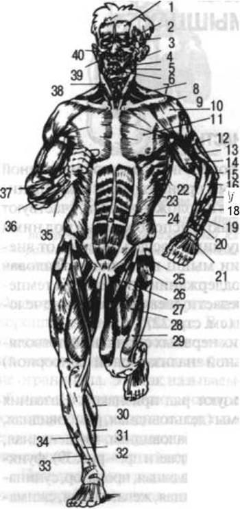
Fig. 37. Muscles (general view from the front):
1 — frontal belly of the occipitofrontalis muscle, 2 — orbicularis oculi muscle; 3 — temporalis muscle; 4 — masseter muscle; 5 — buccinator muscle; 6 — orbicularis oris muscle; 7 — depressor anguli oris muscle; 8 — trapezius muscle; 9 — infraclavicular fossa, 10 — deltoid muscle; 11 — pectoralis major muscle; 12 — triceps brachii muscle; 13 — brachialis muscle; 14 — biceps brachii muscle; 15 — brachioradialis m.; 16 — extensor carpi radialis longus; 17 — extensor carpi radialis brevis; 18 — extensor digitorum; 19 — abductor pollicis longus m.; 20 — extensor pollicis brevis; 21 — extensor pollicis longus; 22 — serratus anterior m.; 23 — external oblique m. of the abdomen; 24 — rectus abdominis m.; 25 — gluteus medius m.; 26 — tensor fasciae latae m.; 27 — sartorius m., 28 — rectus femoris m.; 29 — vastus lateralis m. of the thigh; 30 — gastrocnemius m.; 31 — soleus m.; 32 — tibialis anterior m.; 33 — extensor hallucis longus; 34 — extensor digitorum longus; 35 — flexor carpi ulnaris; 36 — extensor carpi ulnaris; 37 — extensor digitorum; 38 — sternocleidomastoid m.; 39 — zygomaticus Major and minor m.; 40 — levator labii superioris alaeque nasi m.
Muscles located on the trunk are flatter in shape than those on the limbs. Limb muscles are characterized by a relatively greater length, a fusiform shape (Fig. 41), and span across one (monoarticular), two (biarticular), or several (polyarticular) joints (Fig. 42).
Differences in muscle shape are related to their function. Long, thin muscles with a small attachment area on bones (e.g., long flexors of the fingers or toes) typically participate in wide-amplitude movements. In contrast, short, thick muscles (e.g., quadratus lumborum) participate in low-amplitude movements but can overcome significant resistance.
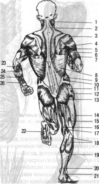
Fig. 38. Muscles (general view from the back):
1 — sternocleidomastoid m.; 2 — trapezius m.; 3 — deltoid m.; 4 — teres minor m.; 5 — infraspinatus m.; 6 — teres major m.; 7 — triceps brachii m.; 8 — latissimus dorsi m.; 9 — external oblique m. of the abdomen; 10 — lumbar triangle; 11 — gluteus medius m.; 12 — tensor fasciae latae m.; 13 — gluteus maximus m.; 14 — vastus lateralis m. of the thigh; 15 — biceps femoris m.; 16 — semitendinosus m.; 17 — semimembranosus m.; 18 — gastrocnemius m.; 19 — soleus m.; 20 — calcaneal (Achilles) tendon; 21 — fibularis (peroneus) longus m.; 22 — flexor digitorum brevis; 23 — brachioradialis m.; 24 — flexor carpi radialis; 25 — palmaris longus m.; 26 — flexor carpi ulnaris
P. F. Lesgaft proposed dividing muscles into two main types: strength muscles and dexterity muscles. He wrote: "Muscles that are predominantly strong originate and attach to large surfaces; as the attachment surface increases and moves further away from the fulcrum of the lever upon which they act, they can exert considerable force with little tension, which is why they do not fatigue easily. They act predominantly with their entire mass and cannot produce subtle nuances of movement; they manifest their strength at a relatively low speed and most often consist of short muscle fibers. Muscles of the second type, distinguished by dexterity in their actions, originate and attach to small surfaces close to the fulcrum of the lever on which they act; they act with greater tension, fatigue more quickly, most often consist of long fibers, and can act through their individual parts, producing various nuances of movement. These are muscles that primarily allow for dexterous and rapid movements." According to P. F. Lesgaft, Examples of strength muscles include the extensors of THE Vertebral Column, the gluteus maximus, and the quadriceps femoris; examples of dexterity muscles include the muscles of THE EYE AND face. Transitional forms exist between these extreme types of dexterity and strength muscles.
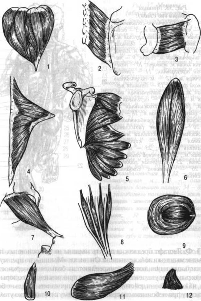
Fig. 39. Muscle shapes:
1 — deltoid m.; 2 — rhomboid m.; 3 — quadratus femoris m.; 4 — trapezius m.; 5 — serratus anterior m.; 6 — soleus m.; 7 — piriformis m.; 8 — lumbrical mm.; 9 — orbicularis oculi m.; 10 — pyramidalis m.; 11 — teres major m.; 12 — triangular m. (now called the depressor anguli oris muscle)

Fig. 40. Direction of muscle fibers:
1 — rectus abdominis m.; 2 — external oblique m. of the abdomen; 3 — transversus abdominis m.; a — muscular part; b — aponeurosis
All large muscles consist of several individual muscles that are united into a single whole only anatomically, but have independent innervation and can contract in isolation. Therefore, they can be considered functionally independent muscles that often perform opposite, "antagonistic" work. Only small muscles represent a unified entity both anatomically and functionally. For example, large muscles such as the pectoralis major and serratus anterior cause completely opposite movements when their upper and lower sections contract in isolation (the upper part of the pectoralis major participates in flexion of the humerus, i.e., forward movement, while the lower part participates in depression); the anterior part of the deltoid muscle moves the arm forward when contracted in isolation, the posterior part moves it backward, and the middle part moves it laterally. The ability to isolate the contraction of individual PARTS OF THE same muscle or the contraction of a single muscle (e.g., the rectus abdominis or biceps brachii) without the involvement of neighboring muscles depends on training. Usually, muscle contraction has a summative character, meaning an entire group of muscles contracts simultaneously.
3 P. F. Lesgaft, Fundamentals of Theoretical Anatomy, part 1, 1905, pp. 249–250.
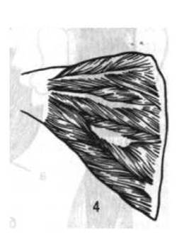
Fig. External and Internal Structure of muscles:
1 — fusiform m.; 2 — unipennate m.; 3 — bipennate m.; 4 — multipennate m.
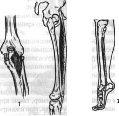
Fig. 47 Uni-, bi-, and multiarticular muscles:
1 — uniarticular (brachialis) muscle, 2 — biarticular (rectus femoris) muscle; 3 — multiarticular (flexor hallucis longus) muscle
Muscle Structure. Each Skeletal Muscle is composed of bundles of striated muscle fibers (Fig. 43). These bundles are bound together and surrounded by loose Connective Tissue, with layers known as the internal perimysium. The outer surface of the muscle is covered by the external perimysium.
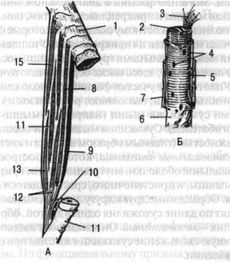
Fig. 43. Structure of a striated muscle
(A — muscle between two bones. B — individual fiber at high magnification):
1, 11 — periosteum; 2 — fiber cross-section; 3 — myofibrils; 4 — muscle fiber nuclei; 5 — CONNECTIVE TISSUE Cell Nucleus; 6 — sarcolemma; 7 — endomysium; 8, 13–15 — muscle fiber bundles surrounded by the internal perimysium; 9 — external perimysium; 10 — synovial bursa; 12 — tendon fibers (after Braus)
Muscle fibers form the middle, fleshy part of the muscle — its belly or body, which attaches to bones via tendinous ends. Tendons are particularly well-developed in long muscles (see Fig. 41).
Tendons are formed from Collagen fibers and exhibit high tensile strength. Broad muscle tendons are called aponeuroses (see Fig. 40) or tendinous expansions. The term aponeurosis is also used to describe thickened fascias located beneath the Skin of the palmar surface of the hand and the plantar surface of the FOOT. Some muscles (such as the rectus abdominis) feature tendinous intersections that divide the entire muscle into individual segments, enabling their isolated contraction.
Fascias covering muscles are fibrous sheaths that encase not only individual muscles but also groups of muscles. The Role of fascia in The Musculoskeletal System is extremely significant: by covering muscles and attaching to bones, they serve as a functional extension of the Skeletal System. Some fascias serve as sites of muscle origin or insertion. Thickened bands formed by fascia between individual muscle groups are called intermuscular septa, which also serve as sites of origin for muscle fiber bundles. Condensed areas of fascia located over the tendons of long muscles act as ligaments and are termed tendon retinacula (e.g., flexor or extensor retinacula). Tendons characterized by significant mobility (primarily in the hand and foot) are enclosed within synovial sheaths composed of two layers of the synovial membrane: the visceral layer, adhering to the muscle tendon, and the parietal layer, fusing with surrounding Tissues. These continuous layers of the synovial membrane transition into one another along the length of the tendon, forming a tendon mesentery, or mesotendon. They secrete synovial fluid, which facilitates the gliding of the tendon During muscle contraction or relaxation.
Synovial sheaths are enclosed within fibrous canals, and in certain areas (such as the PALMAR ASPECT OF the finger Phalanges) within osteofibrous canals, though they occasionally extend beyond them. Together with the surrounding fibrous layer, synovial sheaths form tendon sheaths.
Muscle gliding is facilitated by synovial bursae containing synovial (mucous) fluid. They may have a single cavity (simple) or multiple compartments (compound). Synovial bursae are located not only between muscles but also, in certain areas, between a muscle and a bone, as well as between skin and bone.
Fascias, synovial bursae, tendon sheaths (including the synovial and fibrous layers), osteofibrous canals, and sesamoid bones are collectively referred to as the auxiliary apparatus of muscles.
Participating in motor activity, muscles are directly linked to life-support systems (vascular, digestive, respiratory, etc.) and regulatory systems (nervous, endocrine). Blood Vessels and nerves enter the muscle at the so-called hilum (or neuromuscular hila) and branch out within the muscle through connective tissue layers (perimysium and endomysium). Through The Vascular System, the muscle receives nutrients, oxygen, and Hormones, and releases Metabolic waste products (carbon dioxide, Water, salts, etc.).
A nerve supplying a muscle contains Three types of fibers: motor, autonomic, and sensory. Motor fibers transmit impulses from the Central Nervous system that trigger muscle contraction. Autonomic fibers conduct impulses from respective autonomic centers that influence adaptive-trophic Functions (METABOLISM, vascular wall tone, muscle growth, and development). Sensory fibers transmit impulses from the muscle to the Brain. (Some of these fibers conduct impulses resulting from thermal and painful stimuli, while others signal the state of the muscle: tension, shortening, relaxation, etc. These fibers are called proprioceptors. Their role is particularly crucial for athletes, as they provide awareness of body segment positions and spatial orientation, ensuring the so-called "feel for the water," "feel for the opponent," "feel for the track," etc.)
Muscle states. Morphologically, any muscle can be in one of three states: resting (initial), lengthened, and shortened. Functionally, muscles can be in either a contracted (tensed) or relaxed state. Combinations of these states yield several possible variants:

It should be noted that combining elongation with relaxation is difficult to achieve, as muscle elastic properties become increasingly pronounced when the muscle is stretched.
I. 1. The muscle is lengthened and tensed. Its points of origin and insertion are moved apart, the muscle is stretched and firm to the Touch.
II. 1. The muscle is in its initial state and tensed. The points of origin and insertion are unchanged, the muscle is firm to the touch.
III. 1. The muscle is shortened and tensed. Its attachment points are brought closer together, the belly is thickened, and the muscle is firm to the touch.
II. 2. The muscle is in its initial state and relaxed. Muscle tension is minimal and serves merely to maintain natural muscle tone.
III. 2. The muscle is shortened and relaxed. Its points of origin and insertion are brought very close together; the muscle is soft to the touch and sags under its own weight, despite constant natural tone.
Between these defined states lie transitional states, which depend on the degree of muscle contraction or relaxation, as well as the extent of its shortening or lengthening.
Possessing the ability to shorten and stretch, a muscle is characterized by a specific condition — constant involuntary tension, known as tone, which enables the muscle to resist stretching. The degree of muscle tone is typically judged by its consistency.
Muscle tone is regulated by the central nervous system and is of a reflex nature, i.e., it depends on impulses (proprioceptive) originating within the muscle itself, especially when it is stretched. When the nerves supplying a muscle are severed, it becomes paralyzed and its tone decreases.
To this, it should be added that the active state of a muscle during contraction is also of two kinds: in isometric contraction, the muscle contracts, but no movement occurs, its length remains unchanged, and the muscular work is of a static nature; in isotonic contraction, movement occurs, the muscle length changes, and the work is of a dynamic nature.
Muscle strength depends (apart from fatigue, the state of The Nervous System, training conditions, etc.) on the cross-sectional area perpendicular to the course of all muscle fibers making up a given muscle (see Fig. 41). In the so-called fusiform muscle, the direction of the fibers is parallel to the length of the muscle. The cross-sectional area of the fibers is perpendicular to the length of the muscle. In a pennate muscle, determining the cross-sectional area is somewhat more difficult. Due to the fact that its characteristic feature is the presence of a tendon running down the middle (bipennate) or along the edge (unipennate muscle), the cross-sectional area of each fiber runs obliquely relative to the length of the muscle. By summing up the cross-sections of individual fibers, it is easy to see that their total area significantly exceeds the cross-sectional area of a fusiform muscle (at the same belly circumference). Therefore, pennate muscles possess a considerably greater lifting force. On the other hand, they have a comparatively smaller magnitude of shortening.
The magnitude of shortening by which a muscle can contract is very significant and in some cases reaches a third or even half the length of the muscle bundles. However, the Skeletal structure does not allow for the full utilization of this potential contraction capability. This explains the state of natural muscle tension that is characteristic of it even in a state of complete relaxation.
The anatomical cross-section of a fusiform muscle, corresponding to a section perpendicular to its length, is identical to the physiological cross-section perpendicular to the course of all its fibers, whereas in a pennate muscle, the physiological cross-section is larger than the anatomical one. To determine the physiological cross-section of a muscle, its volume is divided by the average length of a single fiber. Pennate muscles have significant layers of Cytology/practical/45.html">Dense connective tissue; therefore, they are difficult to stretch and can perform greater static work than fusiform muscles. Fusiform muscles have almost no layers of dense connective tissue. In them, states of contraction and relaxation easily alternate (e.g., the sartorius muscle). Determining the magnitude of the lifting force of a particular muscle is easily accomplished by direct observation in animals under experimental conditions by suspending a load of a definite magnitude from one end of the muscle while simultaneously stimulating it in one way or another to elicit a contraction.
It is difficult to accurately determine the strength of any muscle group in humans, let alone an individual muscle. Various Research Methods are employed for this purpose. For example, to determine the lifting force of all muscles that flex the foot, the following method is used. If a person is in a standing position and rises onto their toes, then by loading them with some additional weight, one can balance the force of the foot-flexing muscles by finding the threshold load beyond which rising onto the toes becomes impossible. Here, the body weight supplemented by the weight of the load serves as an indicator of the rotational moment exerted by the foot-flexing muscles. Knowing the distance from the heads of the Metatarsal Bones to the line of action of the net resultant force of these muscles, and assuming that the weight of the load and the body is transmitted to the foot through the BONES OF THE lower leg, one can approximately determine the strength of all foot-flexing muscles. However, using this method and modifying it According to the Structural Features of the given limb segment yields only approximate values. If these observations are supplemented by observations on a cadaver, one can determine the cross-sectional area of each muscle individually, the magnitude of its lifting force, and the percentage ratio of this force to the strength of the muscles of the entire given group.
The strength of a muscle with a cross-sectional area of 1 cm2 is 8—10 kg. Based on this figure, the muscle strength is approximately 160 kg for the forearm flexors and 480 kg for the leg flexors. At first glance, these figures may seem exaggerated, since the weights a person can lift by flexing the forearm or leg are much smaller. However, one should not forget that the lifted weight has a point of application on the limb that is usually located at a considerable distance from the joint where the movement takes place, due to which the moment of this force is very large. At the same time, the muscles producing the given movement run close to the joint and in many cases attach in its immediate vicinity, which reduces their force moment, since The Effect of rotational movement depends not only on the magnitude of these forces, but also on the distance at which the forces act.
Points of origin and insertion of muscles. When we speak of the "point of origin or support" and the "point of insertion" of a muscle, or of the "fixed" or "mobile" point of a muscle, this should be understood conditionally. Such convention is associated with THE CONCEPT OF the most frequently observed movements caused by the contraction of the given muscle. For example, the brachialis muscle, passing anterior to the elbow joint, is usually described as a forearm flexor. Its point of origin, or fixed point, is conventionally considered to be the humerus, and its point of insertion, or mobile point, to be the ulna. However, if the forearm or hand is fixed, as, for example, during a pull-up on a horizontal bar, the brachialis muscle flexes the arm at the shoulder. Thus, the fixed point, or point of Origin of the muscle, and the mobile point, or point of insertion, may interchange depending on which body segment is more mobile in a specific case. In most cases, the distal segment is more mobile than the proximal one. At the same time, the force with which a given muscle pulls the proximal segment toward the distal one and simultaneously the distal one toward the proximal one always remains equal, in accordance with Newton's third law of action and reaction (Fig. 44).
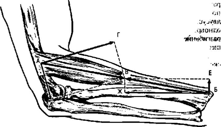
Fig. 44. Diagram illustrating The Significance of the angle at which muscle pull acts on a bone:
AB — direction of pull of the brachioradialis m.; AB and BV — two halves of the pulling force of this muscle; point A is pulled toward point B with the same force as point B toward point A; AG and AD — components of AB; BE and BZh — components of BV. As can be seen from the diagram, the brachioradialis m. approaches the forearm at a significantly smaller angle than the humerus. At the same time, AG is considerably greater than BE, whereas BZh is greater than AD
Paradoxical action of muscles. Multi-joint muscles can cause movements in several joints across which they pass. When bi-articular muscles contract, they create force moments of opposite directions in the joints, while the length of the muscles changes very little. Uni-articular muscles cause movement in only one joint; however, indirectly, they influence movements in joints located proximally and distally relative to the given joint. For example, flexion at the elbow joint is usually accompanied by some simultaneous extension at the shoulder joint. This indirect action of uni-articular muscles constitutes the so-called paradoxical action of muscles. Extension at the shoulder joint is facilitated by the fact that the center of mass of the entire arm during elbow flexion continues to remain beneath the shoulder joint, since the displacement of one part of the arm's mass forward is compensated by the displacement of another part backward. By virtue of this, the entire arm is shifted slightly backward, thereby preserving its equilibrium position. Another reason why extension at the shoulder joint occurs is that when the forearm is flexed, the triceps brachii muscle, located on its posterior surface, is stretched, and its tone increases. Since this muscle is bi-articular and its long HEAD originates from the scapula, simultaneously with the increase in tension it produces some extension at the shoulder joint across which it passes. Its extensor action increases as flexion at the elbow joint progresses.
Chain of segments. Movement usually occurs simultaneously in several body segments that are inextricably linked to one another. If the chain of segments is closed, then every muscle, even a uni-articular one, exerts an indirect effect on the segments of The Human Body that make up this chain, causing them to displace in space. When a person stands on the ground with both legs, the contraction of the popliteus muscle, for example, causes Movements of the lower leg and thigh, and via a roundabout path, movements of the pelvis, thigh, and lower leg of the other side of the body. If the chain is not closed, the displacement occurs mainly in the distal segment. Thus, when a person rests on the ground with one leg—i.e., there is no closed "leg—pelvis—leg—ground" system—contraction of the popliteus muscle on the other leg can displace only the lower leg and foot. Associated movements are also possible in these cases, but they are much less noticeable. They may even be absent if the proximal part of the body is fixed (for example, if a person, while flexing the lower leg, sits on some stationary support surface).
Antagonists and synergists. There is no true antagonism in Muscle Action, since muscles of both cooperative and opposite action work in coordination, jointly ensuring the execution of a given movement (Fig. 45). However, individual muscles or muscle groups participating in directly opposite movements are conventionally called antagonists. For example, the muscle group that flexes the foot is an antagonist to the group that extends it; i.e., the muscles located on the posterior and anterior surfaces of the lower leg are antagonists. Muscles that perform common work by participating in the same movement—i.e., muscles located on the same side of a given joint axis—are synergists. Uni-articular muscles of Uniaxial joints always perform only a single function with respect to these joints. For example, the brachialis muscle is a permanent flexor of the forearm at the elbow joint and a permanent antagonist to the anconeus muscle. With respect to multiaxial joints, especially ball-and-socket joints, the function of the same muscles (both multi- and uni-articular) can vary depending on the initial position of the articulating bones. Thus, the adductor muscles of the thigh turn out to be its flexors if it was previously extended. They can also work as pronators of the thigh if it was excessively turned outward, and conversely, they can assist supination if the thigh was strongly turned inward.
Muscles that are synergists for one movement can become antagonists for another. For example, during wrist flexion, its ulnar and radial flexors act as synergists. During movements of the wrist around the anteroposterior axis of the radiocarpal joint, however, these muscles act as antagonists: the ulnar flexor participates in wrist adduction, while the radial flexor participates in its abduction.
Much more complex is the work of muscles located at a considerable distance from one another. They form cooperatively working complexes that make the execution of a given movement possible. For example, the external oblique muscle of the abdomen on one side of the body and the internal oblique on the other, working cooperatively, take part in the Rotation of the trunk around its vertical axis. No less complex complexes are formed by muscles such as the trapezius and serratus anterior, participating in the rotation of the scapula with its inferior angle outward, or the pectoralis minor and the lower part of the rhomboid major muscle, causing opposite movements. In every movement, as a rule, not just a single muscle or even a single muscle group participates, but several cooperatively acting muscle groups. Among them, it is always possible to distinguish muscles that produce the given movement directly and muscles that contribute to stabilizing those parts of the body upon which the active segment rests.
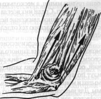
Fig. 45. A pair of antagonists located on opposite sides of the transverse axis of a uniaxial joint. The dot in the circle shows the PROJECTION OF THE transverse axis of the joint. Arrows indicate the direction of pull of: the flexor m. (left) and the extensor m. (right), which perform opposite functions. Contracting simultaneously, these muscles help stabilize the joint and immobilize the bones articulating within it
While synergistic muscle groups make the execution of a given movement possible, other muscles inhibit this movement through their tension. The learning of movements, especially those having the character of a jerk or push, and training proceed along the lines of developing a more isolated contraction of those muscles and muscle groups that are necessary for the given movement. For the execution of smooth movements, however, the action of antagonists is necessary, since without their regulatory influence, the contraction of synergists alone can cause jerky, abrupt movements. The initial period of learning movements is usually associated with the contraction (to a greater or lesser extent) of all muscles in the given region—both those necessary for the movement and those that inhibit it. This period is characterized by the fact that a process of excitation occurs in the cortical region of the motor analyzer within the Cerebral Cortex, along with the irradiation of this excitation. With such an unlearned movement, the active muscle group must overcome internal resistance from other muscles.
Characteristics of muscle work. Muscles can perform overcoming, yielding, and holding work. In overcoming work, a muscle overcomes the weight of a given body segment or some resistance when the force moment of the muscle or muscle group is greater than the gravity moment. In yielding work, the muscle, remaining tense, gradually relaxes, yielding to the action of gravity or resistance; the force moment of the muscle is in this case less than the moment of gravity or resistance. In holding work of muscles, the action of resistance is balanced, the force moments are equal, and As a result, movement is absent. Thus, the deltoid muscle during abduction of the arm to the side, holding it in a horizontal position, and during its slow adduction to the trunk is tense, but its work is not identical: in the first case it is overcoming, In the second — holding, and in the third — yielding. Yielding muscle work is very important for athletes, as it allows for an increase in both the strength and speed of movement. The stretching of muscles that occurs during yielding work leads to the accumulation of elastic strain energy within them, which is subsequently utilized by the body to execute a "return" movement, and to a greater extent if muscle tension follows immediately after the preliminary stretching of the muscles, without a pause. It has been established that during bouncing on the toes with straight legs, 45 J of energy is accumulated in the gastrocnemius muscle and the calcaneal tendon; when running at a speed of 3.9 m/s, 46–50 J is accumulated in the Muscles of the Lower Limb; during deep knee bends with a load in the same muscles, 730 J; and without a load, 394 J. The property of muscles to accumulate elastic strain energy depends on The ratio of fast and slow fibers in them; the higher the percentage of slow fibers, the better the elastic strain energy is utilized. Preparatory phases of movements, starting positions (squatting before a jump, winding up before throwing a projectile, etc.) facilitate the stretching of muscles performing the main movement. Yielding work is also referred to as relaxation.
A distinction is also made for ballistic muscle work — a sharp, rapid, overcoming contraction following preliminary muscle stretching (for example, in the upper limb during throwing). In this case, the muscle gives a push to the segment and relaxes, and the subsequent movement of the given segment continues by inertia.
Direction of pull. Oversimplified, the direction of a muscle's pull is considered to be the straight line connecting the centers of its points of origin and insertion. To refine the course of this line, it is necessary to make cross-sections of the muscle at different levels. The line connecting the centers of the cross-sections will be the resultant of the muscle forces of all fibers (the muscle centrode); it is usually somewhat curved.
Addition of forces acting in the same direction. To determine the magnitude and point of application of the resultant of a group of synergist muscles whose vectors are parallel, one should successively add the forces of all muscles in the given group. If, in the simplest case, it consists of two muscles, the resultant will be equal to the sum of the forces of these two muscles, and its point of application will lie on a line perpendicular to the direction of the resultants of these two muscles, at a distance inversely proportional to the strength of each of these muscles. If the synergist muscle group consists of not two, but a larger number of muscles, their resultant is likewise equal to the sum of the forces of all muscles. The point of application of this resultant is a point located between the attachment sites of the given muscles.
When adding forces that exert an effect on the movement of a specific body segment, the addend can be not only the force of the muscles, but also the force of gravity of the given segment.
Subtraction of forces. If muscles are attached to a bone that pull it in opposite directions, movement in this case occurs under the action of the difference in forces. The resultant in force subtraction is equal to the difference between them and is directed toward the greater force. When the forces of the muscles moving a given bone in different directions are equal, they balance each other and the bone remains stationary. Only a few muscles pull the bones to which they are attached in diametrically opposite directions. Most Muscles Attached to the same bone from its different sides form lines of pull directed at some angle to one another. However, these pulls can be resolved into components in such a way that they may turn out to run in opposite directions and participate in opposite movements.
Forces acting at an angle. When muscles pull a bone in two different, yet not diametrically opposite directions, the resultant force is represented by the diagonal of a parallelogram constructed on the vectors of these forces. For instance, the line of pull of each of the major muscles that adduct the arm—the pectoralis major and latissimus dorsi (Fig. 46)—does not coincide with the direction of movement during adduction.
Moreover, there is simply no muscle whose line of pull completely coincides with the direction of movement during arm adduction. Instead, the resultant force of these two muscles replaces the missing muscle force required to perform this specific movement.
The parallelogram of forces applies not only to two, but to several muscles pulling a given bone in various directions. In such cases, to determine the total resultant force, one must first construct the parallelogram of the resultants for every pair of muscles, and then construct parallelograms between the diagonals of the preceding ones, until the ultimate resultant of the entire muscle group is found.
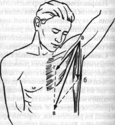
Fig. 46. Parallelogram of forces: ag — line of pull of the pectoralis major m.; ab — line of pull of the latissimus dorsi m. Working together, these muscles pull the humerus in the direction of ab, i.e., along the diagonal of the force parallelogram
Couple of forces. As is well known, every rotation results from the action of a force couple (Figs. 47, 48, 49); consequently, rotation in all joints can also be viewed as the result of a force couple. In most cases, one force in this couple is the muscle pull, while the other is the resistance offered to the given bone by the adjacent articulating bone. The resistance force is directed parallel and opposite to the muscle pull. The shortest straight line between the lines of action of these two forces constitutes the lever arm of the force couple, and the product of this lever arm and the magnitude of the force (specifically, the muscle pull) gives the torque of the force couple. If we were to assume that a given joint is disrupted—meaning one of the forces of the couple (namely, the resistance from the adjacent bone) is eliminated—then instead of rotating, the given skeletal segment would simply shift in the direction of the resultant force of that muscle group.
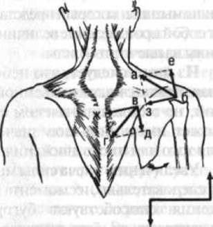
Fig. 47. Diagram showing that the upper and lower parts of the trapezius m., working together, facilitate the rotation of the scapula with its inferior angle laterally; these two parts form a force couple: ba — line of pull of the upper part of the trapezius m.; vg — line of pull of the lower part of this m., be and vd — their components forming the force couple; b v — lever arm of the force couple; bz and vzh — components contributing to scapular adduction
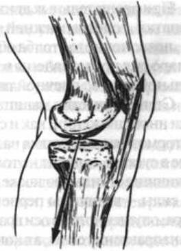
Fig. 48. Force couple producing extension at the knee joint: the upward arrow indicates the direction of muscle pull of the quadriceps femoris m.; the downward arrow indicates the direction of resistance of the Femur relative to the Tibia
During rotational movements of the scapula, head, Mandible, and spinal column, both forces of the couple are largely represented by muscle pulls.
The degree of a muscle's participation in a given movement, as well as its braking effect on joint motion, depends not only on its absolute strength, but also on its lever arm—the length of the perpendicular dropped from the axis of rotation to the line of muscle pull—and ultimately on the muscle torque, which is the product of its force magnitude and its lever arm.
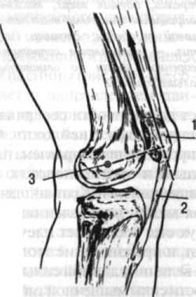
Fig. 49. Diagram illustrating the Significance of the Patella as a sesamoid bone that increases the lever arm (dashed line perpendicular to the arrow) of the quadriceps femoris m. force (arrow pointing up and to the left):
1 — patella; 2 — patellar ligament; 3 — projection of the transverse axis of the knee joint at one of the moments of movement within it
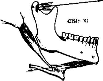
Fig. 50. Force couple formed by the lateral pterygoid (upper arrow) and digastric (lower arrow) mm. attached to the mandible
It follows from this that a small muscle with modest lifting capacity, but with a large lever arm, can play a significant role in a particular movement.
The lever arms of muscles, and consequently their torque, are enhanced by tubercles, tuberosities, crests, and roughnesses where muscles attach, as well as by sesamoid bones. These are embedded within the tendons of certain muscles near their passage across a joint, slightly proximal to the joint space, thereby increasing the lever arm of the muscle pull. The largest sesamoid bone is the patella, which enlarges the lever arm of the quadriceps femoris muscle (Fig. 50).
In many areas, as muscles course toward their attachment points, they wrap around bony prominences that effectively increase their lever arms (e.g., the medial and lateral malleoli). Pulleys and fibrous loops also serve to increase muscle lever arms (for instance, the fibrous loop anchoring the tendon of the digastric muscle to the Hyoid bone). The torque of these muscles increases correspondingly.
Laws of the lever and muscle action. Levers are traditionally classified into two orders—First and Second. When two forces are located on opposite sides of the fulcrum of a rigid body capable of rotation, and act in the same direction, the body functions as a lever of the first class. When forces are applied exclusively to one side of the fulcrum and act in opposite directions, the body constitutes a lever of the second class. Muscle forces may be applied to a lever either perpendicularly or at an angle. If a given force is applied to a lever at an acute or obtuse angle, its mechanical effect can be determined by resolving it into components: one directed along the longitudinal axis of the lever, and the other perpendicular to it. The first component can be disregarded when analyzing the movement itself, as its effect merely causes compression of the lever—increasing or decreasing the pressure between articular surfaces—or, if the lever is inadequately stabilized, a longitudinal displacement. The second component is the effective (useful) force that actually produces the movement.
First-class lever. In the context of the human Locomotor Apparatus, a first-class lever is also referred to as a "balance lever" (Fig. 51). This type of equilibrium characterizes the relationship between all superior body segments and the inferior ones (e.g., the head relative to the vertebral column, or the pelvis relative to the thigh). In the first example, the primary forces contributing to forward head flexion are gravity, muscular tension, and ligamentous pull.
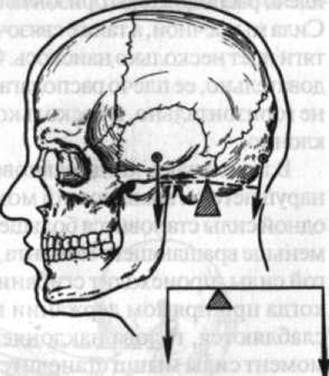
Fig. 51. Skull as a first-class lever (balance lever):
The left arrow indicates the direction of the force of gravity, the right arrow shows the direction of muscle pull; the point at the apex of the wedge indicates THE POSITION OF the transverse axis of the atlanto-occipital joint; the dashed lines denote the lever arm of gravity (left) and the lever arm of muscle pull (right)
When the head is held erect, the vertical line of its center of gravity, which lies slightly posterior to the sella turcica, passes anterior to the transverse axis of the atlanto-occipital joint. The resultant force of the muscular and ligamentous tension, applied to the Occipital bone, passes posterior to this axis. A state of equilibrium is maintained when the torque of these two forces is equal.
Since the force of gravity always acts in a vertical direction, its lever arm is horizontal.
The force of muscular and ligamentous tension runs somewhat obliquely. Consequently, its lever arm is not horizontal, but rather inclined.
In cases where equilibrium is disrupted and the torque of one force becomes greater or less than that of the other, flexion or extension of the head occurs. For example, when the Neck Muscles relax during an erect head posture, the head tilts forward because the muscular torque becomes less than the torque of gravity. Conversely, if the tension of the occipital muscles increases and their torque exceeds the gravitational torque of the head, it tilts backward.
Head tilt occurs not only due to The Influence of gravity, but also with some—albeit minor—participation of muscles located anterior to the cervical spine. These muscles include not only all those attached to the hyoid bone from above and below, but also, primarily, the muscles lying directly on the anterior surface of the spinal column (the longus capitis and longus colli). Therefore, it would be more accurate to speak not simply of the torque of gravity, but of the torque of the forces contributing to forward head flexion.
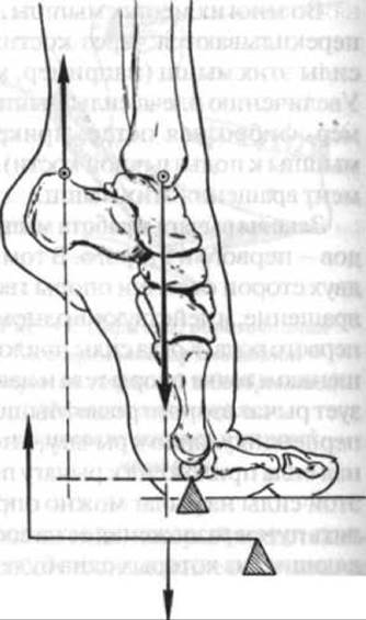
Fig. 52. The Foot as a force lever: the downward arrow indicates the direction of the force of gravity acting from the lower leg onto the foot; the upward arrow indicates the direction of pull of the triceps surae muscle
Second-class lever. There are two varieties of this type of lever. The first is commonly referred to as a "force lever." It is characterized by the fact that the lever arm of the muscular force is longer than the lever arm of the force of gravity. An example of such a lever is the foot during a rise onto the forefoot (Fig. 52). The fulcrum in this case is primarily the heads of the metatarsal bones, through which the axis of rotation of the entire foot passes. The muscular pull force—if its direction is represented as a straight line extending from the calcaneus in the direction of pull of the triceps surae muscle (as the most powerful plantarflexor)—has a longer lever arm than the force of gravity. The latter is transmitted through the bones of the lower leg to the foot and presses directly on the talus, facilitating the lowering of the foot. Movements of this lever type are rather limited; There is a mechanical advantage in force at the expense of a disadvantage in range of motion and speed. The second variety of the second-class lever (Fig. 53) is conventionally called a "speed lever." It is characterized by the fact that the muscular force is applied close to the axis of rotation and has a significantly shorter lever arm than the opposing force of gravity or any other resistance. For example, during forearm flexion, muscles contract whose resultant force passes anterior to the transverse axis of the elbow joint. The lever arm of this resultant force is approximately 2 cm, while the lever arm of gravity—if a person holds a 16 kg load in their hand with a flexed forearm—is approximately 20 cm; that is, the resistance lever arm is about 10 times greater than the muscular lever arm. Equilibrium requires the torques of these two forces to be equal. Hence, it is clear why with a lifting force of the forearm flexors equal to approximately 160 kg, an untrained person can only hold about 16 kg with a flexed forearm. Indeed, 160x2 = 16x20, meaning that each torque equals 320.
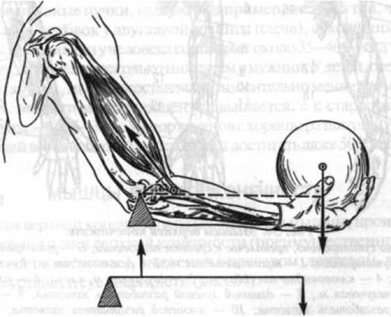
Fig. 53. The forearm as a speed lever: the downward arrow shows the action of gravity; the arrow running along the biceps brachii shows the direction of this muscle's pull; the dashed line perpendicular to it represents the lever arm of the muscular pull; the point at the top of the triangle indicates the Location OF THE transverse axis of the elbow joint
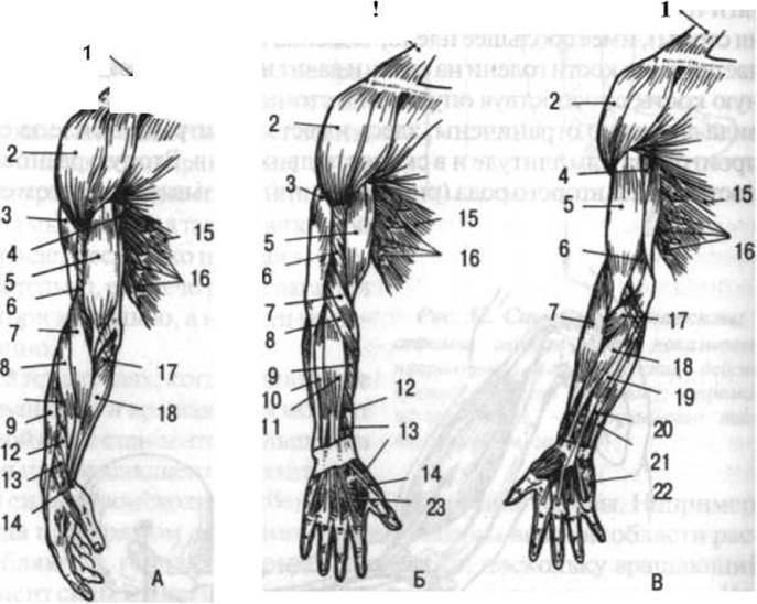
Fig. 54. Muscles of the upper extremity:
A — arm supinated, forearm in neutral position; B — arm pronated; C — arm supinated: 1 — trapezius m.; 2 — deltoid m.; 3 — triceps brachii m.; 4 — coracobrachialis m.; 5 — biceps brachii m.; 6 — brachialis m.; 7 — brachioradialis m.; 8 — extensor carpi radialis longus m.; 9 — extensor carpi radialis brevis m.; 10 — extensor carpi ulnaris m.; 11 — extensor digitorum m.; 12 — abductor pollicis longus m.; 13 — extensor pollicis brevis m.; 14 — extensor pollicis longus m.; 15 — external oblique m. of abdomen; 16 — serratus anterior m.; 17 — pronator teres m.; 18 — flexor carpi radialis m.; 19 — palmaris longus m.; 20 — flexor digitorum superficialis m.; 21 — flexor carpi ulnaris m.; 22 — abductor digiti minimi m.; 23 — dorsal interosseous m.
With this type of second-class lever, there is a disadvantage in lifting force, but a substantial advantage in range of motion and movement speed. Indeed, during flexion at the elbow joint, the hand—and even more so the fingertips—can perform movements with a significantly greater range and speed than the heel when rising onto the toes. However, when rising onto the toes, a person lifts the weight of the entire body, which may also be increased by an additional load, whereas the hand lifts a considerably lighter weight.
The degree of muscular development varies extremely among different individuals. It depends on age, sex, occupation, and other factors. Individual variations in muscular system development are also quite common. In addition to these peculiarities, usually referred to as muscle variants, Developmental anomalies of the muscular system can occur. For instance, certain muscles may be absent (even such a large muscle as the pectoralis major) or, conversely, additional muscles may be present (such as the sternalis); sometimes muscles feature extra fascicles, heads (e.g., three, four, or even five heads in the biceps brachii instead of two), tendons, etc.
The total muscle mass in humans accounts for about 35–40% of body weight. In women, this percentage is slightly lower than in men. In children, especially newborns, muscles constitute a relatively smaller mass—20–22%. With age, this percentage increases, and in old age it decreases again, down to 25–30%. In athletes with well-developed musculature, the total weight of muscle mass can even reach 50% of body weight.
Last update: 08/08/2026
Editorial and Educational Adaptation: This material has been compiled based on the primary/original source text. The project team performed an editorial review, corrected technical inaccuracies, structured sections, and adapted the content for an educational format.
What was processed:
- elimination of formatting defects (OCR errors, structural breaks, corrupted characters);
- editorial organization of content;
- standardization of terminology in accordance with academic sources;
- verification of factual statements against the original source text.
All mentions of the author, publication year, and origin of the primary text have been preserved in accordance with the source.