TEXTBOOK OF PEDIATRIC GYNECOLOGY - 2013
Chapter 7. DEVELOPMENTAL ANOMALIES OF THE REPRODUCTIVE ORGANS IN GIRLS: CLINICAL FEATURES, DIAGNOSIS, AND MANAGEMENT
EMBRYOGENESIS OF THE REPRODUCTIVE SYSTEM
The Development of the Gonads during Embryogenesis is determined by the set of sex Chromosomes formed after egg Fertilization. The 46,XX karyotype directs ovarian development, whereas the 46,XY karyotype determines testicular development. Primary sex differentiation is The process of gonadal development that begins at 6–7 weeks of embryonic life. Under METABOLISM/18.html">The Influence of Transcription factors WT1 (Wilms Tumor suppressor 1), steroidogenic factor 1 (SF1), and DAX1, the primordial gonad is formed. It consists of cortical and medullary components. In the presence of two X chromosomes, the cortical component transforms into an Ovary. The Y chromosome promotes the Development of the medullary component of the primordial gonad into a Testis. Furthermore, sexual differentiation is a highly specific process controlled by genes such as SRY, SOX9, and DSS. The SRY Gene plays a fundamental role in Sex Differentiation and can induce male Sex Determination. By the 8th week of gestation, the Testes are capable of secreting Sex Hormones, which dictate the development of the external genitalia. Male-type Differentiation of the external genitalia is driven by testosterone secreted by embryonic Leydig Cells. Alongside testosterone, the anti-Müllerian hormone (AMH)—secreted by Sertoli cells—plays a crucial role in sex differentiation by suppressing the development of the Müllerian ducts. If normal interactions occur between AMH and its receptors in a male embryo, the Müllerian ducts undergo regression. Thus, a normal male phenotype requires a Y chromosome carrying the SRY gene, functional Leydig and Sertoli cells, appropriate secretion of testosterone and AMH, and the presence of receptors for these hormones in target cells. A defect at any stage of this process leads to female-type development. In the presence of a 46,XX karyotype, primordial Germ Cells develop into Ovaries. The genes that inhibit the development of the primordial gonad into a testis include DAX1, SOX3, and Wnt4. The transformation of the primordial gonad into an ovary occurs at 17–20 weeks of embryogenesis, when primordial cells differentiate into oocytes (the functional equivalent of Sertoli cells). Their number reaches a peak (6.7 million) by the 5th week of gestation. By 7 years of age, this count declines to 300,000. When only a single X chromosome is present (45,X0), oocytes undergo degeneration even before birth, as the second X chromosome is essential for maintaining ovarian mass and its further development. The Müllerian ducts give rise to the FEMALE REPRODUCTIVE Organs: the Uterus, fallopian tubes, and upper third of the Vagina. The lower two-thirds of the vagina develop from the urogenital sinus. Consequently, The formation of female reproductive organs proceeds independently of ovarian endocrine function.
Differentiation of the primordial gonad into an ovary is a passive process induced by specific molecules homologous to the H-Y antigen in males. The X chromosomes play a leading role in ovarian differentiation.
An anomaly in one or more sex determinants, or a disruption in the complex mechanism that triggers sexual development, can lead to anatomical and functional deviations from the norm, resulting in various Clinical forms of disorders of sex development (DSD). Various Chromosomal aberrations and Gene Mutations causing hormonal imbalances or altered hormone reception during the Embryonic period can trigger congenital anomalies of sexual development. Alongside chromosomal aberrations (structural and numerical) and gene mutations, congenital DISORDERS OF SEXUAL development may also stem from various embryonic factors (intoxications, infections, trauma, medications) and maternal hormonal imbalances during Critical Periods of reproductive tract formation.
The specific type of disorder of sex development depends on the cause and the timing of its occurrence during ontogenesis. Gonadal agenesis occurs during the Cytology/cytology/16.html">Early stages of embryogenesis (6–10 weeks). If the process of gonadal differentiation and formation is disrupted for any reason, the Organism may develop without gonads and their functional elements (gonadal agenesis). Most frequently, gonadal agenesis results from sex chromosome pathology. Occasionally, other damaging factors (intoxications, infections, radiation) prevent proper gonadal formation, in which case a normal male or female set of sex chromosomes is observed. Regardless of the underlying cause, the Clinical presentation is largely similar. Instead of gonads, streaks of Connective Tissue devoid of functional elements are found in girls. Subsequently, a female phenotype develops with marked hypogonadism, compounded by the absence of fetal gonads and any autonomous tendency of the fetus to develop along female, "neutral" lines. Therefore, gonadal agenesis represents the earliest pathology of sexual development, encompassing Turner syndrome and "pure" gonadal agenesis.
Pathology of sexual development arising in the later stages of embryogenesis is termed gonadal dysgenesis. Gonadal dysgenesis is a broad concept encompassing a range of syndromes caused by impaired embryonic gonadal development resulting from chromosomal aberrations, gene mutations, or embryotoxic factors. These include ovarian dysgenesis syndrome and testicular dysgenesis syndrome. In many cases, ovarian dysgenesis is associated with a 45,X/46,XX mosaic karyotype, which impedes normal ovarian differentiation. Even with a normal female karyotype, gene mutations or other noxious factors can lead to the formation of a defective, dysgenetic ovary. The phenotype of girls with ovarian dysgenesis is invariably female, and the ovarian impairment manifests only at Puberty as more or less pronounced hypogonadism.
Impaired Formation of the reproductive tract can also be linked to hypoplasia and dysgenesis of the testes, sharing similar etiologies. Dysgenetic testes fail to ensure the regression of the paramesonephric ducts and normal masculinization of the external genitalia, thereby permitting the persistence of paramesonephric duct derivatives.
Genetically determined disorders of sex differentiation in testicular feminization syndrome occur during embryogenesis. Due to impaired specific reactivity of target organs to androgens, a female phenotype develops in a genetic and gonadal male fetus. Internal reproductive organs are male, whereas external genitalia are female. In incomplete forms of the syndrome, mild masculinization of the external genitalia is observed. One chromosomal syndrome associated with impaired sexual development is Turner syndrome, the typical clinical picture of which manifests with a 45,X0 karyotype. Various mosaic variants also exist: 45,X0/46,XX; 45,X0/46,XY; 45,X0/47,XXX, and less commonly 45,X0/47,XXY. Birth weight in children with Turner syndrome is typically below average. Newborns may exhibit lymphedema of the feet, lower legs, and hands, as well as redundant Skin folds on the neck. Growth retardation becomes evident at any age, occasionally from birth, and is particularly pronounced during puberty. Stature rarely reaches 150 cm.
Physical examination reveals generalized dysplastia: children tend to be stocky (low-set HEAD, short neck, barrel-shaped chest with widely spaced nipples and a sternal depression—a "shield-like chest"). Characteristic Features of patients with Turner syndrome include a short, webbed neck with a low posterior hairline, and pterygium colli (the so-called "sphinx neck"). Facial dysmorphism may include micrognathia, retrognathia, ptosis, epicanthus, and malformed, low-set ears. The neurocranium is relatively larger than the viscerocranium. In some cases, cubitus and Genu Valgum, shortening of the metacarpals and metatarsals, and deep-set nail deformities are observed. The skin shows numerous pigmented nevi and vitiligo, along with hypoplastic, rounded fingertip pads. Congenital Malformations of Internal Organs—such as coarctation of the aorta, ventricular septal defect, patent ductus arteriosus, aortic or pulmonary stenosis, Horseshoe Kidney, double renal pelves and Ureters, renal malrotation, and Renal Hypoplasia—may also be present. Intellectual disability can occur.
Sexual infantilism becomes apparent during puberty: secondary sex characteristics are absent, breasts fail to develop, pubic and axillary Hair is sparse or absent, and the labia, vagina, and uterus remain hypoplastic. The vulvar and vaginal mucosa is dry with a thinned epithelial lining. Patients with Turner syndrome most frequently seek medical attention due to short stature. Ultrasound reveals an infantile, hypoplastic uterus and streak gonads. Radiography of the hand (showing bone age delayed by 2–4 years) demonstrates osteopenia/Osteoporosis. Cranial radiography reveals hyperpneumatization of the sphenoid sinus. Pneumopelvimetry demonstrates a markedly hypoplastic uterus and streak-like adnexa. Gonadotropic activity, particularly FSH, is elevated during puberty (ages 9–13), reaching peak functional activity at 16–17 years.
LH secretion follows a similar pattern, though its levels typically range between 1/2 and 1/6 of FSH levels. There is a sharp increase in LH and FSH with frequent chaotic, desynchronized fluctuations, while feedback mechanisms within the pituitary-gonadal axis are preserved. Estrogen excretion is low, and vaginal smears are of an atrophic type. A slight elevation in Growth Hormone (GH) levels may occur, and tissue resistance to THYROID HORMONES is possible. Glucose tolerance is reduced, with a borderline or diabetic glycemic curve. Cytogenetic analysis reveals a 45,X0 karyotype or 45,X0/46,XX mosaicism. Sex Chromatin is absent or markedly diminished. These patients require hormone replacement therapy.
The classic variant of Klinefelter syndrome presents with a triad of symptoms: hypogonadism, gynecomastia, and dysgenesis of the seminiferous tubules. The condition is caused by one or more extra X chromosomes in the karyotype, resulting in a 47,XXY or 48,XXXY chromosomal Complement. Occasionally, chromosomal mosaicism such as 47,XXY/46,XY; 47,XXY/46,XX; 48,XXXY/46,XY; or 47,XXX/47,XXY is detected. Each additional X chromosome exacerbates intellectual impairment, whereas in mosaic forms, the external manifestations of the syndrome may be mild or muted. The primary consequence of the abnormal sex chromosome complement is interstitial Cell dysfunction leading to aspermatogenesis. This dysfunction manifests as general androgen deficiency on the one hand, and localized hyalinization of the seminiferous tubules on the other. Clinical Diagnosis of Klinefelter syndrome in the neonatal period is impossible, as newborn boys with a 47,XXY karyotype exhibit no distinct physical abnormalities. Buccal smear analysis reveals chromatin-positive interphase nuclei. Gonadal hypoplasia—a mandatory sign of the disorder—is rarely identifiable in the prepubertal period. Occasionally, signs of delayed mental development appear during this time, prompting sex chromatin testing. A presumptive clinical diagnosis is possible only during the prepubertal or pubertal periods. Despite the paucity of early signs, the clinical presentation exhibits marked polymorphism. Depending on somatic development, types of Klinefelter syndrome include the dysplastic (asthenic) type, pseudomuscular type, and eunuchoid type, characterized by fat deposition on the thighs, chest, and lower abdomen. Patients with the eunuchoid body habitus are typically tall due to long lower extremities. A tendency toward narrow shoulders with a relatively wide pelvis, a flat and narrow chest, poor Muscle Development, and a stooped posture is frequently noted. Adolescents may exhibit cyanosis and excessive sweating of the hands and feet.
Gynecomastia develops during the prepubertal and pubertal periods (ages 12 to 16), typically bilaterally. The breast tissue consists of ducts surrounded by dense fibrous tissue proliferation, and the glands are generally non-secretory. The development of gynecomastia is driven by increased estrogen production by testicular interstitial cells. The external genitalia are male. The testes are located in the Scrotum, firm, and markedly reduced in size—about 1.5 cm (compared to the normal 5 cm)—while internal reproductive organs are significantly smaller than average. Facial hair growth is sparse; however, findings include a low anterior hairline, sparse axillary hair, and a female-pattern pubic hair distribution (evidently due to quantitative and qualitative androgen deficiency, as well as reduced tissue sensitivity to androgens). Elevated gonadotropin levels are also characteristic.
Testicular feminization syndrome is transmitted via pathogenic genes from female carriers, resulting in sparse pubic hair and delayed menarche. Its Pathogenesis is explained by reduced activity or instability of the 5-alpha-reductase enzyme, which is responsible for converting testosterone into its potent metabolite, 5-alpha-dihydrotestosterone, essential for the male development of external genitalia. In this syndrome, androgen receptors are deficient or entirely absent in the cells of certain target Tissues. In the absence of cellular response to sex hormones, tonic gonadotropin secretion increases.
In testicular feminization, Blood levels of LH are markedly elevated. Gonadotropins interacting with specific receptors on cell membranes not only bind to protein receptors but also regulate their synthesis. High concentrations of LH downregulate its own receptors. Despite tissue insensitivity to androgens, normal masculinization of the Central Nervous system centers regulating gonadotropin secretion occurs. Pathogenetically, the condition is rooted in a genetically determined tissue insensitivity to androgens coupled with preserved sensitivity to estrogens. Fetal testes possess anti-Müllerian activity, leading to the regression of the paramesonephric ducts, which explains the absence of the uterus, fallopian tubes, and upper third of the vagina in these patients.
Internal genitalia are male, the prostate is absent, and a blind vaginal pouch derived from the urogenital sinus is present. With complete tissue insensitivity to androgens or impaired testosterone Biosynthesis, masculinization of the external genitalia fails to occur, and they retain a female, neutral conformation. Feminization phenomena during puberty are attributed to increased estrogen production by the testes driven by hyperstimulation from gonadotropins.
Depending on the predominance of estrogenic versus androgenic activity, two forms of testicular feminization are distinguished. The complete form is characterized by an absolute lack of androgen responsiveness, resulting in a female phenotype, normal body proportions, and breast development. Pubic and axillary hair is absent or sparse, whereas scalp hair is thick, and the overall appearance is distinctly feminine. The incomplete form is characterized by partially preserved target-organ sensitivity to androgens, manifesting as incomplete masculinization of the external genitalia at birth. During puberty, masculine features and an intersexual body habitus appear. The core features of testicular feminization syndrome include a genetic and gonadal male sex, absence of the uterus, fallopian tubes, and upper third of the vagina, incomplete masculinization of the external genitalia, and the development of feminization at puberty. Pubic hair and menstruation are absent. Cytogenetically, sex chromatin is negative and the karyotype is male (46,XY). Basal testosterone levels fall within the normal age-adjusted range for boys. Basal estradiol levels exceed the normal range for boys, occasionally approaching normal values for girls. The functional test with human chorionic gonadotropin (hCG) yields ambiguous results, and LH levels are elevated. An increase in gonadotropic activity is observed at ages 12–13. Testosterone and estradiol levels rise by ages 13–14. By ages 16–17, testosterone levels reach the normal range for boys, while estradiol levels are elevated.
Hermaphroditism Syndrome
This is a group of disorders characterized by ambiguous, dual-sex structures of the external genitalia.
In true hermaphroditism, or true intersexuality, the karyotype is usually 46,XX, less commonly 46,XY, or various chimeras/mosaics. The phenotype can be male or purely female. The external genitalia are ambiguous. On one side, the labioscrotal fold forms a hemiscrotum containing a testis, while on the contralateral side, it forms a labium majus. A uterus, and occasionally fallopian tubes, are present in all affected individuals. Ovaritesticular (mixed) gonads are identified. Diagnosis is established on The basis of ambiguous gonads and internal female organs coexisting with secondary male characteristics.
Treatment involves assigning the individual as female through reconstructive genital surgery combined with estrogen therapy to promote the development of female secondary sex characteristics.
Disorders of sex differentiation may result not only from chromosomal aberrations but also from gene mutations and various embryotoxic factors. Clinical practice encounters a considerable number of syndromes accompanied by various signs of impaired sexual development, and numerous distinct syndromes are known to be rooted in these developmental anomalies.
Perrault syndrome: sensorineural Hearing loss of any degree, ovarian aplasia, hypoplasia or dysgenesis, hypogonadism; uterine aplasia, hypoplasia, or unicornuate uterus; proportional dwarfism; ataxia; intellectual disability; short neck; Scoliosis, etc. It is inherited in an autosomal recessive manner.
Woodhouse syndrome: oligophrenia, deafness, hypogonadism, intellectual disability, Diabetes Mellitus, scalp hypotrichosis, eyebrow aplasia and hypoplasia, uterine aplasia and hypoplasia, unicornuate uterus, ovarian aplasia, hypoplasia, and dysgenesis, breast hypoplasia, etc. Inherited in an autosomal recessive manner.
de Grouchy syndrome (oligophrenia — dwarfism — sex reversal): intellectual disability; microcephaly; uterine aplasia and hypoplasia, unicornuate uterus; sex reversal, XY dysgenesis; proportionate dwarfism; hirsutism, thick eyebrows, prominent Nose; ovarian aplasia, hypoplasia, and dysgenesis; labial hypoplasia, etc. Inherited in an autosomal recessive manner.
Mainzer syndrome (polydactyly — urogenital anomalies): renal aplasia and hypoplasia; uterine aplasia and hypoplasia, unicornuate uterus; ovarian aplasia, hypoplasia, and dysgenesis; gonadal tumors; prenatal hypoplasia, etc. Mode of inheritance is undetermined.
Tibi syndrome (oligophrenia — obesity — hypogonadism): intellectual disability; gynecomastia; hypogonadism; obesity; ovarian arachnodactyly; stooping posture; knock-knees (genu valgum), etc. Inherited in an autosomal recessive manner.
Ulnar-mammary syndrome: nipple hypoplasia; hypogonadism; aplasia or hypoplasia of the 4th and 5th fingers or toes; microcephaly; uterine aplasia and hypoplasia, unicornuate uterus; proportionate dwarfism; obesity; eunuchoid habitus, etc.
Hajime syndrome (deafness — nephropathy — hypogonadism): deafness, hearing loss; nephropathy, proteinuria; hypogonadism; onset of the disease after 5 years of age; uterine aplasia and hypoplasia, unicornuate uterus; ovarian aplasia, hypoplasia, and dysgenesis; Amenorrhea, etc. Inherited in an autosomal recessive manner.
Al-Awadi syndrome (hypotrichosis — hypogonadism): uterine aplasia and hypoplasia, unicornuate uterus; testicular aplasia and hypoplasia; hypogonadism; focal alopecia; body hypotrichosis; ovarian aplasia, hypoplasia, and dysgenesis; amenorrhea, etc. Inherited in an autosomal recessive manner.
Bernard-Wahl syndrome (neurological anomalies — hypogonadism): ataxia; dysarthria, dyslalia; hypogonadism; high arched FOOT (pes cavus); onset of symptoms after 10 years of age; amenorrhea, etc. Inherited in an autosomal recessive manner.
Fraser syndrome (gonadal dysgenesis — nephropathy): nephropathy; ovarian aplasia, hypoplasia, and dysgenesis; sex reversal, XY dysgenesis; amenorrhea, etc. Mode of inheritance is undetermined.
Bengtstad syndrome (oligophrenia — dwarfism — hypogonadism): ataxia; intellectual disability; microcephaly; hypogonadism; proportionate dwarfism; diabetes mellitus, etc. Inherited in an autosomal recessive manner.
Brosneck syndrome (oligophrenia — dwarfism — sex reversal): intellectual disability; trigonocephaly; atresia or stenosis of the ear canals; sex reversal, XY dysgenesis; proportionate dwarfism; uterine aplasia and hypoplasia, unicornuate uterus, etc. Inherited in an autosomal recessive manner.
Malouf syndrome (cardiopathy — hypogonadism): eyelid ptosis; broad, high nasal bridge; cardiopathy; ovarian aplasia, hypoplasia, and dysgenesis; hypogonadism, etc. Inherited in an autosomal recessive manner.
Neuman syndrome (multiple endocrine gland hypoplasia): aplasia and hypoplasia of the Pituitary Gland, Thyroid Gland, and Adrenal Glands; aplasia, hypoplasia, and dysgenesis of the ovaries and testes. Inherited in an autosomal recessive manner.
Weinstein syndrome (deafness — cataract — dwarfism — hypogonadism): cataract; deafness, hearing loss; hypogonadism; dwarfism; obesity.
Frisk syndrome (facial anomalies — hypogonadism): intellectual disability; enophthalmos; broad nasal bridge; progenia; hypogonadism; eunuchoid habitus, etc. Inherited in an autosomal recessive manner.
Fitch syndrome (dwarfism — hand anomalies — hypogonadism): hypogonadism; dwarfism; isodactyly; aplasia and hypoplasia of the hand Phalanges, etc. Inherited in an autosomal recessive manner.
Famizimi syndrome (acromegaly — acanthosis nigricans): broad nasal bridge, thick Lips, macrogenia; hypogonadism; dwarfism; brachydactyly; Insulin resistance. Inherited in an autosomal recessive manner.
It should be noted that the prevalence of the above-mentioned syndromes in populations is very low, ranging from 1 : 200 000 to 1 : 500 000.
Uterine developmental anomalies
Mayer-Rokitansky-Küster syndrome — uterine aplasia in the presence of fallopian tubes and functional ovaries. Occasionally, a small muscular ridge may be present in place of the uterus. This defect is associated with partial or complete Aplasia of the vagina, specifically its upper part. This pathology is characterized by primary amenorrhea. The diagnosis is established during puberty; secondary sex characteristics are well-developed, and the phenotype is female. Gynecological examination, ultrasound, and diagnostic laparoscopy are of diagnostic importance. Treatment is surgical and consists of vaginal construction.
Uterus didelphus — duplication of the uterus and vagina with separate positioning.
Uterus duplex et vagina duplex — duplication of the uterus and vagina, connected in a certain area by a fibro-muscular layer.
Uterus bicornis bicollis — bicornuate uterus with two cervices.
Uterus bicornis unicollis — bicornuate uterus with a single cervix.
Arcuate uterus — presence of a rudimentary internal septum within the uterus.
Hymenal Stenosis
In some women, the opening of the hymen is too small, which causes dyspareunia. In such cases, the hymen is thick and fibrous. Its opening can be enlarged via a small incision under local anesthesia. If necessary, incisions are made at "the 2, 4, 8, and 10 o'clock positions" (or alternatively at 2, 4, 8, and 10 o'clock), starting from the edge of the hymen and extending along the longitudinal axis of the vagina. These incisions are then closed transversely using absorbable sutures (Fig. 15).
Class="center">
Fig. 15. Surgical correction of hymenal stenosis: a — infiltration of the hymen with a vasoconstrictor solution (0.1% epinephrine or vasopressin solution); b — excision of part of the hymen with a vertical incision; c — approximation of the hymenal edges with interrupted sutures
Imperforate Hymen
This is a congenital anomaly. Patients may present with primary amenorrhea and a bulging mass in the vaginal vestibule. Rectal examination reveals hematocolpos. The hymen must be widely incised and sutured in the same manner as for hymenal stenosis (Fig. 16). The vagina is then irrigated with antiseptic solutions.

Fig. 16. Imperforate hymen. Incisions are made at the 3 and 9 o'clock positions, followed by 12 and 6 o'clock; the hymenal flaps are excised, and the wound edges are approximated with interrupted 3-0 Vicryl sutures (a-c): 1 — Clitoris; 2 — Urethra; 3 — Labia minora; 4 — incision lines; 5 — hymenal flaps; 6 — anus
Vaginal Septum
Vaginal septa can be either transverse or longitudinal (Fig. 17).

Fig. 17. Longitudinal vaginal septum
A longitudinal vaginal septum results from the incomplete fusion of the paired Müllerian ducts. The proximal end of the septum may terminate near the cervix or between a duplicated cervix. The septum can be complete or partial, spanning a short distance. Occasionally, it may lead to the occlusion of one vaginal hemi-canal, resulting in hematocolpos.
Most cases of longitudinal vaginal septum are asymptomatic, only occasionally causing dyspareunia or coital bleeding. Once identified, surgical management is indicated. Undiagnosed longitudinal septa can result in severe Hemorrhage during childbirth. Excision of the septum is a relatively straightforward Procedure (Fig. 18). As with transverse septa, attention must be paid to preventing postoperative reduction of the vaginal caliber.

Fig. 18. Removal of a longitudinal vaginal septum: a — localization of the septum; b — excision of the septum using gentle traction; c, d — repair of the vaginal mucosal defect
A transverse vaginal septum can be located at any level between the hymen and the cervix. Prior to Surgical treatment, the thickness of the septum should be determined. If incomplete, it can sometimes be dilated digitally by the surgeon. In some cases of a complete septum, approximately 1/3 of the vagina is absent. If the septum is relatively thin, it can be incised and sharply divided (Fig. 19). Preserving the vaginal diameter during the procedure is crucial. In the postoperative period, The Use of vaginal dilators is recommended for several weeks or months. When a significant segment of the vagina is absent (greater than 1 cm), the septum is excised, and dilators are used to prevent vaginal stenosis. Vaginal reconstruction and McIndoe vaginoplasty improve outcomes in selected patients.
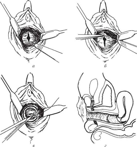
Fig. 19. Transverse vaginal septum: a — initial vertical incision of the septum; b — sharp dissection of the septum from the vaginal mucosa; c, d — repair of the mucosal defect with interrupted absorbable sutures
Developmental anomalies of the Vagina
1. Vaginal agenesis — primary complete absence of the vagina.
2. Vaginal aplasia — primary absence of a portion of the vagina.
3. Vaginal atresia — secondary absence of the vagina, characterized by complete or partial obliteration resulting from intrauterine inflammatory processes.
Forms of atresia:
— hymenal, retrohymenal;
— vaginal;
— cervical;
— uterine;
— tubal.
4. Vaginal septum — complete, incomplete.
Anomalies of the External Genitalia
1. Clitoral agenesis, hypoplasia, hypertrophy.
2. Hymenal atresia — imperforate hymen.
3. Aplasia of the labia and hymen.
Gynatresia — developmental Disorders of the genital tract: hymen, vagina, or uterus (Fig. 20).
Fig. 20. Gynatresia: a — haematocolpos in imperforate hymen; b — haematocolpos, haematometra, and haematosalpinx
Gynatresia can be congenital (developing prenatally due to embryonic developmental defects) or acquired (secondary), resulting from inflammatory processes in childhood. In adulthood, vaginal atresia may develop due to birth trauma, cervical atresia — following diathermocoagulation, and uterine cavity atresia or fusion — after excessive curettage during abortion. With hymenal atresia, upon the onset of first menstruation, blood begins to accumulate in the vagina, stretching it (haematocolpos). The girl complains of pain. After such a concealed menstruation ceases, the blood hemolyzes, its liquid part is absorbed, the volume decreases, and the pain subsides until the next period. If the patient does not seek medical attention, the blood accumulates further, collecting in the uterine cavity (haematometra), and subsequently in the fallopian tubes (haematosalpinx). Due to the distensibility of the vagina, pain syndrome typically appears when the haematocolpos reaches a significant size. Haematometra and haematosalpinx are less common in this pathology.
Atresia of the hymen is most frequently diagnosed during puberty. The clinical presentation is characterized by periodic lower abdominal pain in girls over 12 years of age in the absence of menstruation (false amenorrhea). The intensity of the dull aching pain increases, and with large haematocolpos, urination and defecation difficulties may occur. Secondary sexual characteristics correspond to the patient's age. During a pain attack, urinary retention occurs. Examination of the vaginal vestibule reveals the absence of an opening in the hymen, cyanosis, and bulging. Rectoabdominal examination reveals a tense-elastic tumor-like mass in the vaginal area, with a small, dense uterus located at its apex.
Gynatresia is not diagnosed before puberty; after the onset of menstruation, diagnosis is straightforward thanks to the classical Anamnesis and clinical presentation. In hymenal atresia, a cruciate incision of the hymen is performed, and its edges are sutured with interrupted stitches. Blood clots are removed from the vagina.
Vaginal atresia occurs in its upper, middle, and lower sections and varies in size. Blood accumulates above the site of atresia, gradually filling the overdistended cervical canal and uterine cavity.
Clinical presentation: periodic pain appears in the lower abdomen and lumbar region, with absence of menstruation. Ultrasound examination and vaginal probing, which helps determine the level of atresia, are of diagnostic importance. Rectoabdominal examination reveals a tense-elastic mass located anterior to the rectum. If vaginal atresia is complicated by haematometra and haematosalpinx, several round, tense-elastic masses are detected in the lesser pelvis.
Treatment is surgical. In atresia of the lower third of the vagina, the Tissues of the vaginal introitus are dissected, the lower vaginal vault is incised, and its mucous membrane is sutured to the vaginal entrance. If the atresia is located in the middle third of the vagina, the tissues between the lower and upper thirds are incised, and the mucous membranes of these areas are subsequently sutured. The most complex procedure is surgery for atresia of the upper third of the vagina, when the fornices are absent and the cervix is located within the pelvic connective tissue. Surgery is effective if the cervical canal can be found and sutured into the upper vaginal vault.
Primary hymenal atresia is diagnosed by medical personnel or the girl's mother during the neonatal period. In hymenal atresia, examination of the external genitalia is of decisive diagnostic importance. Pelvic sonography reveals a sharply dilated, fluid-filled vagina as an echo-negative formation, while the uterus is of normal size and located at the apex of the haematocolpos. It is believed that the stretched vagina fills first, and the uterine cavity does not dilate until the pressure of the accumulated fluid exceeds the force of uterine muscular contraction, after which haematometra and haematosalpinx develop.
The prognosis depends on timely diagnosis of the condition. With a prolonged course of the disease and the development of haematometra and haematosalpinxes, the girl may experience difficulties with Pregnancy in the future, and if destructive changes have developed in the tubes, tubal obstruction may occur. Endometriosis of the internal genital organs frequently develops.
Vaginal aplasia is the absence of the vagina. It occurs due to underdevelopment of the lower (caudal) PARTS OF THE paramesonephric (Mullerian) ducts. In this pathology, the uterus, fallopian tubes, and ovaries are often underdeveloped.
In vaginal aplasia, amenorrhea is true or false, and sexual intercourse is impossible.
It is diagnosed based on anamnestic data (amenorrhea) and gynecological examination of the patient.
Treatment is surgical. To create a vagina, a skin flap, a segment of the sigmoid colon, or pelvic Peritoneum can be used.
The external genitalia develop from the urogenital sinus. If harmful factors, particularly medications, affect the pregnant woman's body during the formation of the genital organs, the process of sexual differentiation may be disrupted.
Based on The Mechanism of development of the genital organs, the following variants of uterine and vaginal malformations are possible (Fig. 21, 22):

Fig. 21. Malformations of the uterus and vagina: a — bicornuate uterus, duplication of the cervix, vaginal septum; b — bicornuate uterus with a single cervix; c — septate uterus (or arcuate uterus / saddle-shaped uterus); d — unicornuate uterus

Fig. 22. Malformations of the uterus and vagina: a — rudimentary right vagina, rudimentary right uterine horn; b — bicornuate uterus, rudimentary right uterine horn, hematometra
1. Both mesodermal ducts develop properly, but fail to fuse along their entire length. This results in complete duplication of the uterus and vagina: the patient has two vaginas separated by a thin septum, each vagina leads to a separate cervix, and There are two uteri; each uterus has a single fallopian tube and a single ovary.
2. Both mesodermal ducts develop properly, but their fusion occurs only over a certain length, while the remaining parts of the uterus and vagina are separated by a septum. There are numerous variants of this malformation: a vaginal septum; a single vagina into which two cervices open; a uterine septum; a bicornuate uterus; a saddle-shaped uterus.
3. One of the Müllerian ducts develops normally, while the other fails to develop entirely. This leads to the formation of a vagina, a unicornuate uterus with a single ovary and a single fallopian tube. The patient's Urinary System should be evaluated, as this anomaly is frequently associated with renal agenesis on the affected side.
4. One of the mesodermal ducts develops normally, while the other is underdeveloped, resulting in a uterus with a rudimentary horn. These cavities may communicate with each other, making pregnancy in the rudimentary horn possible. Such a pregnancy develops as an ectopic one, and its rupture causes massive hemorrhage requiring surgical intervention.
Diagnosis involves an inspection of the external genitalia, speculum examination of the cervix, and bimanual pelvic examination. To clarify the diagnosis, ultrasound, uterine sounding, hysterosalpingography, or contrast-enhanced sonography are sometimes required.
Treatment of developmental anomalies is surgical. Uterine and vaginal duplication that does not impair a woman's sexual and reproductive Functions requires no surgical intervention; however, surgery is necessary in the presence of a rudimentary uterine horn if menstrual blood accumulation (hematometra) or Ectopic Pregnancy occurs. Vaginal septa are usually diagnosed during pregnancy or childbirth. If they obstruct fetal delivery, they are surgically incised.
According to their clinical course, developmental anomalies of the uterus and vagina are classified into two groups:
1. Malformations accompanied by unilateral obstruction to menstrual blood outflow.
2. Malformations without outflow obstruction.
The Clinical presentation of these groups differs.
A characteristic feature of the first group is painful menstruation (dysmenorrhea). Rectal examination reveals a tense-elastic mass closely attached to the uterus or located along the lateral wall of the vagina. Sometimes hematocolpos of the secondary vagina is misdiagnosed as a Gartner's duct cyst of the vagina, and blood accumulation in the uterine horn is mistaken for a uterine or ovarian tumor.
The second group of developmental anomalies is asymptomatic in adolescent girls who are not sexually active. In other cases, patients may experience dyspareunia, recurrent pregnancy loss, or Infertility.
Diagnosing uterine developmental anomalies is quite challenging, especially those with unilateral menstrual outflow obstruction. Ultrasound, hysterosalpingography, and laparoscopy are utilized for this purpose.
Forms of uterine and vaginal duplication that are not accompanied by menstrual outflow obstruction do not require treatment. In cases of unilateral outflow obstruction caused by uterine and vaginal anomalies, surgical treatment is recommended, aiming at maximal excision of the septum or removal of the functioning uterine horn. If pregnancy occurs in a rudimentary uterine horn, laparotomy with resection of the pregnant horn is performed.
Hypoplasia of the genital organs occurs when their development is arrested during intrauterine life or childhood, leaving the anatomical and functional Features of the Reproductive System corresponding to those of a child in adulthood.
Disorders of genital differentiation According to the timing of intrauterine development
1. Unicornuate uterus and renal agenesis — 2nd–4th week.
2. Absence or hypoplasia of the fallopian tube, rudimentary horn — 4th–5th week.
3. Duplication of the reproductive system — 6th–8th week.
4. Various variants of partial uterine duplication — 7th–18th week.
5. Impairment of uterine and tubal histogenesis — 28th week of pregnancy.
The clinical presentation of congenital reproductive tract anomalies during puberty depends on the specific type of defect. All patients with Various Forms of congenital malformations of the female reproductive organs (CMFRO) exhibit a female phenotype with well-developed secondary sexual characteristics and normal ovarian function. Numerous malformations that do not cause menstrual blood retention remain asymptomatic during puberty.
Aplasia of the uterus and vagina may be clinically suspected in cases of primary amenorrhea at age 15 or older, or upon unsuccessful attempts at sexual intercourse. According to A. G. Kurbanova, surgical exploration in patients with Rokitansky-Küster syndrome reveals either two rudimentary uteri located parietally or a single uterus situated centrally in the pelvic cavity. A functioning rudimentary uterus in Rokitansky-Küster syndrome combined with vaginal aplasia typically manifests at ages 17–19, marked by progressively worsening monthly pain. Rectal examination reveals a painful, round-shaped uterus located high in the pelvic cavity, parietally, at a distance of 9–10 cm from the anus.
Among the numerous genital malformations, only those that obstruct the outflow of menstrual blood present with pronounced clinical symptoms during puberty. Such CMFRO variants include: hymenal atresia; aplasia of a portion or the entire vagina in the presence of a functioning uterus (aplasia of the lower, middle, or two-thirds of the vagina, or total vaginal aplasia); duplication of the vagina and uterus with partial aplasia of one vagina; and an accessory closed functioning uterine horn.
Uterine developmental anomalies result from a congenital defect involving the improper fusion of the paramesonephric ducts. If these embryonic structures fail to unite, uterus didelphus is formed, characterized by two uteri, two cervices, and two vaginas (Fig. 23).
Fig. 23. Duplication of the uterus and vagina: a — complete; b — incomplete; c — left-sided hematocolpos in incomplete uterine duplication with aplasia of the left vagina
These variants of CMFRO typically manifest clinically with the onset of menarche. The first Two Types of genital malformations are characterized by a combination of pseudoamenorrhea and pelvic pain, whereas the third and fourth types (involving unilateral retention of menstrual blood) present with algomenorrhea.
Vaginal absence (aplasia vaginae) is a developmental defect that precludes normal menstrual, sexual, and reproductive functions in women. It develops either primarily (prenatally) or secondarily As a result of vaginal obliteration following severe inflammatory processes in early childhood (smallpox, diphtheria, scarlet fever), and very rarely following severe birth trauma.
Partial vaginal aplasia is associated with more intense pain, particularly when a smaller portion of the vagina is formed. The pain is cramp-like, progressively increasing in intensity, initially occurring periodically, and eventually becoming constant. The size of the hematocolpos in these girls is typically smaller than in hymenal atresia, while hematometra and hematosalpinx develop more frequently.
Ultrasonography reveals widening of the uterine cavity due to fluid accumulation. A diagnostic ultrasound criterion for partial vaginal aplasia is the presence of its characteristic shape, as well as the extent of the hematocolpos.
In cases of a normally functioning uterus combined with total vaginal aplasia, patients typically present with lower abdominal pain starting at ages 14–15. A fibrous cord is palpated in place of the vagina, while the pelvic examination reveals an enlarged, dense, and tender uterus (hematometra). Occasionally, elongated masses with a firm-elastic consistency (hematosalpinx) are palpated laterally to the uterus. Ultrasonography demonstrates an enlarged uterus in the center of the pelvic cavity with a fluid-distended cavity, flanked on both sides by elongated, fluid-filled fallopian tubes (hematosalpinx). The ovaries are of normal size and in their usual anatomical position.
Treatment of this specific pathology—total vaginal aplasia with a functioning uterus—presents significant challenges, especially if the uterus lacks a cervix or if the cervical canal is absent.
Management is surgical, involving vaginoplasty using a segment of the sigmoid colon. Alloplasty has also become widespread in recent years. Attempts to construct a vagina using various alternative Methods rarely yield optimal results, as the procedure is frequently complicated by infection, leading to pyometra and pyosalpinx.
Duplication of the uterus and vagina without obstruction of menstrual outflow is often identified incidentally during routine examinations in puberty. Most uterine and vaginal anomalies manifest clinically during this period and are characterized by complete or partial retention of menstrual blood due to the presence of a second, closed rudimentary vagina and a secondary rudimentary uterine horn.
The formation of a closed accessory rudimentary uterine horn is associated with a marked developmental lag in one of the Müllerian ducts. If the rudimentary horn possesses a cavity, it may communicate with the main uterine cavity. Pain is most pronounced when an accessory closed horn is present. Cramp-like pain appears 3–6 months after menarche, accompanies every menstruation, and intensifies with each successive cycle.
In the presence of an accessory closed functioning horn, a small, dense, and acutely tender mass is palpated laterally to a uterus of normal age-related size. Vaginoscopy reveals no structural deviations from the norm.
The clinical course of the disease is pivotal in diagnosing an accessory closed functioning uterine horn. Ultrasonography reveals a Structure resembling a uterine tissue profile that is closely associated with the main uterus but positioned asymmetrically relative to the uterine fundus. These sonographic features indicate the presence of a second rudimentary uterine horn.
In girls with duplication of the uterus and vagina accompanied by partial aplasia of one of them, pain accompanies every menstruation, with algomenorrhea typically manifesting several months after menarche (following the accumulation of a specific volume of hematocolpos). The character of the pain is identical to that observed in lower-third vaginal aplasia, though it is usually localized to the side of the hematocolpos.
In some girls, menstrual pain is mild; however, they experience purulent or spotting intermenstrual discharge, indicating the presence of an opening in the septum separating the closed and primary vaginas. Vaginoscopy reveals bulging of one of the vaginal walls caused by the hematocolpos of the partially aplastic second vagina, making the uterine cervix not always fully accessible for inspection. Occasionally, pinpoint openings leading into the secondary vagina can be identified. Rectal examination reveals a doughy, ill-defined tumor-like mass located above the vaginal introitus and displaced laterally.
Transverse ultrasound scanning reveals two uteri located symmetrically or asymmetrically. The uteri are identical in size and acoustic properties, corresponding to age-related norms.
In some girls, on the side of the closed vagina, the uterine length is increased and the cavity is dilated due to fluid accumulation, indicative of hematometra.
Ultrasonography demonstrates that the relative positioning of the uteri can vary: they may be located parietally, separated by a distance, closely apposed along their entire length, or connected only at the level of the cervices, with two distinct cervical canals visualized. Two ovaries are located adjacent to the uteri, and their dimensions correspond to age norms.
Ultrasound examination of the vaginal region reveals expansion due to fluid accumulation, frequently containing a fine-particulate suspension. The cavity of the closed rudimentary vagina is located low, posterior to the Urinary Bladder, and close to the pelvic wall. The dimensions of the hematocolpos depend primarily on the height of the vaginal aplasia and the volume of accumulated content. Hematometra and hematosalpinx may also be visualized on the side of the aplastic vagina.
Following surgical restoration of patency in the closed vagina, follow-up pelvic ultrasound generally demonstrates complete drainage and decompression of the previously obstructed vagina. In some patients, a residual cavity is detected, indicating The Need for a secondary surgical intervention.
Rare forms of genital malformations include developmental defects of the reproductive organs combined with anomalies in other body systems, most commonly the urinary system. Uterine and vaginal aplasia is occasionally associated with pelvic Kidneys. In 100% of cases involving duplication of the uterus and vagina with partial aplasia of one of them, sonograms reveal renal agenesis on the side of the obstructed vagina. The contralateral kidney is either enlarged with a normal structure or exhibits sonographic signs of Pyelonephritis with dilation of the pelvicalyceal system, and occasionally with its duplication.
Due to the frequent association between reproductive organ malformations and urinary system anomalies, it is advisable to evaluate both systems concurrently.
Last update: 08/08/2026
Editorial and Educational Adaptation: This material has been compiled based on the primary/original source text. The project team performed an editorial review, corrected technical inaccuracies, structured sections, and adapted the content for an educational format.
What was processed:
- elimination of formatting defects (OCR errors, structural breaks, corrupted characters);
- editorial organization of content;
- standardization of terminology in accordance with academic sources;
- verification of factual statements against the original source text.
All mentions of the author, publication year, and origin of the primary text have been preserved in accordance with the source.