TEXTBOOK ON PEDIATRIC GYNECOLOGY - 2013
Chapter 8. TUMORS OF THE REPRODUCTIVE ORGANS IN GIRLS. CLINICAL FEATURES, DIAGNOSIS, AND TREATMENT
The morphological varieties of tumors in girls are determined by the specific anatomical and histological Structure OF THE Ovaries.
The structure of the Ovary consists of the following layers:
1) the epithelial covering, represented by a single layer of cuboidal epithelium;
2) the tunica albuginea;
3) the cortical layer, which contains a significant number of primordial follicles enclosing oocytes;
4) the medullary layer, containing Blood Vessels and nerves.
The follicle contains a Connective Tissue capsule (the theca), which consists of external and internal parts. The inner surface of the follicle is lined with follicular epithelium, forming the granulosa layer closely associated with oocyte maturation. Together with the thecal tissue, it participates in the expression of estrogenic Hormones. After the oocyte leaves the follicle, the Cells of the granulosa layer transform into luteal cells, which undergo regression at the end of the Menstrual cycle. The interstitial tissue of the cortical layer contains hilus cells that secrete androgens.
Tumor-like or retention lesions as well as true ovarian tumors can develop from ovarian tissue.
OVARIAN CYSTS
Retention lesions of the ovary (cysts) are not capable of proliferation (they are referred to as pseudotumors) and form As a result of fluid retention or excessive secretion within preformed cavities.
Ovarian Cysts can originate from the follicle, corpus luteum, parovarium (epophoron), or endometrium implanted on the ovarian surface. Depending on their origin, cysts are classified as follicular, corpus luteum, parovarian, and endometrioid. An increase in cyst volume due to transudation can lead to Atrophy of the ovarian tissue and The formation of a large ovarian cyst.
The Diagnosis of follicular cysts and corpus luteum cysts is performed via rectoabdominal examination, which reveals a unilateral, small, smooth-walled, mobile mass. The presence of a true ovarian tumor is an indication for Surgical Treatment. Surgery is performed in cases of significant cyst mobility (as Torsion of the cyst stalk may occur) and when pain is present. Laparoscopy is the preferred approach.
Parovarian cysts are somewhat larger than ovarian cysts and have limited mobility because they are located between the leaves of the broad ligament. They are rare in adolescent girls. A characteristic feature specific to children and adolescents is the detection of parovarian cysts with papillary proliferations on the inner surface of the capsule. The onset of menarche and menstrual function is generally not impaired in the presence of parovarian lesions. Parovarian cysts are an indication for surgical intervention, which involves cyst enucleation, predominantly via laparoscopy; however, under no circumstances should the ovary and fallopian tube on the affected side be removed.
Endometrioid cysts of the ovary are rare in individuals under 20 years of age. The presence of endometrioid cysts in adolescence is typically associated with congenital Anomalies of the internal reproductive Organs.
On the inner surface of endometrioid cysts, condensed areas of tissue structurally similar to the endometrium are found. These lesions react to cyclic menstrual changes with Hemorrhage and promote adhesion formation. They frequently form a conglomerate comprising the tumor, matted uterine appendages, and adhesions to the parietal Peritoneum and omentum. Diagnosis of endometrioid ovarian cysts is often challenging. Girls experience lower abdominal and back pain that intensifies during menstruation. A combination of such Complaints with tumor enlargement in the premenstrual and menstrual periods, in the absence of fever or blood test abnormalities, indicates the presence of ovarian Endometriosis.
A clear picture is obtained through diagnostic laparoscopy. Laparoscopic surgical treatment involving the enucleation of endometrioid cysts is preferred.
True ovarian tumors are divided into benign and malignant, The Nature of which depends on the tissue from which the tumor originates (Fig. 24).
Class="center">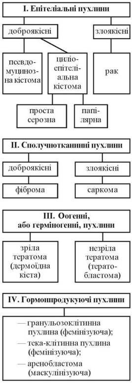
Fig. 24. Classification of ovarian tumors
Benign epithelial ovarian tumors are termed cystomas. Like true tumors, cystomas possess the capacity for proliferation. Pseudomucinous and cilioepithelial cystomas are distinguished. Ovarian cystomas are typically detected with the onset of menstruation.
Pseudomucinous cystoma (or glandular cystoma) is externally covered by germinal epithelium, followed by a layer of Fibrous connective tissue, while the inner surface of the tumor capsule is lined with single-layer epithelium. The cystoma is a multilocular formation with chambers filled with pseudomucin. Pseudomucinous cystomas are classified into those with a smooth inner surface lacking mitoses and proliferating pseudomucinous cystomas featuring a velvety, villous epithelium covering papillary proliferations within the chambers, accompanied by minor mitoses in the epithelium. The tumor reaches significant size, is mobile (has a stalk), is usually unilateral, rarely located between the ligaments, is not accompanied by ascites, and relatively infrequently undergoes malignant transformation into Cancer. Endoscopy is preferred for their diagnosis. In cases of pseudomucinous cystoma in girls, the tumor-affected ovary is removed if the contralateral ovary appears macroscopically normal. During surgery, an urgent histological examination of the removed tumor must be performed before the Procedure is completed, and the inner surface of the resected cystoma should be inspected to rule out malignancy. An exploration of the Abdominal cavity (Liver, Stomach, omentum, peritoneum) is mandatory in all cases.
Cilioepithelial cystoma, similar to pseudomucinous cystoma, contains a connective tissue capsule lined with germinal epithelium; from the inside, the cystoma is lined with single-layer cuboidal and ciliated epithelium capable of forming papillae. The cystoma is filled with serous content, is most commonly bilateral and of small size, is frequently located between the ligaments, may be accompanied by ascites, and relatively often undergoes malignant transformation into cancer due to the high proliferative activity of its cells. Numerous papillae line the capsule and occasionally grow through it to the outer surface. The epithelium covering the papillae is usually single-layered. In some cases, it is compressed by fluid and undergoes atrophy, losing its capacity for proliferation, thus forming a variant of cilioepithelial cystoma known as a serous cystoma. In other instances, the cyst contents are scant or absent, and the tumor is a conglomerate of papillary proliferations (papillary cilioepithelial cystoma). For simple serous cystoma, The Scope of surgical intervention is the removal of the ovary. Papillary cilioepithelial cystoma requires radical surgical intervention due to pronounced proliferative processes. When one ovary is affected and papillary proliferations are confined to the inner surface of the tumor capsule, unilateral salpingo-oophorectomy is possible. If both ovaries are affected or papillary proliferations extend onto the outer surface of the cyst capsule, subtotal Hysterectomy with bilateral salpingo-oophorectomy and omentectomy, followed by Chemotherapy, is performed.
Ovarian fibroma (a connective tissue tumor) is a unilateral mass, occasionally of considerable size, and occurs rarely in girls. An ovarian fibroma is an indication for its removal.
Dermoid cyst (benign oogamous tumor) is a mature teratoma classified among ovarian tumor-like formations (cysts). It is characterized by the absence of proliferative elements in the dermoid cyst capsule. Malignant transformation occurs very rarely.
Mature teratoma (dermoid cyst) is one of the most common ovarian tumors in children and may even occur in newborns.
Ovarian dermoids are characterized by slow growth and are typically asymptomatic. Occasionally, patients experience transient dull lower abdominal pain and urinary symptoms caused by the mechanical pressure of the dermoid on the bladder. Dermoid cysts are highly mobile. The tumor size varies, generally not exceeding 12-15 cm in diameter. The tumor is usually unilateral, round, with an unevenly firm-elastic consistency, smooth surface, and yellowish (or whitish with various shades) color. As a rule, dermoid cysts have a stalk and are rarely located between the ligaments. A dermoid is predominantly a unilocular cyst (multilocular structures are rare), filled with adipose tissue—Tissues of ectodermal origin—and Hair (Figs. 25-27).

Fig. 25. Opening of the ovary during dermoid cyst enucleation. The cyst capsule is intact.
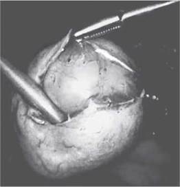
Fig. 26. Aquadissection of the cyst from the ovarian tissue. If necessary, the incision of the ovary is slightly enlarged to facilitate cyst enucleation.

Fig. 27. Bipolar coagulation of the cyst "stalk" close to the ovarian hilum.
Distinctive features of dermoid cysts include their typical Location anterior and lateral to the Uterus, as well as significant mobility.
Treatment for mature teratomas is surgical, with laparoscopic intervention being the preferred approach. The prognosis is favorable. Malignant transformation is observed in 1.7% of cases.
Teratoblastoma is an immature tumor belonging to the group of malignant ovarian neoplasms, ranking first among malignant tumors in girls. The tumor reaches significant sizes of up to 20 cm in diameter, with an irregular ovoid or round shape. The color is whitish, the consistency is soft, though it may be firm in places, and the surface is nodular or smooth. Cystic cavities vary in size and contain fluid contents. Macroscopically, the tumors may contain bone, Cartilage, and adipose tissues, Teeth, and occasionally hair.
The tumor is characterized by rapid growth, invades the capsule and peritoneum, and metastasizes to retroperitoneal Lymph Nodes, Lungs, liver, and Brain. Girls with immature ovarian teratomas typically complain of lower abdominal pain, general weakness, lethargy, rapid fatigue, and reduced performance. Menstrual function is not impaired. In later stages, cachexia, ascites, and fever are observed. Blood tests reveal changes typical of malignant tumors.
Surgical treatment involves Supravaginal amputation of the uterus with appendages, followed by Radiation therapy. The prognosis is unfavorable.
Dysgerminoma is predominantly a malignant tumor developing from primordial Gonads (synonyms: seminoma, genocytoma, embryocytoma). In almost half of the cases, dysgerminoma develops in childhood.
The tumor varies in size, has a round shape, often nodular or with a smooth surface, and a well-defined capsule. Its color is whitish, and the consistency may be firm, moderately elastic, doughy, or medullary. Dysgerminoma most commonly arises in one of the ovaries. Bilateral tumors occur in 8-12% of cases.
When dysgerminomas develop in girls, hypoplasia of the genital organs and underdeveloped secondary sexual characteristics are typically observed, almost always accompanied by delayed menarche. Tumor growth is accompanied by menstrual irregularities. Prolonged Amenorrhea (4-6 months) may alternate with uterine bleeding.
Complaints include dull, aching lower abdominal pain accompanied by frequent urination, general malaise, weakness, and drowsiness. Girls fatigue easily. In early-stage cases, the blood picture is usually unremarkable. With tumor necrosis and rapid growth, a slight evening Temperature elevation and increased ESR are observed, along with a left shift in the leukocyte formula. Metastases commonly spread to para-aortic lymph nodes.
Treatment is surgical, followed by radiation therapy. For unilateral tumors without signs of metastasis beyond the affected ovary, treatment is limited to its removal. The tumor must be handled with extreme care during surgery; compromising the integrity of the capsule significantly worsens the prognosis. If the tumor metastasizes beyond a single ovary, radical surgery—removal of the appendages and uterus followed by radiotherapy—is recommended. Both the primary tumor and metastatic nodes respond well to radiotherapy.
Granulosa Cell tumor develops from granulosa tissue, which structurally resembles the granular epithelium of a mature follicle. This is accompanied by elevated estrogen levels (feminizing effect), leading to precocious Puberty in girls. The tumor is usually unilateral and benign, though frequently malignant as well. Treatment for granulosa-cystic tumors involves removing the affected ovary and conducting careful follow-up monitoring of the young patient.
Thecoma (theca cell tumor) is extremely rare in girls and exerts a feminizing effect on the body. The tumor consists of cells resembling maturing theca cells and atretic follicles. Its size ranges from a pea to an adult's HEAD; it is typically unilateral and firm.
The presence of the tumor leads to elevated estrogen levels. Histological examination of uterine curettage specimens reveals glandular-cystic endometrial hyperplasia. Treatment for theca cell tumors is strictly surgical.
Arrhenoblastoma, which exhibits a masculinizing effect, can arise from ovarian hilus cells that excrete androgens. The resulting excess androgens suppress pituitary function, thereby lowering estrogen levels in the body. The primary clinical sign is defeminization (amenorrhea occurs against a Background of complete health, Mammary Glands decrease in size), followed by the appearance of male characteristics—deepening of the voice, male-pattern hair growth, and male contours of the body and face. Treatment for arrhenoblastoma is surgical; in girls, it may be limited to simply removing the tumor.
Ovarian tumors in children are characterized by diverse clinical symptoms. Asymptomatic tumors are typically discovered during routine preventive examinations. In such cases, the ovarian tumor is differentiated from a floating Kidney and tumors of other abdominal and retroperitoneal organs.
In the presence of acute pain, Differential diagnosis is performed with acute appendicitis, intussusception, intestinal obstruction, Ovarian Apoplexy, and Developmental anomalies of the hymen and vaginal septum leading to hematocolpos and hematometra. Diagnostic laparoscopy or diagnostic laparotomy is recommended to clarify the cause of the "acute abdomen."
Complications of ovarian tumors include torsion of the tumor pedicle, rupture of the cyst or cystoma capsule, adhesion formation between the cystoma and adjacent organs, and malignant transformation. The most frequent complication in girls is torsion of the pedicle of a tumor or tumor-like lesion. Occasionally, torsion of structurally normal adnexa occurs, presenting with a classic picture of an acute abdomen.
Torsion of a cyst or cystoma pedicle. Anatomically, the pedicle of a cystoma is formed by the stretched infundibulopelvic ligament, ovarian ligament, and broad ligament (mesovarium). The pedicle contains the vessels supplying the tumor (the ovarian artery, a branch of the ascending uterine artery), Lymphatic vessels, and nerves. There is also the surgical pedicle, which refers to the structures that must be transected during tumor removal. Most frequently, the surgical pedicle includes the anatomically normal structures along with an overstretched fallopian tube. Torsion of the tumor pedicle is triggered by sudden movements, changes in body position, or physical exertion; it can be acute or gradual, and complete or partial. Complete torsion severely disrupts the Blood supply to the tumor, resulting in hemorrhage and necrosis. Clinically, this manifests as an "acute abdomen" syndrome: sudden sharp pain, rigidity of the anterior abdominal wall Muscles, a positive Blumberg's sign, often accompanied by nausea or vomiting, intestinal paresis, and constipation with the retention of gas. The young patient's condition deteriorates rapidly, marked by Skin pallor and cold sweat. Body temperature rises, the pulse accelerates, and arterial pressure drops. The tumor increases in size due to edema and hemorrhages, and its Palpation elicits severe pain. Treatment is surgical. During surgery, untwisting the tumor pedicle is not recommended, as thrombi within the Blood Vessels of the pedicle can lead to thromboembolism. Delaying surgery leads to tumor tissue necrosis, secondary infection, adhesion formation with adjacent organs, and Peritonitis.
In cases of partial tumor pedicle torsion, when the aforementioned symptoms are less pronounced and signs of ischemia and tissue necrosis are absent, laparoscopic untwisting of the pedicle is feasible if less than 6 hours have passed since the onset of torsion.
In complete torsion of the cystoma pedicle, the clinical picture is typically so classical that it readily allows for a definitive diagnosis.
To refine the diagnosis, additional investigative Methods are employed:
— gastrointestinal tract imaging, including barium enema (irigoscopy), which helps clarify the Nature of the ovarian tumor, assess the condition of the bowel, and determine its spatial relationship to the tumor;
— pelvic X-ray, which AIDS in detecting calcifications characteristic of dermoid cysts;
— urography;
— laparoscopy;
— gas and double-contrast pneumopelvigraphy;
— in the absence of laparoscopy, diagnostic laparotomy (particularly when other diagnostic modalities cannot be utilized for any reason).
Diagnosing tumors and tumor-like lesions of the uterus and its adnexa in pediatric patients presents considerable challenges. Gynecologists must always keep in mind the possibility of tumors in adolescent girls. Complaints of lower abdominal pain or behavioral changes are absolute indications for a pediatric gynecologist consultation.
The Anatomical Features of uterine and adnexal localization in children often result in a high-riding tumor positioned outside the true pelvis, even when the tumor is relatively small; therefore, rectoabdominal examination under anesthesia should be performed when necessary.
Cytology/practical/136.html">DIFFERENTIAL DIAGNOSIS OF adnexal tumors and tumor-like lesions in girls must be conducted with acute appendicitis, especially in cases of tumor pedicle torsion; thus, a joint evaluation by a pediatrician, pediatric gynecologist, and pediatric surgeon is highly recommended.
Surgical interventions for tumors of the reproductive organs in adolescent girls should be as conservative (organ-sparing) as possible to preserve reproductive potential. True tumors should be enucleated (excised), leaving the healthy ovarian tissue intact. Nevertheless, girls who undergo tumor removal must remain under the continuous dispensary observation of an obstetrician-gynecologist.
Removal of the adnexa is indicated only in cases of tumor pedicle torsion where the ovarian tissue is so extensively damaged that its preservation is impossible.
Intraoperative frozen-section histological examination of the excised tissue is of paramount importance for young patients, as it dictates the appropriate scope of surgery. Radical surgical intervention must be justified by histological confirmation of a malignant neoplasm.
Last update: 08/08/2026
Editorial and Educational Adaptation: This material has been compiled based on the primary/original source text. The project team performed an editorial review, corrected technical inaccuracies, structured sections, and adapted the content for an educational format.
What was processed:
- elimination of formatting defects (OCR errors, structural breaks, corrupted characters);
- editorial organization of content;
- standardization of terminology in accordance with academic sources;
- verification of factual statements against the original source text.
All mentions of the author, publication year, and origin of the primary text have been preserved in accordance with the source.