BOTANY VOLUME 2 - PLANT PHYSIOLOGY - 2007
8. PHYSIOLOGY OF MOVEMENTS
8.3. Movements of Living Organs
If a bending movement of an attached Organism or its individual Organs is induced by a one-sided stimulus and determined by its direction, such a movement is called a tropism (see 8.3.1). Bending typically occurs due to varying growth rates on opposite sides of the organ, while turgor changes are very rarely the cause. Conversely, if the manner and direction of movement are determined solely by The Structure of the responding organ, with the stimulus acting merely as a trigger (regardless of whether it acts unilaterally or from all sides), it is referred to as a nastic movement (nasty). Nasties (see 8.3.2) are mostly based on reversible turgor changes, and less frequently on different growth rates of the opposite sides of the organ. Both nasties and Tropisms are contrasted with autonomous organ movements (see 8.3.3), which are controlled by internal mechanisms.
Both tropisms and nasties can be further subdivided According to the type of stimulus that induces them.
In positive tropism, the response is directed toward the source of the stimulus, whereas in negative tropism, it is directed away from it. If the movement occurs at a specific angle to the stimulus source, it is called plagiotropism; if this angle is 90°, the movement is termed diatropism (also known as transversal, or transverse, tropism). Since tropism is based on differential growth, it manifests exclusively in growing organs, and this capacity disappears as growth ceases. Tropistic reactions primarily involve elongation growth and, much less frequently—such as in the upward bending of horizontally tilted stems—also involve Cell Division. In positive tropism, the side of the organ facing away from the stimulus source generally grows more intensively; this is observed in both Higher Plants and certain single-celled systems (e.g., the sporangiophores of Phycomyces and Pilobolus). However, in Cells with pronounced apical growth, such as fern chloronema and pollen tubes, lateral stimulation can inhibit apical growth and induce a new apex that faces the stimulus source, continuing growth with a sharp bend. Thus, in the Examples given, during positive tropism, the side facing the stimulus grows faster.
8.3.1.1. Phototropism and Scototropism
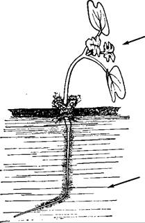
As a result of unilateral illumination, many plant organs shift into a position that confers an advantage, such as optimizing light capture for Photosynthesis. Positive phototropism is exhibited by most shoots (Fig. 8.12) and many leaf petioles, the single-celled sporangiophores of certain Mucorales Fungi (e.g., Phycomyces and Pilobolus), and the fruit bodies of some Coprinus species. Negative phototropism is less common, occurring, for example, in clinging and aerial roots (such as ivy and aroids), liverwort rhizoids, fern prothalli equipped with appressoria, wild grapevine tendrils, mistletoe hypocotyls, and, in exceptional cases (e.g., Sinapis, Fig. 8.12), embryonic roots; however, most roots do not exhibit phototropism. Many lateral branches react plagio-phototropically, while leaf blades (see Fig. 8.12) and liverwort thalli react diaphototropically. Sometimes the phototropic response type changes during development. Flower stalks (e.g., Linaria cymbalaria, Cyclamen persicum, Tropaeolum maius) show positive phototropism before Fertilization and negative phototropism after fertilization, so that the fruits end up tucked into fence crevices or similar microhabitats suitable for germination.
Class="center">Fig. 8.12. Mustard seedling in Water culture, unilaterally illuminated from the right (arrows). The SHOOT exhibits positive phototropism, while the ROOT shows negative phototropism (which is an exception!). The leaf blades are positioned perpendicular to the direction of light, showing diaphototropism.
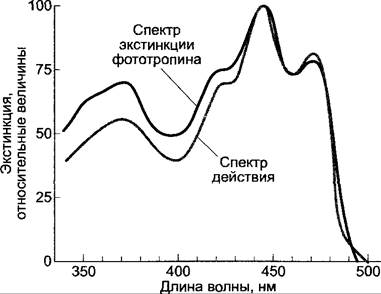
In moss protonemata and fern chloronemata, where the phototropic response is based on the Displacement of the growth point, the photoreceptor is the Phytochrome system. Experiments with linearly polarized light led to the Conclusion that, much like chloroplast rotation (see 8.2.2), photoreceptor molecules are arranged in the ectoplasm with a high degree of order. All phototropic responses based on growth differences between the illuminated and shaded sides share an identical action spectrum, featuring a single peak in the ultraviolet region (370 nm) and three peaks with maximum activity in the blue region (Fig. 8.13). Because the UV peak resembles flavin absorption and the blue region peaks resemble carotenoid absorption (see Figs. 6.46; 6.47), the exact Nature of the absorbing chromophore remained unclear for a long time. Nevertheless, photoreceptors have recently been identified through the successive use of mutants, first in Arabidopsis thaliana and subsequently in other species.
Fig. 8.13. Action spectrum of phototropism (gray curve, first positive curvature of oat coleoptiles) and extinction spectrum of recombinant oat phototropin (cloned under a strong promoter in Escherichia coli) after The addition of the FMN chromophore (in vitro reconstitution)
Researchers successfully isolated Arabidopsis thaliana mutants which, although lacking the blue light-dependent photomorphogenic response (inhibition of coleoptile growth in etiolated seedlings under bright light, cry1 mutant, see 7.7.2.1), still retained normal positive phototropism. Another mutant lacked phototropism under weak light while still exhibiting normal hypocotyl elongation inhibition (nph1 mutant, derived from non-phototropic hypocotyl). This led to the conclusion that at least two distinct blue-light receptors exist: one responsible for triggering photomorphogenesis and the other for triggering phototropism; consequently, the receptor responsible for phototropism cannot be identical to cryptochrome (see Table 7.8). It is now known that phototropism is controlled by two structurally related blue-light receptors: phototropin 1 (encoded by the NPH1 Gene) and phototropin 2 (encoded by the NPL1 gene, from NPH-like). Phototropin 1 responds to low-intensity light, while phototropin 2 responds to high light intensity. The double mutant (nph1/npl1) lacked phototropism entirely. In gymnosperms, phototropins also control chloroplast movements and stomatal guard cell movements.
The phototropism-controlling receptors phototropin 1 and phototropin 2 are Chromoproteins; their apoproteins have a molecular mass of approximately 120 kDa and contain non-covalently bound flavin mononucleotide (FMN) as a chromophore. The absorption spectrum of phototropin corresponds with high precision to the action spectrum of phototropism, suggesting that carotenoids are likely not involved (see Fig. 8.13).
Phototropins function as protein Kinases and undergo autophosphorylation in the presence of ATP (likely at Serine and Threonine residues) in a reaction directly dependent on blue light. This state is generally considered to correspond to the activated receptor state. The subsequent signaling pathway is not yet fully understood. It is hypothesized that the activated state of phototropin regulates auxin distribution within the organ, and that differential growth on the shaded side occurs under METABOLISM/18.html">The Influence of this asymmetric auxin distribution (see below; for auxin, see 7.6.1).
The phototropic response has been studied particularly intensively in cereal coleoptiles, seedling hypocotyls and epicotyls, and Phycomyces sporangiophores. In all cases, a complex dependence of the response on the received light dose (the product of light intensity [W · m-2] and illumination time [s]; W · m-2 · s = J · m-2) has been revealed. In the best-studied oat coleoptiles (Fig. 8.14), which serve as a typical example here, low light doses (approximately in the range of 10-1 to 102 J · m-2) induce a positive phototropism. As the light dose increases further, the response intensity drops back to zero before showing a sharp transition to positive phototropism once the value of 104 J · m-2 is exceeded (referred to as the First and Second positive phototropic responses, respectively). At very high light doses, a second indifference zone occurs, followed by a third positive phototropic response, which only manifests under experimental conditions and plays no role under natural light intensities. The second positive response range is relevant for phototropism under natural daylight; seedlings in the soil may respond under certain conditions to very weak, transiently incident light within the range of the first positive phototropic response, which has been best studied experimentally.
Fig. 8.14. Dose-response curve of the phototropic reaction in oat coleoptiles (dose-response curve based on B. Steyer; phototropin hypothesis based on M. Salomon, M. Zacherl, W. Rüdiger). Plants were illuminated for 1 to 120 s at 8 · 10-2 W · m-2 (black segment of the graph) or from 1 s to 3 h at 3.5 W · m-2 (gray segment of the graph). Light dose in J · m-2 = W · m-2 · s. Schematic diagrams illustrate the current understanding of the phosphorylated state of the phototropism receptor, phototropin (hatched), as a function of the applied light dose (regions a to d). For further explanations, see text
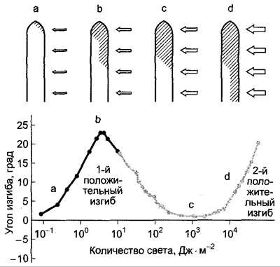
The stimulation threshold for the first positive response in oat coleoptiles lies at approximately 10-1 J · m-2, and an approximate proportionality between the achieved phototropic curvature and the applied light dose is maintained up to 102 J · m-2. The reaction time (depending on environmental conditions such as Temperature) is 25–60 min, and the duration of the response (from the onset to the completion of bending) is approximately 24 hours (Fig. 8.15).
Fig. 8.15. Coleoptile phototropism: A — time course of phototropic bending under unilateral illumination (arrow). Left — first positive response ("apical response") of an oat coleoptile, right — second positive response ("base response"); B — unilateral illumination of a coleoptile. Light strikes perpendicular to the plane of the paper on one half of the organ, while the other half remains in darkness (hatched). The object bends not toward the light source (toward the observer), but within the plane of the paper in accordance with the brightness differences between the illuminated and unilluminated halves (stronger growth on the shaded side)
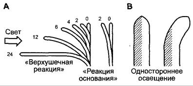
When examining phototropism in cells with apical growth (see above), the positive phototropic curvature is brought about by enhanced growth of the shaded side relative to the illuminated side. The region of maximum light sensitivity typically lies apical to the bending zone. To
induce the first positive response in a coleoptile (Fig. 8.15, A), illumination of the very tip (0.25 mm) is required. Because the response occurs even when only this specific region is illuminated, signal transmission from the site of light perception to the site of the bending response is necessary. The first positive curvature begins at the apex and gradually extends downward to the base ("apical response"). The second positive curvature occurs initially near the Base of the coleoptile ("base response"); while the coleoptile tip is also particularly sensitive in this case (~0.5 mm), all underlying PARTS OF THE organ exhibit a lower degree of sensitivity. Phototropin 1 is involved in both cases; the phototropin 2-mediated response is only possible at higher light doses (third positive curvature).
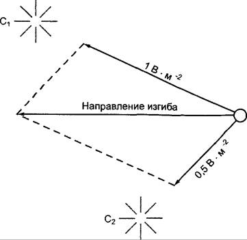
Older plants perceive unilateral illumination at the shoot apex, most commonly via the blade of young leaves. In Tropaeolum, the leaf petioles are phototropically sensitive. If an organ capable of a phototropic response receives light from two lateral sources at different angles and of different intensities, a curvature toward the resultant vector usually occurs, which can be derived from the parallelogram of forces—based on the direction and magnitude of the stimulus (Fig. 8.16).
Fig. 8.16. Phototropic curvature according to the law of the resultant vector under simultaneous illumination by light sources of different intensities (C1, C2). The illumination forces directed from each source exclusively onto the object (viewed from above here) are represented as the vectors of a force parallelogram.
However, phototropism does not perceive the direction of light itself, but rather the difference in light intensity between the illuminated and shaded sides. This can be demonstrated, for example, by means of so-called unilateral illumination (see Fig. 8.15, B). The necessary differences in light intensity between the two sides arise due to light scattering or absorption ("shadow pigments," such as carotenoids in the coleoptile tip) within the organ. The varying light intensity at different sites of the organ could directly correlate in coleoptiles with the degree of phosphorylation of phototropin, which occurs predominantly in the coleoptile tip and to a significantly lesser extent at the coleoptile base (see Fig. 8.14). The phosphorylation hypothesis also offers an explanation for the complex dependence of the phototropic response on light energy. Very low light intensity and a short irradiation time are sufficient only to adequately activate phototropin 1 on the illuminated side in the region of the coleoptile tip. Here, a phototropin phosphorylation gradient is established. With increasing light intensity or duration, phototropin is increasingly phosphorylated also on the shaded side of the coleoptile tip, causing the phosphorylation gradient to disappear. If the light intensity is increased further, phototropin 1 located at the base of the coleoptile—which is present in a much lower concentration or is more dispersed (details are still unknown)—finally responds, first on the illuminated side and subsequently, with a further increase in intensity or duration, on the shaded side as well. The forming (and ultimately disappearing again) phosphorylation gradient regulates the second phototropic curvature ("basal response," Fig. 8.15, A). It is now assumed that phototropin regulates auxin transport either directly or via a signal Transduction cascade. Accordingly, the phototropin phosphorylation gradient causes an asymmetrical distribution of auxin within the organ, which in turn leads to different growth rates on the illuminated and shaded sides. Experimental Evidence for the auxin hypothesis of phototropism has been provided for coleoptiles; for other organs of higher plants, the situation is not yet fully understood, and the mechanisms of phototropism in lower plants remain entirely unstudied.
Fig. 8.17. Transverse auxin transport during coleoptile phototropism. Arrows indicate the direction of illumination. A — demonstration of the necessity for unobstructed lateral transport. A Glass plate placed perpendicular to the direction of light (b) blocks transport and curvature, whereas one placed parallel to the direction of light (c) does not; B — collection of auxin diffusing from isolated coleoptile tips using Agar blocks in the control (a), with an obstruction (b), and without an obstruction (c); numbers represent relative auxin levels. Unilateral illumination under conditions of unobstructed transverse transport leads to an enhanced efflux of auxin on the light-exposed side. Supplying the apex with radioactive IAA externally and subsequently measuring radioactivity in the blocks yields a comparable result; C — signal transmission via longitudinal auxin transport: a — unilateral illumination induces transverse auxin transport in the isolated apex; b — the apex is replaced onto the base; c — the asymmetrical auxin distribution is transmitted to the base, leading to curvature there (cf. the phototropin phosphorylation model — Fig. 8.14).
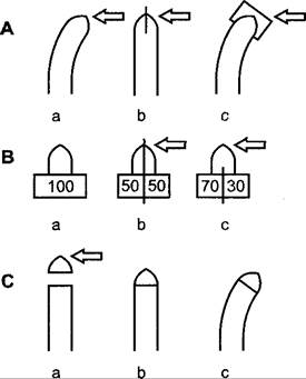
Unilateral illumination of etiolated cereal seedlings in the region of the first or second positive curvature leads, on the one hand, to a lateral translocation of auxin from the illuminated side to the shaded side of the organ in the coleoptile tip region and, on the other hand, to the suppression of basipetal auxin transport (see 7.6.1.3) on the illuminated side (Fig. 8.17). The resulting Asymmetry in auxin distribution within the coleoptile tip is then amplified via polar auxin transport down to the base, resulting in enhanced growth on the auxin-rich shaded side. A growth difference of merely 2% between opposing sides of the organ is already sufficient to cause a 10° curvature. Signal transmission in phototropism thus consists in asymmetrical auxin transport; consequently, inhibitors of polar auxin transport (such as 2,3,5-triiodobenzoic acid, TIBA) also disrupt the phototropic response.
In tropical lianas (e.g., the aroid plant Monstrera gigantea), shoots directionally seek out their supporting tree trunk through a growth curvature toward the darkest sector of the horizon. Because shoots eventually encircle the support trunk from all sides, this is not negative phototropism, but rather growth toward the shadow, i.e., skototropism. Once the shoot reaches the supporting trunk, its skototropic sensitivity shifts to positive phototropism, which guides the plant upward toward the light in the canopy region. The Mechanism of skototropism remains unknown.
Many plants are able to reorient their organs in a specific direction relative to the acceleration vector of gravity ($g = 9.81\text{ m} \cdot \text{s}^{-2}$) via growth curvatures. This response is termed gravitropism (formerly referred to as geotropism). For instance, trees on steep slopes grow such that the longitudinal axis of the trunk aligns with the direction of gravity rather than perpendicular to the local land surface. Axial organs displaced from their normal orientation, such as flower stalks, bend until they resume a vertical position; cereal stems that have lodged due to weather conditions can re-erect themselves through curvature at the node regions.
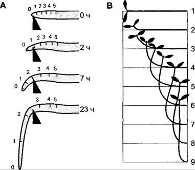
Positively gravitropic, i.e., growing toward the center of the Earth, are primary roots (Fig. 8.18, A) as well as the rhizoids of Algae, liverworts, or fern prothalli. Negative gravitropism is exhibited by main shoots (Fig. 8.18, B), the sporangiophores of Mucoralean fungi, and the fruiting bodies of many agaric fungi. First-order lateral roots grow predominantly horizontally (diagravitropism) or obliquely downward at a defined angle (plagiogravitropism). Many lateral branches and leaves, as well as rhizomes, also respond in a diagravitropic or plagiogravitropic manner. Second-order lateral roots are generally insensitive to the direction of gravity (agravitropic), much like the lateral branches of "weeping" plant forms (e.g., weeping willow). Similar to phototropism, gravitropism in certain plants can switch during development or as a result of changing environmental conditions.
Fig. 8.18. Gravitropism: A — positive gravitropism of an embryonic root; time is indicated in hours from the experimental displacement into a horizontal position. Marks 0–5 on the root indicate the elongation growth of individual root segments during the response. The overall response takes from two to several hours depending on the plant species; B — negative gravitropism of an embryonic shoot. The sequence of numbers denotes individual Stages of the response, which takes from two to several hours depending on the plant species.
For example, the upper part of the flower stalk in a poppy bud is positively gravitropic ("nodding bud"), but gravitropism shifts to negative before flower opening. In many species (Holosteum umbellatum, Calandrinia, Arachis1, etc.), the flower stalks are negatively gravitropic, but upon developing into fruit stalks, they become positively gravitropic, whereas in Lilium martagon, the exact opposite occurs. If, for instance, the negatively gravitropic main shoot of a spruce or fir is cut off, the upper lateral branches—originally dia- or plagiogravitropic—reorient themselves in a negatively gravitropic manner. Typically, one of them assumes the function and position of the main shoot, while the others return to their original orientation (apical dominance, see 7.6.1.4).
1 In peanuts, fruit burial into the soil is driven by the growth of the Ovary base rather than the flower stalk. — Ed. note.
Lower winter temperatures, for instance, render the shoots of certain field weeds diagravitropic (Senecio vulgaris, Sinapis arvensis, Lamium purpureum, etc.), whereas they are negatively gravitropic in summer. In this way, they ensure protection beneath the snow cover. Diagravitropic rhizomes of Adoxa or Circaea become positively gravitropic upon exposure to light, enabling them to re-enter the soil. For underground shoots of Aegopodium podagraria, a 30-second exposure to red light is sufficient to induce this shift. Diagravitropic shoots of Vinca, Lysimachia nummularia, and others turn negatively gravitropic when shaded.
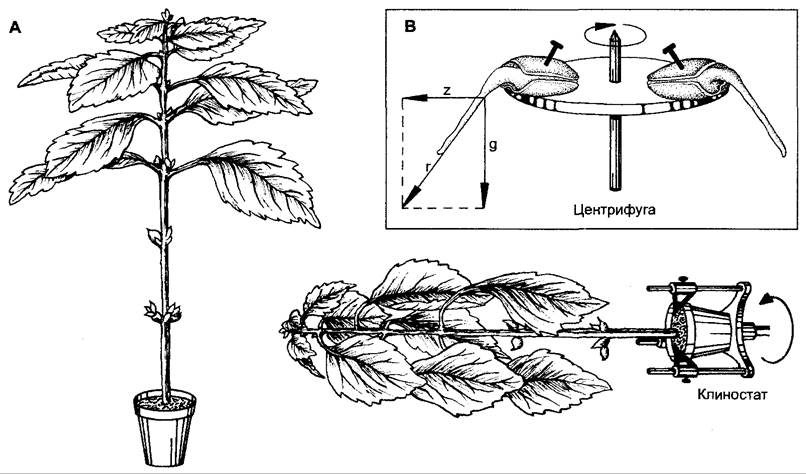
That gravitropic curvatures are responses to mass acceleration, normally elicited by the unilaterally acting force of gravity, can be demonstrated in various ways. First, the acceleration generated by a centrifuge ($z$) acts in the same manner as gravitational acceleration ($g$, Fig. 8.19, B). Because both forces are of the same order of magnitude, the law of the resultant vector applies here as well (see Fig. 8.16): gravity and centrifugal force are perceived by the plant as equivalent. Alternatively, gravitropic curvature can be abolished if a plant initially grown in an orthotropic orientation is rotated slowly around its longitudinal axis (on a clinostat, Fig. 8.19, A). When the rotation speed is high enough to preclude the unilateral perception of gravity, yet low enough to prevent the centrifugal force from becoming effective (a few revolutions per minute), the action of the gravitational field is compensated.
Fig. 8.19. Demonstration that mass acceleration is the primary stimulus in gravitropism: A — a Coleus plant normally oriented (left) and grown on a clinostat with slow rotation (a few revolutions per minute) about its longitudinal axis (right). Upon elimination of the unilaterally acting force of gravity, the negative gravitropic shoot curvature does not occur, and leaf epinasty arises, which would otherwise be compensated by negative gravitropism; B — applicability of the resultant force law under the simultaneous action of centrifuge-generated acceleration ($z$) and gravitational acceleration ($g$). The direction of growth coincides with the direction of the resultant force ($r$).
Gravitropic curvatures, like phototropic ones, are typically based on differing growth rates of opposing organ sides. Consequently, in these cases too, growth-competent zones respond: the growth zone located immediately behind the root apex, or the corresponding zone of shoots, hypocotyls, or seedling epicotyls (see Fig. 8.18). Because the elongation zone is short, the course of root curvature is relatively straightforward. In shoots, the curvature begins in the upper region and subsequently extends further in a basal direction; meanwhile, the upward gravitropically induced curvature overshoots the vertical, followed by a counter-curvature. Bending continues until the shoot, after a few oscillatory movements, settles precisely in the vertical position. These oscillatory movements are only partially caused by a renewed (counter-directed) gravitropic stimulus; in part, they occur independently of gravity (for example, also on a clinostat), and the underlying regulatory mechanism remains unknown.
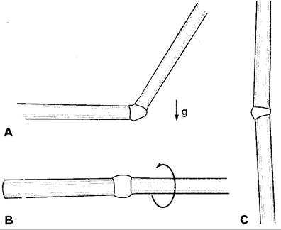
In certain instances, previously matured plant parts can resume growth following a gravitropic stimulus: for example, in cereal stems displaced from the vertical, the lower sides of the nodes begin to grow vigorously, causing the stem to rise again (Fig. 8.20). On a clinostat, nodal growth is uniformly enhanced, meaning that gravity stimulation is still perceived in this case. Tree trunks, branches, and roots are also capable of exhibiting gravitropic responses (albeit very slowly), expressed as enhanced longitudinal and radial growth involving the cambium. During this process, the gravitropically stimulated cambium produces a specialized, anatomically distinct "reaction wood"—on the lower side in conifers (compression wood) and on the upper side in dicot trees (tension wood). Reaction wood formation also occurs in the absence of longitudinal growth and bending (e.g., after removal of the apical bud); thus, its formation is not induced by compressive or tensile deformations. Rather, The formation of reaction wood is the underlying cause of gravitropic curvature.
Fig. 8.20. Gravitropic upward bending of a horizontally inclined (A) cereal node compared to one rotated horizontally around its longitudinal axis on a clinostat (B), shown alongside an unstimulated control (C). The experiment is conducted on an excised shoot segment. Comparing B and C reveals that a horizontal position combined with rotation around the longitudinal axis stimulates longitudinal growth of the node. Gravitropic stimulation (A) leads to strong elongation of the lower side of the node, while the upper side remains shortened.
The presentation time for gravitropism can be very short, lasting a matter of minutes (e.g., 3 min for the hypocotyl of Helianthus). Reaction times can likewise fall within the minute range (oat coleoptile — 14 min, cress roots — less than 20 min). Shoots often begin to respond only after more than an hour, whereas cereal nodes take several hours. The stimulation threshold for continuous stimulation lies at a mass acceleration of approximately $10^{-2}g$ ($g$ being gravitational acceleration); the summation of sub-threshold stimuli can, as in phototropism, lead to a visible response.
For stimuli slightly exceeding the threshold, the law of stimulus quantity applies (equation 8.1), much as in phototropism. That is, within certain limits, it is equivalent whether a stronger stimulus acts briefly or a weaker one acts over a longer duration: the crucial factor is the stimulus quantity $R$, i.e., the product of stimulus intensity $I$ and exposure time $t$. At low stimulus quantities, a proportionality is also observed between the stimulus quantity and the response intensity. This can be easily investigated by applying metered centrifugal forces (see Fig. 8.19) or by deviating the plant from the vertical at an angle less than a right angle. In the latter case, only a fraction of the gravitational force acts, which is proportional to the sine of the angle of deviation from the vertical (sine law). In many cases, deviations from the vertical of as little as 1 to 2 degrees are corrected by the gravitropic growth response. Trees, for instance, grow vertically not only on steep slopes but also on very gentle ones, i.e., parallel to the vector of gravitational acceleration rather than perpendicular to the local land surface.
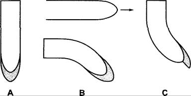
Perception of the force of gravity occurs in coleoptiles (which are also well studied) within the parenchyma of the apex (-3 mm), in roots within the central part of the root cap (calyptra), and in shoots, presumably, within the elongation zones of all still-growing internodes (in this case, within the Cells of the starch-sheath). Removal of the root cap slightly enhances root elongation, but completely abolishes gravitropic responsiveness (Fig. 8.21, A, B); this indicates that gravitropism in this case relies on an inhibitory effect. This is further evidenced by the bending of non-gravitropically stimulated roots in which the calyptra has been removed from only one side (Fig. 8.21, C). In Arabidopsis, the scarecrow mutant lacks starch sheaths and endodermis (in the root-neck region, the starch sheath transitions into the endodermis). The shoot of this mutant is agravitropic, but the root responds normally.
Fig. 8.21. The root cap as the gravity-perceiving organ in which the root growth inhibitor is produced. Compared to non-stimulated roots (A), roots lacking root caps (B, top) show a slight increase in elongation growth, but no gravitropism, which is dependent on the presence of the root cap, or calyptra (B, bottom). C — removal of part of the root cap on one side leads to a curvature of the root tip. The side lacking the calyptra grows faster than the intact side, suggesting the presence of a factor or complex of factors produced by the root cap that inhibits root elongation growth
Cells or Tissues involved in the perception of gravity acceleration typically exhibit a pronounced asymmetry in the Intracellular Distribution of Organelles: "light organelles" with lower specific density (such as vacuoles) are located in the upper part, whereas the specifically denser "heavy organelles" (cell nuclei and especially amyloplasts, or barium sulfate crystals—"statoliths" or "shiny bodies"—in Chara rhizoids) occupy the physically lower side. Such specific heavy particles are called statoliths (in plants with Plastids, these are the aforementioned amyloplasts), cells bearing statoliths are termed statocytes, and the tissue comprising them is called statenchyma; the sedimentation of statoliths within The Cell is associated with the perception of mass acceleration (Fig. 8.22). Even in plants that accumulate little or no starch due to prolonged dark adaptation or genetic defects, the gravitropic response is not completely abolished, although it is much weaker. Such "starch-free" plants still exhibit a certain degree of leucoplast sedimentation acting as statoliths. Consequently, "statolith starch" should not be dismissed as a component important for the cell's ability to perceive mass acceleration; it may contribute to increasing the specific density of amyloplasts and, accordingly, to improving gravitropic sensitivity.
Fig. 8.22. Location of statenchyma: A — in a shoot (starch sheath), B — in the root cap, C — in the coleoptile tip. Statocytes contain amyloplasts as statoliths. The central tissue of the root cap surrounding the statocytes is also called the columella. In Embryogenesis, like the quiescent center, it originates from the basal cell of the two-celled embryo, whereas the peripheral root cap and the rest of the embryo descend from the apical cell1
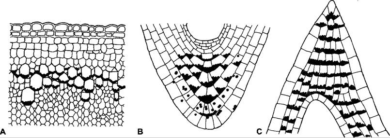
1 This refers exclusively to the embryonic root, not to lateral or adventitious roots. — Ed. note.
The exact nature of the perception mechanism remains unclear. Various hypotheses have been proposed.
• Topographic model: The asymmetrical distribution of statoliths within the cell is decisive.
• Kinetic model: The longitudinal sliding of statoliths during their displacement within The Cell as a result of gravitropic stimulation is decisive.
• Deformation model: Pressure on or stretching of cellular structures is decisive.
It is not yet possible to make a definitive choice between these models. However, in the case of Chara rhizoids—cells with extremely intensive apical growth—much evidence favors the topographic model (Fig. 8.23), whereas for most cells, especially statocytes of higher plants, the deformation model appears more adequate.
Fig. 8.23. Ultrastructural diagram of a Chara foetida rhizoid exhibiting positive gravitropism (A). Secretory vesicles (Golgi vesicles) pinching off from Golgi dictyosomes, containing Cell wall and membrane components, migrate toward the peripheral region in an apical direction, surrounding a group of "shiny bodies" (approximately 50 barium sulfate (BaSO4) particles acting as statoliths) and ensuring uniform surface growth at the tip from all sides. B — Horizontal position of the rhizoid: the displaced statoliths block the movement of Golgi vesicles to the lower side, which consequently lags in growth behind the rapidly growing upper side. The result is positive gravitropism
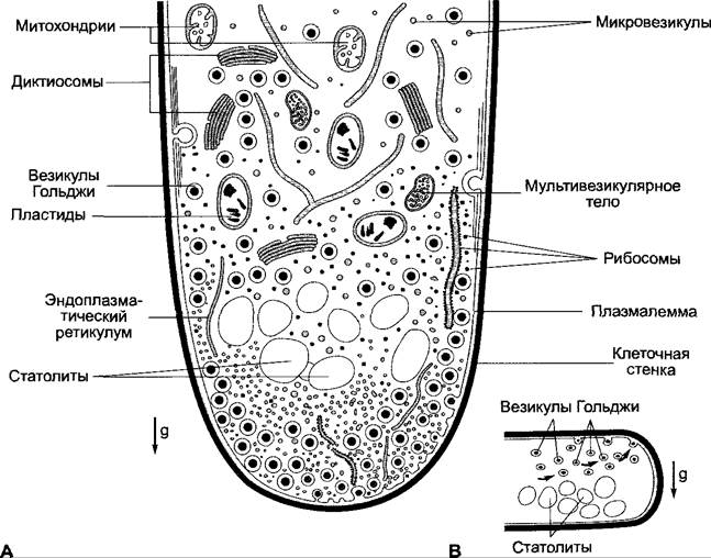
According to the topographic model in Chara, the arrangement of statoliths ("shiny bodies") directs the flow of secretory vesicles (Golgi vesicles) budding from dictyosomes, which supply membrane and cell wall material to the apical region of the cell, resulting in uniform surface growth of the tip. Displacement of statoliths upon deviation from the vertical (horizontal position in Fig. 8.23, B) accordingly causes a shift in the vesicle flow toward the physically upper side, thereby stimulating its growth.
Various deformation models have been proposed to explain The Link Between mass acceleration and cellular metabolism.
✵ The pressure exerted by sedimenting organelles, particularly statoliths, on cellular structures such as The Endoplasmic reticulum (in certain root tips, e.g., cress), and the corresponding reduction in pressure through statolith displacement during gravitropic stimulation, regulates the biochemical process of primary gravity perception.
✵ Statoliths are "suspended" on Cytoskeleton filaments, stretching or relaxing their tension as they move within the cell; the primary biochemical response is regulated by the cytoskeleton via mechanical coupling.
✵ The entire protoplast acts as a statolith, stretching The cell membrane (Plasmalemma) that envelops it; the primary biochemical response is triggered by plasmalemma tension. Statoliths act as ballast, enhancing the gravitropic sensitivity of the cell (plasmalemma-control model).
To date, the plasmalemma-control model appears to be the most consistent with experimental data. It accounts for the facts that there are cells lacking visible statoliths yet responsive to mass acceleration (e.g., Chara internode cells and Phycomyces sporangiophores) and that, as already mentioned, starchless mutants exhibit a noticeable, albeit much weaker, gravitropic response. Furthermore, the total mass of the protoplast significantly exceeds the mass of all statoliths combined, and correspondingly higher is the kinetic energy available to trigger the cellular response.
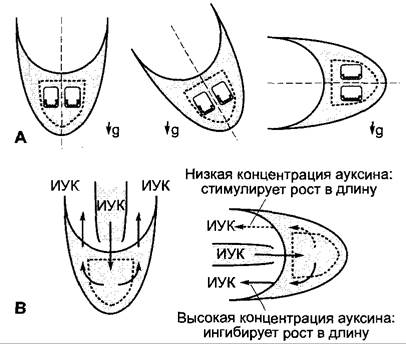
In many cases (e.g., in roots), the gravitropic response occurs only in the presence of extracellular Ca2+ ions. It is debated whether cell membrane deformation affects the cellular calcium level (for instance, the opening of mechanosensitive channels in a stretched cell membrane could lead to an enhanced influx of Ca2+ ions into the cell on the physically lower side). In the vertical position, a radially symmetrical organ (root, shoot) must be symmetrical with respect to its longitudinal axis; however, upon deviation from the vertical, an asymmetry likely arises, which can be corrected by a compensatory growth response (Fig. 8.24, A). It is hypothesized that this involves a shift in auxin flux direction (dependent on Ca2+ ions), similar to phototropism. The details of this scheme remain highly hypothetical, but there is abundant evidence for the involvement of auxin in the growth responses of higher plants.
Fig. 8.24. Lateral polarization during root gravitropism: A — Hypothetical model of the process in the root tip directed along the gravity vector (g). This process occurs when the longitudinal axis of the organ is oriented parallel to the gravity vector, maintaining Symmetry relative to the longitudinal axis (left). When the organ deviates from the vertical (center and right), this can lead to asymmetry in molecular distribution. The involvement of ion transport (possibly Ca2+) is postulated, triggered by the pressure of statoliths on the physically lower part of the cell and, correspondingly, on the plasmalemma (statolith distribution in two columella cells is shown). This should result in a redirection of auxin transport (see text). According to this concept (B), the elevated auxin concentration in the basal part of the organ already exceeds the optimum, leading to an inhibition of elongation growth. For further details, see the text
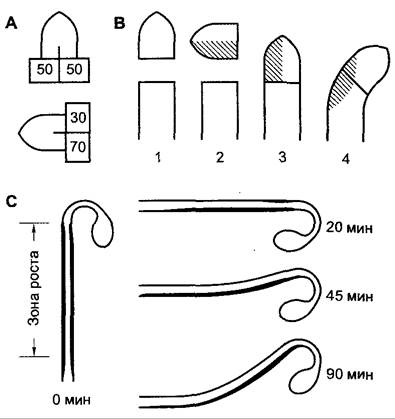
According to the auxin hypothesis of gravitropism, in higher plants, gravitropic stimulation causes the auxin flux to shift toward the physically lower side. In coleoptiles, this has been directly demonstrated experimentally (Fig. 8.25, A, B); the shift is detectable in the coleoptile tips. An enhanced auxin influx to the physically lower side in the coleoptile growth zone leads to increased elongation growth, whereas a reduced auxin influx to the physically upper side decreases its growth rate. The shift of auxin flux in shoots can be demonstrated indirectly (e.g., in soybean hypocotyls, Fig. 8.25, C). Plants possess A number of genes whose activity is rapidly and strongly induced by auxin (SAUR genes, small auxin up-regulated). Using in situ Hybridization with radioactively labeled RNA containing a base sequence complementary to the mRNA under investigation (antisense RNA), it has been shown that in vertically growing seedlings, SAUR mRNA is evenly distributed in the parenchyma of the growth zone. As early as 20 min after placing the seedlings in a horizontal position, preparations reveal a marked increase in mRNA quantity on the physically lower side and very little SAUR mRNA on the physically upper side. After 45 min, a noticeable gravitropic reaction begins (Fig. 8.25). These results indicate that the relocation of auxin to the physically lower side of the organ precedes the onset of differential lateral growth. The involvement of auxin in shoot gravitropic reactions is further supported by the fact that a number of Arabidopsis thaliana mutants exhibiting an auxin-resistant phenotype (e.g., aux1, axr2, auxin resistant), which fail to respond to auxin, are agravitropic. The AUX1 gene has been shown to encode a protein similar to amino acid carriers, which is presumed to be the auxin transporter required for the directed transport of the phytohormone.
Fig. 8.25. Evidence supporting the auxin hypothesis of gravitropism in the shoot region: A — Demonstration of longitudinal and lateral auxin transport during coleoptile gravitropism. Horizontal orientation of coleoptile tips leads to auxin redistribution toward the physically lower side. The quantities of auxin (determined by bioassay) diffusing into agar blocks from coleoptile tips incubated horizontally (bottom) and vertically (top) are given in relative units; B — Demonstration of auxin transport from excised (1), subsequently horizontally incubated (2), and reimplanted (3) coleoptile tips. During incubation (2), the asymmetrically distributed auxin within the tips (shaded) determines the unequal growth rates of the coleoptile sides (4); C — Indirect proof of the asymmetric distribution of auxin concentrations in a gravitropically stimulated soybean hypocotyl. This was demonstrated by quantifying the mRNA transcribed from the SAUR gene (see text). To this end, hypocotyls of vertically oriented plants, as well as plants placed horizontally for 20, 45, and 90 min, were cut longitudinally, the cut surfaces were applied to a nylon membrane for mRNA transfer, and membrane-bound SAUR mRNA was quantified via hybridization with radiolabeled complementary RNA (so-called antisense RNA). In vertically growing plants, SAUR mRNA is uniformly distributed in the growth zone parenchyma, but after only 20 min in a horizontal position, significantly more SAUR mRNA was detected on the lower side of the organ. The negative gravitropic curvature became noticeable only after 45 min. These results are interpreted as follows: as a result of lateral auxin Transport from the upper half of the organ to the lower half, SAUR genes are upregulated on the lower side and downregulated on the upper side of the hypocotyl (it is postulated here that the mRNA itself is not transported!)
It is likely (although not yet conclusively proven by experiment) that the positive gravitropic response of the primary root is also largely regulated by auxin (see Fig. 8.24, B), driven by auxin redistribution toward the physically lower side of the organ. In seedling roots, polar auxin transport occurs within the central cylinder toward the root tip. This auxin flow then redirects in the root cap, so that indole-3-acetic acid (IAA, see 7.6.1.3) is transported backward through the root cortex from the tip toward the base. According to this model, gravistimulation directs an increased flux of IAA to the lower side of the organ. Because roots are highly sensitive to exogenous IAA and respond to overdose with profound growth inhibition (see Fig. 7.31), it is hypothesized that the elevated endogenous IAA concentration on the lower side inhibits cell elongation within the growth zone.
Both plagio- and diagravitropic responses of lateral branches and leaves result from the combination of negative gravitropism (which promotes growth on the lower side) and epinasty (which promotes growth on the upper side). Autonomous epinasty can be observed, for example, when gravitropic stimulation is eliminated on a clinostat (see Fig. 8.19); therefore, it is not caused by mass acceleration.
External factors capable of inducing tropic responses in experiments—such as electrical stimulation (galvanotropism), mechanical wounding (traumotropism), or temperature stimulation (thermotropism)—play no natural role or are of only minor importance for plant organ orientation. However, tactile stimulation (thigmotropism) and chemical stimulation (chemotropism) are indeed significant, at least for certain plant groups.
A vast number of plants are sensitive to mechanical stimulation via Touch. Many seedlings (especially etiolated ones) respond to contact on one side (induced experimentally, for example, by rubbing with a rough wooden stick) with a growth curvature toward the stimulus. Thousands of species of twisting and climbing plants thrive across the globe. In these plants, various organs are tasked with perceiving contact stimuli and thereby finding necessary support (often another plant) to climb upward. This allows them to reach the light more efficiently and grow rapidly over long distances (such as lianas) without needing to develop massive mechanical tissues. Contact-sensitive structures include petioles (e.g., in species of Tropaeolum, Clematis, or Fumaria), leaf tips (Gloriosa), aerial roots (Vanilla), stems (Ipomoea), inflorescences (Vitis, Parthenocissus), and leaves or axillary shoots (such as tendrils in Fabaceae or Cucurbitaceae). Tendril thigmic reactions are particularly remarkable. However, these movements are generally not thigmotropic, but rather thigmonastic. Therefore, we will discuss them in the context of nasties (see 8.3.2.4).
Chemotropic responses refer to growth curvatures induced by the inhomogeneous distribution of dissolved or gaseous chemical compounds in the environment of a growing organ, where the direction of curvature is determined by the concentration gradients of these substances. Frequently, a substance that triggers chemotropism acts as an attractant at lower concentrations and as a repellent at higher ones.
Numerous examples of chemotropic responses can be found among lower plants, particularly during gametangiogamy in Molds. Here, gamones (gamete attractants) secreted by sexual partners cause the mating partners to grow chemotropically toward each other; for instance, volatile compounds in Mucor (Mucorales) and the steroid antheridiol in Achlya (Oomycota), which also plays a role in sex organ differentiation. Many fungal hyphae, especially during the germination stage, exhibit positive chemotropic growth along nutrient gradients (effective substances include sugars, Amino Acids, Proteins, ammonium ions, and phosphate), while reacting negatively to acids and their own metabolic byproducts ("stemi-inhibiting substances"). The growth of Spirogyra conjugation tubes toward one another (see 11.2, Fig. 11.104, B) is also likely based on chemotropism. Chemotropic responses induced by an organism's own metabolites are termed "autochemotropism." This reaction underlies the avoidance response of Phycomyces sporangiophores away from adjacent solid surfaces, which occurs without physical contact and is presumably based on the reduced diffusion ("congestion") of gaseous Ethylene in the immediate vicinity of an obstacle. Ethylene is produced in large quantities by the sporangiophores.
In shoots of higher plants, chemotropic responses play a role only in exceptional cases. For instance, Cuscuta seedlings grow in an oriented manner toward their host plants. Volatile compounds released by the hosts (alcohols, esters, Essential Oils, and possibly water vapor) likely exert a chemotropic effect. Chemotropic responses may also be important when parasitic haustoria search for specific host tissues (such as sieve tubes).
It is generally assumed that the growth of pollen tubes through the stigma and style is predominantly guided anatomically. However, positive hydrotropic responses (toward increasing water concentrations) and negative aerotropic responses (toward decreasing oxygen concentrations) may also play a role during pollen germination. It appears that pollen tubes are guided by chemotropic substances secreted by the ovule only in the immediate vicinity of the ovule itself.
Roots are also capable of positive chemotropism—for instance, toward phosphate ions, increasing partial pressure of O2 (positive aerotropism toward well-aerated soil patches; for soil structure, see 6.2.3), and increasing soil moisture (positive hygrotropism). In this way, tree roots can detect microscopic defects in underground sewer systems and subsequently form clogging "root plugs" inside the water pipes. Besides roots and pollen tubes, positive hygrotropism is exhibited by moss rhizoids, fern prothalli, and Cuscuta seedlings, which use it to locate their hosts via Transpiration. Some parasitic fungi are hygrotropically guided toward Stomata, through which they penetrate leaves. Consequently, infection rates are drastically reduced (by up to 90%) when stomata are closed. Finally, the radially structured sensitive leaf tentacles of Drosera, located on the leaf surface, are positively chemotropic toward ions such as NH+4, causing them to close over prey caught by the adhesive glands (see 9.1.2). The dorsiventrally structured marginal sensitive hairs of these leaves exhibit a chemonastic reaction (see Fig. 8.28, G).
All known chemotropisms are fundamentally based on growth processes. Virtually nothing is known about signal perception and transduction in chemotropism.
Like tropisms, nasties are movements of living organs triggered by specific stimuli, but unlike tropisms (see 8.3.1), their direction and pattern are predetermined by the plant's structural layout. Nasties are classified according to The Nature of the stimulus: thermo-, photo-, thigmo-, chemo-, and seismonasties, as well as hygronasties in the case of stomatal guard cells. Often, though not exclusively, nasties are executed via reversible turgor changes.
Certain flowers (such as tulips, crocuses, and daisies) open when the temperature rises and close when it drops. Sensitivity to temperature is remarkably high: crocus flowers respond to temperature differences as small as 0.5 °C, and tulip flowers to 1 °C. Thermonasty is achieved through differential growth rates between the inner and outer (lower and upper) sides of the perianth leaf base (the temperature optimum for extension growth of the upper side is higher). The extent of the movement is determined by The rate of temperature change: the faster the change, the more intense the movement. Perianth segments retain their responsiveness for a long time; in tulips, they can elongate by up to 7% in a single movement. Over the entire flowering period, cumulative growth driven by repeated thermonastic reactions can exceed 100%.
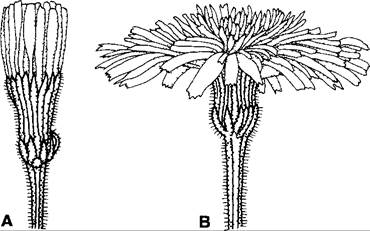
Fluctuations in light intensity can also induce nastic growth movements—primarily, once again, in perianth organs and leaves—as well as turgor-controlled leaf movements in certain species (e.g., Mimosa), which are mediated by pulvini (leaf cushions). Photonasty can be observed in the petals of many water lilies, cacti, and woodsorrels, as well as in the flower heads of numerous Asteraceae (Fig. 8.26), where marginal ray florets function essentially as individual "petals." In most cases, illumination triggers opening, whereas shading (often even a passing cloud shadow) or darkness triggers closing. Night-flowering plants (such as Silene nutans) respond in the exact opposite manner. During their growth phase, leaves perform photonastic movements (e.g., Impatiens species), whereas in fully expanded leaves, such movements occur only if specialized pulvini are present (e.g., Oxalis, Mimosa).
Fig. 8.26. Capitulum of Leontodon hispidus (Asteraceae): A – closed in the dark; B – open in the light
The operational mechanism of pulvini has been only partially elucidated. They function as "osmotic motors," the operational principles of which are presumed to be similar to those of seismonasties (see 8.3.2.4). It is believed that nasties driven by reversible turgor changes—including, for example, the Nastic Movements of stomatal guard cells—share common underlying biochemical principles (see also 8.3.2.5).
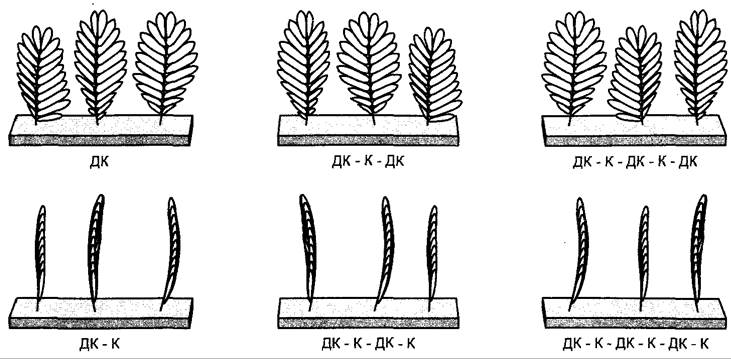
Illumination experiments demonstrate that many photonastic movements proceed with the involvement of the phytochrome system, specifically class II phytochrome (see 7.7.2.4), thereby exhibiting classical red/far-red photoreversibility. In Mimosa (Fig. 8.27), the perception of the night (darkness) state is induced by a brief flash of red light (R), whereas a subsequent flash of far-red light (FR) inhibits this response. Consequently, active phytochrome is required to trigger the movement. The molecular processes that occur during active phytochrome-induced changes in The activity of cellular ion carriers (likely Ion Channels permeable to K+, Cl-, and under certain circumstances also Ca2+)—which serve as prerequisites for turgor movements—have not yet been fully uncovered.
Fig. 8.27. Primary leaflets of Mimosa pudica 30 minutes after transition from white light to darkness. Immediately following white light exposure, the leaflets were irradiated for 2 min with red light (R) and far-red light (FR) in the combinations shown (for phytochrome system action, see 7.7.2.4). Leaflets close only when active phytochrome (Pfr, following red light exposure) predominates upon the onset of darkness.
While the radially structured median glandular hairs of Drosera leaves (Fig. 8.28, G) exhibit chemotropism, the dorsiventrally structured marginal glandular hairs respond to localized mechanical irritation via chemonasty, which involves a bending movement toward the center of the leaf. In this way, they bring the captured prey into contact with other glandular hairs, which may or may not bend subsequently. This process is likewise mediated by Organic compounds secreted by the prey, as well as by NH+4 ions. The response is amplified by the simultaneous transmission of tactile sensitivity among the glandular hairs. Mechanical stimuli typically originate from the prey animal, which is held in place on the leaf surface by the sticky secretions of the HEAD glands on the Hair tips.
8.3.2.4. Thigmonasty and Seismonasty
Thigmonasty, or movement induced by touch (contact stimuli) whose course depends on the structural plan of the organ in question, is widespread in the plant kingdom. It can be divided into two groups.
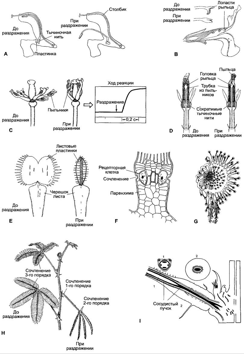
The first group encompasses very rapid movements driven by changes in turgor pressure (Fig. 8.28). Because these movements can also be triggered by non-contact (though significantly stronger) stimuli, such as mechanical Shock, they are likewise referred to as seismonastic movements. In nature, however, contact stimuli generally serve as a more relevant physiological trigger than mechanical shocks. This category of nasties includes the rapid leaf Movements of the sensitive plant Mimosa púdica (Fig. 8.28, H, I); the insect-trapping snap movements of the Venus flytrap Dionaea muscipula (Fig. 8.28, E, F); pollinator-induced stamen movements that serve to dust pollen onto pollinators (e.g., inward movements in Berberis and Opuntia, outward movements in Sparmannia, and the contractile stamen filaments in certain Centaurea species, Fig. 8.28, A, C, D); and the touch-sensitive pistil stigmas (e.g., in species of Mimulus, Catalpa, and Torenia, where the stigma lobes snap shut upon contact, thereby brushing pollen off visiting insects, Fig. 8.28, B).
Fig. 8.28. Rapidly occurring nasties of flowers (A–D) and leaves (E–I). A — Trapdoor mechanism in meadow sage (Salvia pratensis). Both stamen filaments are fused with the corolla tube. One connective forms a plate-like structure with the anthers, while the other is greatly elongated and extends along with its anther into the upper lip. When an insect brushes against the plate in search of nectar, the lever arm of the long connective drops, dusting pollen onto the insect's back. B — Flower of Mimulus luteus (sectioned to show the stamens and pistil stigma in a resting state); above: side view of the unstimulated and stimulated stigma. C — Flower of Berberis vulgaris (perianth removed); in the stimulated state, the anthers lie against the stigma. Diagram: time course of the reaction of the stamen filaments. D — Flower from the central part of the flower head of Centaurea jacea (section). In the stimulated state, the stamen filaments contract by up to 30%. This post-stimulation contraction causes the stamen filaments, fused into a tube, to move downward, while the style—acting as a piston with its stigma—pushes out the pollen contained within the tube, allowing it to adhere to visiting insects. E — Leaf of Dionaea muscipula with 3 sensitive trigger hairs on each leaf blade half. F — Longitudinal section through the base of a trigger hair. G — Leaf of Drosera rotundifolia, top view, left side stimulated; marginal trigger hairs exhibit nastic responses, while inner hairs show tropic responses. H — Shoot of Mimosa púdica; a single leaf has been stimulated. Upon mechanical shock or stimulation of the leaflets of compound leaves, they fold upward in pairs, the secondary petioles draw close together sideways, and finally the primary petiole (rachis) drops downward. Under intense stimulation, the excitation can propagate upward and downward along the shoot for up to 50 cm. Leaves reached by the excitation react in the following sequence: primary pulvinus (at the base of the primary leaf rachis), followed by secondary and tertiary pulvini. I — Longitudinal section through the primary (1st-order) pulvinus of Mimosa púdica and cross sections through the regions labeled 1 and 2. The central vascular bundle strand passing through the pulvinus region facilitates joint movement. The parenchyma on the lower side of the pulvinus (motor tissue), which loses turgor upon stimulation, is also termed the extensor, whereas the parenchyma on the upper side, whose turgor increases, is termed the flexor—since an increase in turgor within this tissue is associated with bending (Latin flexio, bend), whereas turgor increase in the extensor is associated with straightening of the petiole (Latin extendere, to stretch).
The second group comprises slower touch-induced reactions that are not triggered by mechanical shock and that invariably involve growth processes alongside a turgor-governed component. This category primarily includes tendril movements (unless thigmotropisms are concerned, see 8.3.1.3).
A characteristic feature of the rapid, shock-responsive thigmonasties of the first group is that they follow an "all-or-none" principle. If the stimulus
exceeds a threshold value, the full response ensues. Consequently, in most cases, no proportionality is observed between the magnitude of the stimulus and the reaction (Fig. 8.28, C). The reaction time (from the onset of stimulation to THE START OF movement) is 0.02 s under optimal conditions in Dionaea and Berberis, and 0.08 s in Mimosa; seismonastic movements last longer than 0.1 s in Dionaea and Berberis, 1 s in Mimosa, and 6 s in Mimulus. Stimulus perception is invariably linked to the deformation of cellular structures. The mechanoreceptive cells are located at the bases of the stamen filaments, on the stigma lobes, in the "trigger hairs" on the inner surfaces of the trap leaves of Dionaea, or within the leaf pulvini of Mimosa. These trigger hairs act like levers, amplifying the mechanical deformation of the receptor cells located at their bases. Similarly, in Mimosa, the leaflets of the compound leaf can act like levers upon touch, triggering a response. It is likely that shock stimulation invariably triggers a sufficiently powerful lever action (e.g., via the movement of a stamen filament) when the mechanical shock is strong enough, thereby overcoming the stimulation threshold. Responsive tissues (also referred to as motor tissues, designating those that undergo rapid turgor loss following stimulation) are identical to the perceptive cells in stamen filaments and stigma lobes. However, in Dionaea and Mimosa, the sensitive cells and motor tissues are spatially separated: in Dionaea, the tissues responsible for the nastic leaf movement are located along the upper side of the midrib, whereas in Mimosa, they reside in the primary pulvini (at the lower bases of the leaf petioles), secondary pulvini (at the upper bases of the compound leaf rachises), and tertiary pulvini (at the upper bases of the leaflets; Fig. 8.28, H). In these cases, the signal must be conducted from the site of perception to the motor tissue. Signal propagation here is electrical in nature, with a putative chemical component also participating in Mimosa (see below).
Researchers currently proceed from the assumption that all turgor-driven nasties in motor tissues operate via a similar or even identical mechanism. However, experiments have been conducted predominantly on Mimosa. It is hypothesized that KCl efflux occurs from the motor cells—at times very rapidly—followed by an osmotically coupled outflow of water. In the leaf pulvini of Mimosa, the fluid escaping into the apoplast and filling the intercellular spaces can be readily detected by the darkening of the lower side of the primary pulvinus.
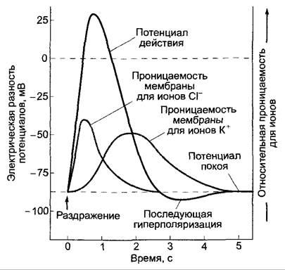
The response of a motor cell begins with a very rapid increase in the permeability of the plasmalemma to chloride ions (Fig. 8.29). As Cl- ions leave the cell, membrane depolarization takes place; that is, the large negative resting electrical Membrane Potential (which ranges from -80 to -100 mV in various objects, with the cell interior being negative relative to the outer surface) shifts by 100 mV or more, occasionally reaching positive
values. This depolarization opens potassium channels, which drive the unidirectional efflux of K+ ions from the cell. This results in the repolarization of the electrical membrane potential back toward its resting level. The effect can be reinforced by the simultaneous closure of voltage-gated K+ channels that mediate K+ influx into the cell, which can only open when the membrane potential is sufficiently negative. The rapid loss of KCl lowers the cellular osmotic potential, causing water to flow out into the apoplast. All CHARACTERISTICS OF THE Action Potential are governed by these electrical processes. It has also been suggested that in systems where the site of stimulus perception is distant from the motor tissue (in Dionaea and Mimosa), signal transmission is mediated by action potentials generated by K+ and Cl- fluxes. These action potentials can propagate from Cell to Cell via symplastic connections through plasmodesmata. Furthermore, impulse conduction through phloem sieve tubes has also been proposed. Signal transmission velocities are considerable in all cases, reaching 3–10 cm·s-1 in Mimosa and 6–20 cm·s-1 in Dionaea. These values already fall within the range of conduction velocities found in the nerves of lower animals (e.g., only 1 cm·s-1 in the freshwater clam Anodonta).
Fig. 8.29. Schematic diagram of action potential generation at the plasmalemma of a stimulated cell. At rest, K+ and Cl- ions continuously enter and exit the cell via diffusion. Ion uptake is powered by the proton motive force (6.1.4.3, 6.1.5). The sum of all ionic fluxes across the plasmalemma constitutes the Resting Potential, which ranges from -80 to -180 mV depending on the cell type and physiological state (being more negative on the inner side of the cell membrane than on the outer). Following stimulation, membrane permeability to Cl- ions increases very rapidly, causing the membrane to depolarize (the potential becomes less negative and, under certain conditions, even assumes positive values). Membrane repolarization occurs through a subsequent increase in membrane permeability to K+ ions accompanied by a concurrent decrease in Cl- conductance. This leads to a transient, slight hyperpolarization of the membrane (the potential becomes more negative than the resting potential). Ultimately, the resting potential is restored and permeabilities return to their baseline levels.
The primary process of mechano-electrical coupling remains unclear. In guard cells (see 8.3.2.5)—which are among the best-characterized plant cells from an electrophysiological standpoint—slower nastic movements are brought about by fundamentally very similar (or identical?) ionic fluxes. Here, it is known that during the initial stage of stomatal closure, the depolarizing chloride flux is carried by plasmalemmal Cl- channels that open in response to an elevation in cytoplasmic calcium ion concentration. In guard cells, calcium release is induced, for example, by the phytohormone Abscisic acid, which is produced during water stress (see 7.6.4). It is probable, though not yet definitively proven, that the depolarizing chloride flux in the perceptive cells of thigmo- and seismonastic organs is likewise triggered by Calcium Ions. Ca2+ ions may enter the Cytoplasm during cell deformation through mechano-sensitive Ca2+ channels located in the apoplast or intracellular stores (endoplasmic reticulum, vacuoles). Such channels are also presumed to participate in the perception mechanism (see 8.3.1.2), but they have yet to be identified at THE MOLECULAR LEVEL.
Nastic movements involve not only motor cells experiencing turgor loss, but also the opposing tissues. This can be observed particularly well in leaf joints or pulvini (e.g., in Mimosa, see Fig. 8.28, I). The loss of turgor in the motor tissue leads to a drop in water potential ( , see equation 6.15) within the cells of the opposite side of the organ, as hydrostatic pressure inside them decreases due to the collapse of the motor tissue. This prompts water to flow into these cells: while the motor tissues release water, the opposing cells take it up and swell, thereby amplifying the nastic movement. If no further stimulation occurs, the organ eventually returns to its original position through active uptake processes that transport ions back into the motor cells, thereby restoring turgor (the turgor recovery time is about 15–20 min in Mimosa, several hours in Dionaea, and about 1 min in the stamen filaments of Berberis or Centaurea). Once this is complete, a new stimulus and response cycle becomes possible. However, insectivorous leaf traps remain closed much longer (for weeks) after a successful catch, a period that in some cases (e.g., Dionaea) is further prolonged by sustained growth processes. Chemonastic stimulation is maintained throughout this time by organic compounds from the captured animals until their remains are completely digested by plant Enzymes. Under certain circumstances, the trap may subsequently fail to reopen at all.
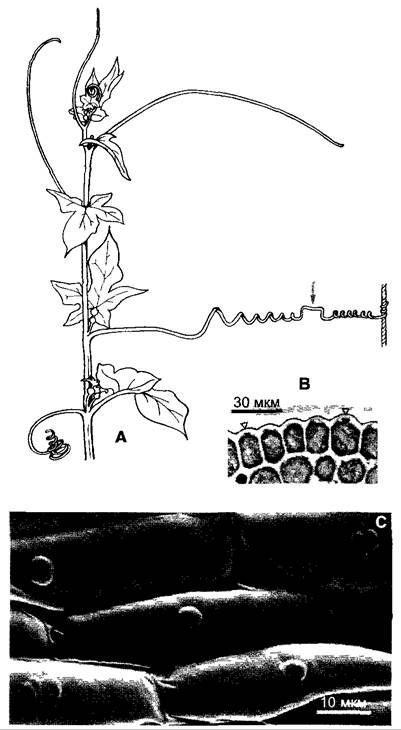
As an example of prolonged thigmonasties that cannot be triggered by seismic stimuli, we must examine the remarkable responses exhibited by the tendrils of climbing plants as they coil around a support. The complex tendrils of cucurbits, particularly white bryony (Bryonia), have been studied in great detail (Fig. 8.30).
Fig. 8.30. White bryony (Bryonia dioica): A — Part of a shoot with tendrils at various Selection/3.html">Stages of development (approx. 1/3 scale). The uppermost (youngest) tendril is still coiled like a watch spring; the middle tendril wrapped around a support approximately one day ago, completing its thigmonastic response. The arrow points to the reversal point where the direction of coiling changes (the only one in this case); bottom left: a tendril showing age-related coiling. "Sensitive pits" in the outer epidermal cell walls; B — in cross section; C — top view.
Cucurbit tendrils are homologous to lateral shoots. In Bryonia, the dorsiventrally structured section of the tendril that executes the movement is homologous to a subtending leaf, while the radially symmetrical and persistently elongated basal part of the tendril is homologous to a lateral shoot. The axillary bud of the tendril is suppressed in Bryonia, but is present in other cucurbits (such as Cucurbita), where it also forms a tendril ("branched tendrils").
Bryonia tendrils, which are coiled like watch springs in early development, uncoil as they grow and become sensitive to mechanical stimuli. Both shoot tips and tendrils perform autonomous circular movements known as nutations. This increases the plant's chances of encountering an obstacle. Typically, the upper third of the tendril is most sensitive to touch. The tendrils of Sicyos and Momordica bend when touched on either their upper or lower sides, with the touched side becoming concave. In contrast, tendrils of Bryonia and Pisum, like those of many other species, respond to touch exclusively on their upper side. However, stimulation of the upper side suppresses the response of the lower side to touch. Thus, the upper side of such a tendril is also sensitive to mechanical stimulation, and these cases unquestionably represent a nastic response. Finally, there are species (such as Cobaea scandens and various Cissus species) whose tendrils are constructed both morphologically and physiologically as radial structures; consequently, they can bend in any direction, with the stimulated side invariably becoming concave. This phenomenon therefore constitutes thigmotropism.
Tendrils respond not merely to pressure, but specifically to frictional stimulation. Water flow, rain, steady pressure, or touching with a smooth glass rod does not stimulate tendril movement, whereas a stream of water carrying suspended gypsum particles or touching with a rough rod does. Bending can even be triggered by the movement of a woolen thread weighing a mere 2.5 × 10⁻⁷ g (0.25 µg)—a stimulus far below the threshold of human touch.
Consequently, the tendril responds not merely to pressure, but to spatial and temporal differences in pressure. Remarkable bubble-like protrusions, visible in microscopic preparations as pore-like structures in the outer walls of epidermal cells ("sensitive pits", see Fig. 8.30), are thought to be associated with stimulus perception. However, these structures are not present in all tendrils, and are sometimes restricted to the lower side even when the upper side perceives the stimulus. Therefore, these structures function more likely as a mechanical amplifier rather than as a primary mechanoreceptor. This is further supported by the observation that "sensitive pits" are characteristic only of the most highly sensitive tendrils.
Upon contacting a support, the tendril of Bryonia—like those of other plants exhibiting nastic responses—bends toward its morphologically lower side. Under favorable conditions, highly responsive tendrils (e.g., in Bryonia, Sicyos, Cyclanthera) can achieve a reaction time of less than 30 seconds, whereas other species (such as Corydalis claviculata) require up to 18 hours. The rapid movements rely on turgor loss on the morphologically lower side coupled with a turgor increase on the opposite side. Following brief stimulation—such as when a tendril fails to secure a proper grip on a support—it straightens out again within 30 to 60 minutes (autotropism) and regains The ability to respond. If a support is successfully grasped, continuous bending leads to multiple coils of the tendril tip around the support. In young tendrils, this response is also supported by rapid elongation growth of the tip, whereas in fully matured organs, it relies solely on turgor-driven bending.
The more basal sections of the tendril likewise undergo coiling (see Fig. 8.30), thereby drawing the entire plant elastically and securely toward the support. In Bryonia, this coiling is achieved by slowing down elongation growth on the lower side of the tendril while growth on the upper side continues or, under certain conditions, even accelerates. Due to mechanical constraints, this process must incorporate one or more reversal points in the direction of coiling (see Fig. 8.30, A) between left- and right-handed helical turns to relieve mechanical stress. Ultimately, mechanical stimulation also induces the formation of strengthening elements and frequently results in localized thickenings (thigmomorphoses), which stabilize the attachment.
Currently, we have only a rudimentary understanding of how signals are transmitted from the stimulus-perceiving tip of a tendril to its base. This process begins almost simultaneously along their entire length as the tendril bends, roughly 1.5 to 2 hours after the tip has successfully grasped a support. The turgor-driven contact bending of the tendril tip around a support may be mediated by a calcium-dependent reaction; the underlying mechanism could be similar to that of rapid thigmo- and seismonastic movements (see Fig. 8.29). Electrical currents have been successfully recorded in the basal regions of stimulated tendrils using surface electrodes. Consequently, under certain circumstances, the signal is transmitted as electrical impulses. Phytohormones are likely involved in inducing basal curvature driven by differential growth rates. Applying ethylene, auxin, or octadecanoids (such as jasmonic acid, see 7.6.6.2) can trigger the thigmonastic response in Bryonia tendrils without any physical touch. There is evidence that mechanical stimulation prompts The production of 12-oxophytodienoic acid (see 7.6.6.2, Fig. 7.66) and that this compound serves as an endogenous inducer1 of the growth response (though it is unlikely to act as the signal carrier).
1 In Russian literature, substances of this class (oxylipins) are traditionally considered secondary messengers that operate within the signaling pathway between the inducer (primary messenger) and intracellular response reactions. — Editorial note.
8.3.2.5. Nastic Movements of Stomatal Guard Cells
This topic is addressed separately not only because stomatal movements play a crucial role in the gas exchange of most land plants, but also because the molecular mechanisms underlying nastic movements have been studied intensively and understood quite well in recent years. It is safe to assume that the molecular processes regulating guard cell turgor are similar to turgor-driven mechanisms involved in Other types of movement (see 8.3.2.1 — 8.3.2.4), and thus they hold general significance for controlling plant cell turgor in response to changing conditions.
In accordance with the function of stomata in regulating leaf diffusion resistance—so that CO2 uptake for photosynthetic or dark fixation remains optimal depending on water availability—stomatal responses can be divided into photonastic and hygronastic. A thermonastic response is also distinguished, which is ecologically advantageous because transpiration-induced water loss increases sharply at higher temperatures. These environment-induced movements are modulated by a circadian rhythm; this means that the readiness to respond to exogenous factors varies throughout the course of the day. Stomatal opening reactions during daylight hours are also endogenously preferred. The timekeeper for this rhythm is the alternation of day and night (see 7.7.2.3).
The immediate cause of movement in each case is a difference in turgor pressure (Section 6.15) between the guard cells and the surrounding epidermal cells, which may possess morphological specializations and are then specifically termed subsidiary cells (see 3.2.2.1, Fig. 3.12). Turgor changes occur primarily as a result of shifts in osmotic potential and associated water fluxes; they are also linked to volume changes in both the guard cells and the adjacent epidermal cells, with the regulatory factors in these two cell types exerting opposing effects on such changes. If the osmotic pressure in the guard cells increases relative to the surrounding environment (i.e., the osmotic potential becomes more negative), this drives water influx, increasing the turgor and volume of the guard cell. If osmotic pressure decreases relative to the environment, water flows out of the cell, turgor drops, and the guard cell shrinks.
Active stomatal movements, which are based on Changes in the osmotic potential of guard cells relative to adjacent cells, are contrasted with passive stomatal movements, which occur as a result of differential water loss or uptake—meaning they are purely hydropassive.
Such volume loss (both absolute and relative to neighboring cells) occurs when transpiration from the guard cells ("peristomatal transpiration") exceeds that of adjacent cells. In this scenario, guard cells act as relative humidity "sensors." This function is supported by the observation that leaves with identical water content exhibit significantly higher transpiration resistance in dry air than in humid air. As a result of this induced stomatal closure, transpiration is more restricted in dry air, and the leaf retains a higher water content than it would in humid air.
Hydropassive processes are frequently the underlying cause of rapid wilting in cut leaves. In such leaves, transpiration-driven water loss occurs more rapidly in epidermal cells than in guard cells, causing the stomata to open.
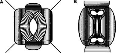
An increase in guard cell volume leads to stomatal opening, whereas a volume decrease causes closure. This is governed by the structural arrangement of the walls in both the guard cells and their neighboring cells—specifically, the orientation of microfibrils within the cell walls, which dictates the direction of cell expansion (Fig. 8.31). The volume change in guard cells can be substantial: the volume of a Vicia faba guard cell is 1.3 pL (picoliter, 1 pL = 10-12 L) when the stoma is closed, and 2.4 pL when it is fully open.
Let us focus exclusively on active stomatal movements, which are subject to physiological regulation. These are fundamentally driven by primary changes in the osmotic potential of the guard cells.
Fig. 8.31. Schematic representation of the orientation of Cellulose microfibrils in the cell walls of stomatal guard cells and adjacent (subsidiary) epidermal cells: A — Kidney-shaped guard cells of commelinids; top view of the cell walls. Cell expansion occurs predominantly perpendicular to the direction of the microfibrils. The kidney-shaped guard cells expand mainly along their longitudinal axis. However, the subsidiary epidermal cells (smaller ones) lying transverse to the longitudinal axis of the guard cells offer greater resistance to the expansion of the guard cells than do the two flanking (larger) neighboring cells. Thus, an increase in the volume of the guard cells during bending causes them to move apart and consequently opens the stomatal pore; B — dumbbell-shaped guard cells of grasses: guard cells shown in optical section, others in top view. Radially oriented microfibrils allow cell volume to increase solely through an increase in their radius. Bulbous swellings are present at the ends of the guard cells; their expansion widens the central pore, thereby opening the stoma
The primary osmolyte in guard cells, as in plant cells generally, consists of potassium ions (K+, Fig. 8.32). The vacuolar potassium concentration can exceed 600 mmol • L-1 when stomata are open, whereas it drops to 100 mmol • L-1 or lower when they are closed. Anions serve as counterions to balance electrical charges. In dicotyledonous angiosperms, the predominant counterion is the dicarboxylic acid malate. The malate anion (malate2-) accumulates in the vacuole alongside potassium (potassium malate). Malate synthesis in guard cells proceeds via The breakdown of starch to phosphoenolpyruvate (PEP) during Glycolysis, followed by carboxylation catalyzed by the enzyme PEP carboxylase and the reduction of the reaction product—oxaloacetate—to malate. We have previously encountered this reaction sequence in the context of CAM metabolism (see 6.5.9, Fig. 6.79). Alongside organic anions, inorganic ions—most notably chloride (CI-)—also act as osmolytes, particularly in monocots. In these plants, malate is replaced as the electrical charge compensator by chloride ions (CI-), either completely (e.g., Allium cepa lacks the ADP-glucose pyrophosphorylase enzyme used in leaves for starch synthesis, see Fig. 6.72) or partially (up to approximately 40% in maize). Chloride is taken up into the guard cells together with potassium.
Fig. 8.32. Distribution of potassium, chlorine, and phosphorus across a cross-section of a closed (left) and open (right) stoma from the lower epidermis of a Vicia faba leaf, expressed in relative concentration units. Measured using an X-ray microprobe. Among the elements examined, only K+ showed a marked increase in concentration within the guard cells upon stomatal opening
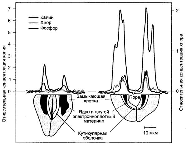
Potassium uptake by guard cells occurs via voltage-dependent K+ channels that transport potassium exclusively into the cell rather than out of it; the degree of channel opening (see 6.1) increases significantly at a sufficiently high electrical membrane potential difference (the inside of the cell is more negative than the outside; this is loosely referred to as membrane hyperpolarization). The membrane potential is generated by the activity of The Plasma Membrane H+-ATPase ("proton pump"), which transports hydrogen ions from the cell into the apoplast (1 H+ per hydrolyzed ATP, see 6.1.5, Figs. 6.4; 6.5). The resulting proton-motive force across the membrane (see 6.4.1.3) also facilitates the uptake of chloride by guard cells, which is likely mediated by a 2 H+/1 CI- symporter (see Fig. 6.5). The efflux of K+ ions from the cell is mediated by a distinct K+ channel regulated through an entirely different mechanism. It allows ions to pass exclusively out of the cell, and the probability of its opening increases when the membrane depolarizes (becoming more positive); during membrane hyperpolarization, this channel remains closed, thereby preventing a "potassium short circuit" during cellular potassium uptake.1 The regulation of stomatal movements can, in principle, be explained by activity changes in these described ion transport systems (Fig. 8.33). Because osmotically active ions ultimately enter the vacuoles of guard cells and correspondingly exit them during stomatal closure, tonoplast-mediated ion transport is also important. However, these processes are not yet as well understood as those of plasma membrane transport.
1 Refers to the unregulated rapid flux of K+ ions across the membrane. — Editorial note.
Fig. 8.33. Simplified reaction sequence: A — during stomatal opening induced by blue light; B — during stomatal closure triggered by ABA. For more detailed explanations, see the text
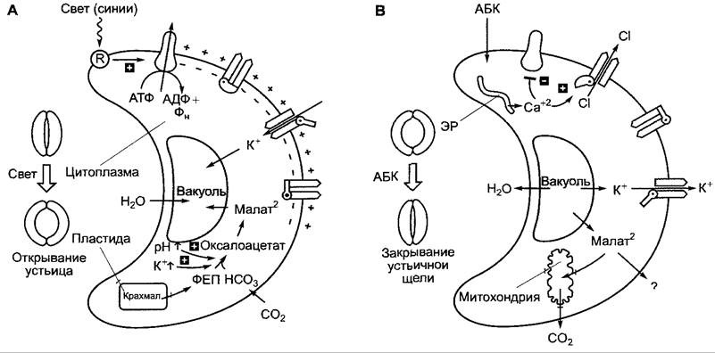
Light typically induces the opening of stomatal pores. The sensitivity of guard cells to light is extremely high; 25 to 30 pmol photons cm-2 • s-1 is sufficient to trigger opening. Action spectra for this photonasty reveal peaks in the red and, particularly, the blue Regions of the spectrum. Stomatal opening driven by red light is powered by photosynthesis. Here, red light does not act as a nastic stimulus, but rather as an energy source for photosynthesis.1 Consequently, the actual regulation is not mediated directly by red light, but rather by the intercellular CO2 concentration—[CO2]—calculated for the guard cells, even though it is determined by mesophyll photosynthetic productivity. Photosynthesis in guard cells, which possess Chloroplasts with few exceptions (e.g., Paphiopedilum), makes only a limited contribution to this [CO2] reduction. Stomatal opening can occur not only upon illumination, but also as a result of dark CO2 fixation (e.g., at night in CAM plants, see 6.5.9) or through experimental reduction of external [CO2] in the light. Conversely, an increase in external [CO2], whether in the dark or in the light, can induce stomatal closure (chemonasty). Within certain limits, alterations in diffusion resistance via stomatal movements maintain the CO2 concentration inside the guard cells—and, proportionally, in the intercellular spaces—at a constant level, or at least prevent major fluctuations. The exact nature of the CO2 sensor and its effect on osmotic potential (when guard cell [CO2] drops, the osmotic potential becomes more negative) remain to be fully elucidated. Blue light is exceptionally effective in driving photonastic stomatal movements. Guard cells lacking chloroplasts respond exclusively to this spectral region (e.g., Paphiopedilum). The blue light receptors are two phototropins—phototropin 1 and phototropin 2. It is also widely accepted that zeaxanthin plays a supplementary role. However, this has been questioned because mutants defective in carotenoid Biosynthesis (such as those known in Arabidopsis thaliana) still exhibit blue-light-dependent stomatal opening, albeit significantly impaired.2 Blue light activates the plasma membrane H+-ATPase, leading to membrane potential hyperpolarization and consequent activation of inwardly rectifying potassium channels. Chloride ion influx into the cell via the 2 H+/1 CI- symporter is also enhanced. H+-ATPase activity can be measured as blue-light-induced acidification of the guard cell apoplast (pH values drop from roughly 7 to 5). The rising K+ concentration and cytoplasmic alkalinization (owing to intensive H+ extrusion raising the cytoplasmic pH) are thought to activate PEP carboxylase, resulting in enhanced malic acid production. Malate, accompanied by potassium, enters the vacuole, while the H+ ions released during the dissociation of the synthesized malic acid into malate are pumped into the apoplast by the H+-ATPase.
1 Experiments conducted in Russia with chlorophyll-deficient pea mutants demonstrate the involvement of the phytochrome system in regulating stomatal movements (see Chapter 7.7.2.4). — Editorial note.
2 The Use of mutants defective in carotenoid biosynthesis is questionable in this context, as zeaxanthin serves as a precursor in ABA biosynthesis. ABA deficiency severely disrupts stomatal movements and produces a "wilted" phenotype. — Editorial note.
The activation of the proton pump by blue light is likely mediated directly by the phosphorylation of the enzyme involving the light-regulated kinase function of phototropin (see 8.3.1.1). It is known that a threonine residue near the C-terminus of the H+-ATPase is phosphorylated (see Fig. 9.15, A), which subsequently leads to the binding of adaptor proteins from the 14-3-3 protein family and a drastic increase in enzymatic activity. Fusicoccin, a toxin produced by the phytopathogenic fungus Fusicoccus amygdali, reduces the dissociation of the H+-ATPase/14-3-3 protein complex, thereby causing an almost irreversible activation of the proton pump. Fusicoccin, a wilt toxin, is particularly potent toward guard cells, causing them to open maximally.
Under conditions of water stress, stomatal opening ceases—meaning the stomatal pores close completely. This response is triggered by the phytohormone abscisic acid (ABA, see 7.6.4, Fig. 7.58), which is synthesized in leaves and roots during water scarcity and transported via the transpiration stream to the guard cells. ABA primarily triggers the release of Ca2+ ions from intracellular compartments within the guard cells. A rise in Ca2+ levels from ~100 nmol • L-1 to over 1 µmol • L-1 initiates the following processes:
✵ it reduces the activity of H+-ATPase, which in turn decreases the transmembrane concentration gradient of H+ and shifts the electrical membrane potential toward a more positive value (depolarization);
✵ it affects Ca2+ binding at chloride-efflux channels, causing them to open; CI- ions passively exit the cell (along their electrochemical gradient), further enhancing membrane depolarization. This Ca2+-triggered chloride efflux contributes to the reduction of the plasma membrane potential (depolarization) even in guard cells where chloride does not serve as the predominant counter-ion to K+ (i.e., in dicotyledonous plants).
Depolarization has two consequences:
✵ K+ inward-rectifier channels, which open solely upon hyperpolarization, are closed, and
✵ K+ outward-rectifier channels, which are particularly active at a depolarized membrane potential, drive a substantial efflux of K+ from the cell. Anions (CI- or malate2-) follow them out, and the cell loses water via osmotic coupling. The expelled ions remain in the adjacent cells, with water osmotically following them. For K+ (and additionally for CI- in monocots), this can be readily demonstrated histochemically; in dicots, the ultimate fate of the malate released by guard cells remains unclear, though a portion of it may be degraded to CO2 during mitochondrial ATP synthesis associated with Respiration.
Movements regulated endogenously rather than by external stimuli are referred to as autonomous movements. They can be mediated by either growth processes or turgor changes.
Turgor-driven movements include daily leaf movements governed by circadian rhythms, as mentioned earlier (see
7.7.2.3), which occur, for example, in Mimosa and Phaseolus. These are executed by pulvini (leaf joints), whose MECHANISM OF ACTION has also been described previously (see 8.3.2.4; Fig. 8.28, 1; however, in Phaseolus, they are located not at the base of the petiole, but at the junction between the lamina and the petiole).
Growth-driven movements involve the oscillatory movements (nutations) of seedlings, young shoot axes, and inflorescences. These arise from temporally uneven growth rates on different sides of an organ, which likely do not represent physiological adaptations, but rather reflect fine-scale regulation of expansion growth within the shoot region.
When an organ traces circular movements, it is said to undergo circumnutations. Besides seedlings, these occur in twining and climbing plants, as discussed above for Bryonia (see 8.3.2.4). The circle traced by the tip of a hop shoot can exceed 50 cm in diameter, and in Hoya carnosa, it can reach over 150 cm. This scanning of the surrounding space facilitates the search for a suitable support.
8.3.4. Turgor-Driven Dispersal and Explosive Movements
While the turgor movements discussed thus far involve reversible bending of an organ driven by turgor changes on a specific side, other movements—primarily associated with the dispersal of propagules—rely on turgor differentials between distinct tissue layers. In most cases, these movements do not represent a typical irritability response; rather, they are the result of normal developmental and ripening processes and are irreversible. A distinction is made between turgor-driven dispersal and turgor-driven ejection mechanisms.
Turgor-driven dispersal mechanisms rely on tissue tension. The Swelling tissue is prevented from undergoing maximal Water uptake and longitudinal extension by a resistance tissue. Once the tension exceeds a specific threshold (which can be significantly amplified by mechanical disturbance), explosive failure occurs, causing the organ to rupture along a pre-formed line of dehiscence.
In species of touch-me-not (Impatiens), the thin-walled parenchymatous cells of the outer fruit wall (the swelling tissue) develop a substantial osmotic potential upon ripening (reaching even more negative values in I. parviflora: -2 MPa). This generates a tensile force that is counteracted by the innermost layers of the inner fruit wall, which consist of elongated fibrous cells (the resistance tissue). As long as the five carpels remain fused into a tube, the fruit remains (meta-)stable despite the prevailing tissue tension. However, as ripening progresses and the middle lamellae along the carpel suture lines dissolve (abscission tissue), a slight touch or even spontaneous trigger can release the stored tension. At this point, the carpels detach at their point of attachment to the pedicel, coil inward like a watch spring, and eject the still-adhering seeds over distances of up to several meters (approximately up to 3 m in I. parviflora and up to 6 m in I. glandulifera). During coiling, the outer parts of the fruit elongate by up to 32%, whereas the fibrous layers shorten by approximately 10%. Seed dispersal mechanisms similar to those in Impatiens are found, for example, in members of the Cucurbitaceae (Cyclanthera explodens) and Brassicaceae (Cardamine impatiens), while pollen dispersal from stamens occurs in a similar fashion in the Urticaceae (Fig. 8.34). In the orchid genus Catasetum, the pollinia (see 11.2, Fig. 11.227, C, D) are discharged over distances of up to 80 cm.
Fig. 8.34. Urtica dioica, longitudinal section through a male flower. The anther of the left stamen remains trapped beneath the rim of the rudimentary ovary, whereas on the right, the filament has already sprung outward and released the pollen (approx. 10×)
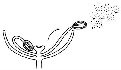
Turgor-driven ejection mechanisms are also widespread. A classic example among higher plants is the squirting cucumber (Ecballium elaterium). Large, thin-walled cells within the interior of the fruit form a swelling tissue in which the osmotic potential reaches nearly -1.5 MPa upon maturation. The outer layers of the fruit wall act as a resistance tissue subjected to intense elastic stretching. Eventually, an abscission zone forms at the pedicel attachment site; this tissue ruptures, and the pedicel is forcefully expelled by the internal pressure of the fruit like a cork from a champagne bottle. Simultaneously, the previously stretched fruit wall contracts, projecting its liquid contents along with the seeds (Fig. 8.35). The seeds are hurled over distances exceeding 12 m, while the emptied fruit hull recoils in the opposite direction.
Fig. 8.35. Ecballium elaterium, squirting cucumber: A — ripe fruit at the moment of pedicel detachment and ejection of the pulpy contents and seeds (approx. 1/2×); B — longitudinal section through an intact, attached fruit (schematic)
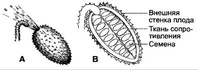
The discharge of fungal spores from a mature ascus occurs through the following mechanism: The cell wall, elastically stretched by turgor pressure (approx. 1 MPa in mature asci), ruptures suddenly at a predetermined point at the apex of the ascus (the operculum, see 11.2, Fig. 11.35). The ascus collapses to half its initial volume, and the resulting contractile force projects the spores over distances ranging from a few millimeters up to a maximum of 60 cm (in Basidiobolus immersus). Crucially in this process—as in other cases (such as pollen release in Urtica)—is that the spores or pollen grains actively cross the boundary layer of stationary air surrounding the surface where they were formed, reaching turbulent air currents where they can be passively dispersed over long distances. In some instances, specific environmental cues are required for ascus rupture; alongside adequate humidity (to maintain turgor), certain species also require light (e.g., in Sordaria curvula, blue light is effective, though the photoreceptor has not yet been identified). Conversely, other ascomycetes (such as Hypoxylon fuscum) discharge their spores at night.
The rupture of sporangia in Pilobolus is based on a similar mechanism. The upper tip of a mature, unicellular sporangiophore (see 11.2, Fig. 11.28) swells into a club-like shape due to turgor pressure, causing the cell wall to stretch elastically by up to 100%. Only the annular zone where the columella invaginates into the sporangium remains inelastic, thereby determining the precise site of rupture. Upon bursting, the entire sporangium is projected at an initial velocity of about 6 m s-1, traveling up to 2.5 m horizontally or up to 1.8 m vertically, guided by positive phototropism toward the incident light (see 8.3.1.1).
Last update: 07/08/2026
Editorial and Educational Adaptation: This material has been compiled based on the primary/original source text. The project team performed an editorial review, corrected technical inaccuracies, structured sections, and adapted the content for an educational format.
What was processed:
- elimination of formatting defects (OCR errors, structural breaks, corrupted characters);
- editorial organization of content;
- standardization of terminology in accordance with academic sources;
- verification of factual statements against the original source text.
All mentions of the author, publication year, and origin of the primary text have been preserved in accordance with the source.