BOTANY VOLUME 2 - PLANT PHYSIOLOGY - 2007
8. PHYSIOLOGY OF MOVEMENTS
8.4. Miscellaneous Movements
Hygroscopic and cohesion movements are widespread in the plant kingdom. Hygroscopic movements are based on uneven Swelling and occur without the direct participation of living Cells. They serve to disperse spores, pollen, seeds, and fruits. These movements are driven by purely physical causes, relying on differences in the stretching or contraction of fibrillar layers during swelling or moisture loss. The swelling pattern of a Cell wall is determined by the predominant orientation of parallel microfibril layers within the secondary cell wall. Stretching or contraction occurs primarily perpendicular to the direction of the microfibrils. If tissue layers with different microfibril orientations and varying cell wall compositions lie on top of one another, fluctuations in tissue moisture (e.g., due to desiccation during maturation, or differing Hydration states in dry versus wet environments) cause twisting. The swelling capacity of the main cell wall components increases in the following order: Lignin < Cellulose < hemicellulose < pectin.
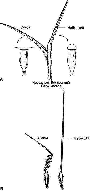
The outer peristome Teeth on the sporangia of bryophytes, which largely consist only of The Cell wall remnants of two adjacent cell layers, bend inward or outward upon drying depending on their fine Structure. Through these movements—driven by fluctuations in atmospheric humidity—they facilitate or hinder spore dispersal. Fig. 8.36 illustrates another example: the movement of a peristome tooth upon drying is governed by the fact that the microfibrils in the outer plate are oriented transversely to the longitudinal axis of the tooth, causing this layer to shorten primarily along the longitudinal axis. Conversely, due to the axial orientation of its fibrils, the inner plate merely wrinkles in thickness without reducing its length. Because it is firmly attached to the outer wall layer, it prevents the tooth from shortening and forces it to curl outward. The cell wall architecture of peristomes within a single moss genus is highly diverse, resulting in various movement directions that serve as adaptations to specific ecological needs. Similar hygroscopic movements are characteristic of the spore elaters of Equisetum (see 11.2; Fig. 11.141, H, J) and the capillitia of certain slime Molds (see 11.2; Fig. 11.17, E).
Many capsule fruits open when the protoplasts of the fruit wall cells die and the cell walls begin to dry out (xerochasy, e.g., in Saponaria); others remain closed when dry and open only upon wetting (hygrochasy, e.g., in species of Mesembryanthemum, Sedum, Verónica). The opening (upon drying) and closing Movements of the cover scales in conifer cones (e.g., pine cones, see 11.2; Fig. 11.203) are also attributable to the anisotropic swelling of individual scale layers.
In the schizocarps of Erodium species (Fig. 8.36, B), desiccation triggers a spiral coiling of the awns. When re-wetted, the awns attempt to straighten out again, and if their free tips encounter resistance, they screw the fruits into the soil. The awns of caryopses in certain grasses (e.g., Stipa) behave similarly. Hygroscopic mobility is also exhibited by the pappus hairs of many seeds and fruits (e.g., dandelion).
Class="center">Fig. 8.36. Hygroscopic movements: A — outer peristome tooth of the moss capsule Orthotrichum diaphanum in dry and swollen states. The inner and outer cell layers of the tooth are shown with a schematic representation of the microfibril orientation. Next to it, a moss capsule with an open (left) and closed (right) peristome (only two peristome teeth are shown in the diagram); B — schizocarp of Erodium gruinum in dry and swollen states
In the North African Brassicaceae plant Anastatica hierochuntica (the "Rose of Jericho"), the dry branches curl inward, while moistened ones spread out widely. It was once believed that the dry, spherical Anastatica plants could be tumbled by the wind to disperse seeds, but this hypothesis was disproven.
Unlike hygroscopic movements, cohesion movements rely on the remarkably strong tensile forces between Water molecules, even within very thin water layers (see 6.3.2.2, Equation 6.34).
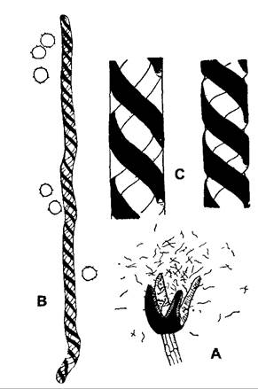
For instance, in the individual Cells of the annulus that arches around a fern sporangium (Fig. 8.37), the inner and lateral walls are thickened compared to the outer walls. As the sporangium matures, these cells begin to slowly lose water. However, because the water permeating the cell walls is tightly held by them, and the water filling the cell lumen initially does not detach due to strong intermolecular cohesion (requiring a negative hydrostatic pressure more negative than -25 MPa!, see 6.3.5), the upper PARTS OF THE anticlinal cell walls draw closer together as the thin outer walls bow inward. This generates a tangential tension on the upper part of the sporangium. As a result, two cells at a predetermined site (the stomium) separate, and the dead wall of the sporangium begins to tear slowly from this point, peeling outward. When the deformation of the arched cells reaches a limit where the cohesive forces of the intracellular water are successively overcome, tension is released in individual cells of the annulus. Each such "jump" in a cell creates a jerk; collectively, these movements cause the bent-back sporangium wall to snap back violently into its original position, thereby scattering the spores. A very similar mechanism governs the opening of anthers, where the fibrous endothelial cells in the anther wall function like annulus cells due to the rigidity of their walls. Comparable mechanisms operate in the sporangial septa and elaters of many liverworts (Fig. 8.38).
Fig. 8.37. Cohesion mechanism in the sporangial annulus of Dryopteris: A — closed sporangium; B — rupture (cells contract due to the cohesive bonding of water, annulus bends outward); C — final state after repeated compression of the annulus (tension released due to The entry of air bubbles)
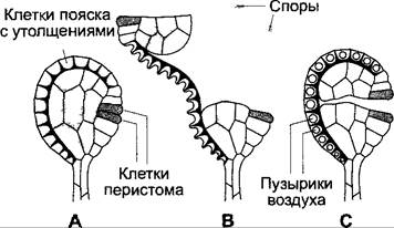
Fig. 8.38. Elaters of the liverwort Cephalozia bicuspidata: A — opened capsule (6x); B — individual elater with spores (100x); C — part of an elater; left — saturated with water, right — after partial evaporation of the contained water (425x)
The trapping mechanism of Utricularia bladders (see Box 4.4, B) is also based on the action of cohesive forces in the water filling the cells. Through the active outward transport of Na+, K+, and Cl- ions from the cell lumen through the bladder wall, followed by osmotic water efflux, the bladder loses up to 40% of its water. This creates a negative hydrostatic pressure relative to the external environment, which is evident from the invagination of the set trap bladder. Touching the trigger bristle causes the trapdoor to open, and the surrounding water, along with the prey, is sucked into the trap (the "suction-trap principle").
Last update: 07/08/2026
Editorial and Educational Adaptation: This material has been compiled based on the primary/original source text. The project team performed an editorial review, corrected technical inaccuracies, structured sections, and adapted the content for an educational format.
What was processed:
- elimination of formatting defects (OCR errors, structural breaks, corrupted characters);
- editorial organization of content;
- standardization of terminology in accordance with academic sources;
- verification of factual statements against the original source text.
All mentions of the author, publication year, and origin of the primary text have been preserved in accordance with the source.