BOTANY VOLUME 2 - PLANT PHYSIOLOGY - 2007
8. PHYSIOLOGY OF MOVEMENTS
8.2. Free Locomotion
There are types of movement by which certain seedlings or rhizomes slowly crawl through the substrate, growing forward at the apex while dying off at the rear (e.g., seedlings of Cuscuta, see 4.2.6, Fig. 4.38). However, free locomotion is found primarily in lower plants (e.g., flagellates, volvocaleans, diatoms, myxomycetes) and Bacteria, in specialized Cell stages such as zoospores of many Algae and Fungi, and in freely motile male Gametes, which occur in ferns and certain gymnosperms (Cycas, Ginkgo, see 11.2).
Locomotion is accomplished through various mechanical principles:
✵ amoeboid movement (crawling on or through the substrate; amoeba and plasmodium stages in myxomycetes);
✵ elastic movement (movement with a push-off) driven by the unilateral secretion of slime: slime Swelling in an aqueous environment pushes The Cell forward across the substrate (Desmidiaceae);
✵ gliding along the substrate (via streaming plasma in the raphe region of pennate diatoms: the caterpillar-track principle);
✵ crawling of many cyanobacteria over secreted slime aided by microfibrils;
✵ swimming by means of cilia or flagella.
The mechanics of movement are known in detail only for swimming propelled by cilia or flagella. The Structure of eukaryotic Cilia and flagella is fundamentally identical (see 2.2.2.3; Figs. 2.16; 2.17). Flagella are referred to as such when they occur singly or in small numbers per cell and reach a considerable length relative to the cell size
while cilia are numerous and short. Bacterial flagella are distinct structures. They are built entirely differently (see 2.3.2; Fig. 2.96) and function on a completely different mechanical principle than eukaryotic cilia or flagella (Fig. 8.1).
Class="center">Fig. 8.1. Mechanics of flagellar and Ciliary movement: A — propeller-like movement of a prokaryotic flagellum, which rotates at its base within the cell body, driven by the force of the proton gradient (see 6.1.4.3). As the rotor turns, H+ ions enter the cell. H+-transporting ATPases consume ATP to "pump" H+ ions back out of the cell to maintain the proton-motive force; B — effective stroke in the flagellar movement of Monas sp. (Chrysomonadales, see 11.2); I — recovery stroke of the flagellum; II — effective stroke. Arabic numerals indicate The sequence of individual stroke phases, arrows show the direction of movement. The cell body is not drawn to scale

Bacterial flagella (structure — see Fig. 2.97) rotate within The cell membrane, driven by the energy of the transmembrane proton gradient (see 6.1.4.3; exception Vibrio alginolyticus: Na+ gradient), much like propeller-driven rotary motors. THE PRINCIPLE OF such a rotary motor was already described when considering the action of chloroplast and mitochondrial ATP synthases (see Fig. 6.63), but the flagellar motor is built differently and has a more complex structure. Driven by the H+ gradient, flagellar motors rotate at frequencies of several hundred hertz and enable Cells to achieve forward swimming speeds of up to 20 µm • s-1; motors driven by the Na+ gradient rotate even faster (in Vibrio, over 1,000 Hz, providing forward cell speeds of up to 200 µm • s-1). A characteristic feature of prokaryotic flagellar motility is the alternation of run and tumble phases. During the run phase, the flagellum (or, in the case of multiple flagella, a bundle formed by the coordinated movements of individual flagella) rotates in such a way that the cell is propelled through the medium. During the subsequent tumble phase, the flagella briefly reverse the direction of their rotation. Due to the low inertia of the cell combined with the high viscosity of the medium, forward motion stops immediately, whereupon the cell assumes a random new orientation in the medium and continues moving in this new direction during the next run phase. Typical time intervals lie between 1 and 0.1 s. During the run phase, Escherichia coli, for example, swims using 4 to 8 flagella inserted at various points and reaches a speed of about 20 µm • s-1. Environmental stimuli affect the frequency of the run/tumble transitions, altering it and thereby converting locomotion into taxis (see 8.2.1). To sustain forward movement via flagella, the cell requires only a limited expenditure of energy: for example, Spirillum consumes about 0.1% of its metabolic energy.
In contrast to the propeller mechanics of prokaryotic flagellar motility, the eukaryotic flagellum acts like an oar. In the simplest case, a single flagellum oriented forward in the direction of swimming (tractive flagellum) performs beating movements within a single plane (e.g., in Euglena, see Fig. 8.6). If there are multiple flagella (e.g., two in Chlamydomonas reinhardtii), their movements must be synchronized to ensure coordinated cell motion (see Fig. 8.7). Pyrrhophyceae, heterokont algae possessing two flagella of unequal length (see Fig. 11.70, A), swim in wide spirals while simultaneously rotating their cell body. In ciliated eukaryotes (e.g., Volvox, fern spermatozoa), the cilia generally beat in a coordinated manner. There are also flagella that, being attached at the posterior pole of the cell, push it through the medium. The flagellar motor is highly efficient. Zoospores of the slime mold Fuligo varians reach speeds of up to 1 mm • s-1, covering a distance per second that is a hundred times their own cell length (approx. 10 µm).
The ciliary and flagellar beating is brought about by dynein-mediated sliding of peripheral microtubule doublets relative to one another within the axonemal complex (see 2.2.2.3, Fig. 2.17; function of dynein — see Fig. 6.6). Because the axonemal complex is anchored in the basal body, the flagellum bends As a result. Microtubule sliding may also involve, in a defined pattern, a changing sequence of two or more peripheral doublets along the entire length or specific segments, resulting in various types of movement. Energy for flagellar beating is supplied by ATP, which is hydrolyzed as a consequence of Conformational Changes in dynein. The axonemal complex responds to The addition of ATP even when isolated from the cilium (e.g., after disrupting the flagellar membrane with a detergent and washing out the Cytoplasm). Naturally, the flagellar membrane regulates the intracellular Ca2+ ion concentration within the flagellum, which plays a major role in controlling movement. For instance, in Chlamydomonas, when the internal Ca2+ concentration exceeds 10-5 M, the flagellum alters its beating pattern so that the cell swims backward. This switch from pushing to pulling occurs, for example, when the cell encounters an obstacle. Mechanical contact opens Ca2+ channels in the flagellar membrane, allowing Ca2+ ions from the external medium to enter the flagellum.
When free locomotion is induced by a stimulus, it is termed taxis. When movement is directed toward the source of the stimulus, it is positive taxis; if directed away from the source, it is considered negative taxis. Directed movement toward or away from a stimulus source is called topotaxis. If a freely moving Organism finds an optimal zone within a stimulus field simply by selecting the "correct" direction over the "incorrect" one while reverse behavior is hindered, it is referred to as phobotaxis or Shock reaction. Such reactions are observed, for example, during flagellar movement as a result of stimulus-dependent changes in beat frequency (see 8.2.1.1, Fig. 8.3). Currently, the term kinesis is also used for phobotaxis, reserving the term taxis strictly for topotaxis. In phobotaxis (kinesis), a cell moving within a stimulus field perceives temporal differences in stimulus intensity, whereas organisms capable of topotaxis respond to spatial differences in stimulus intensity, such as between the anterior and posterior ends of the cell.
Finally, Taxes are classified According to the Nature of the stimulus that triggers them (e.g., chemotaxis, phototaxis). Often, the same cell perceives multiple stimuli, such as light and chemical cues.
Chemotaxis enables freely motile bacteria and fungi to find nutrient sources or hosts and avoid areas containing harmful substances, and also allows gametes to engage in a targeted search for mating partners (Table 8.1). In the former case, many different substances can elicit chemotaxis. More than 30 distinct chemosensors have been identified in bacteria: two-thirds of them are tuned to attractants, and one-third to repellents. In the case of gamones (gamete-attracting substances), soluble compounds act with high Specificity, enabling gametes to locate mating partners of the same species with great precision even in habitats containing closely related species.
Table 8.1. Examples of compounds important for chemotaxis in PROKARYOTES AND EUKARYOTES
Organism (cell type) |
Chemoattractants / repellents |
Locomotion mechanism |
Type of response |
Bacteria |
Acetic acid |
Bacterial flagella |
Negative phobotaxis |
O2, many sugars (e.g., galactose), nitrogenous compounds, phosphate, alkali and alkaline-earth Metal Ions |
Ditto |
Positive phobotaxis |
|
Fungi: |
|||
Myxomycetes (zoospores) |
Malate |
Flagella |
Ditto |
Dictyostelium (myxamoebae in feeding phase) |
Amoeboid |
Positive topotaxis |
|
Dictyostelium (starving myxamoebae) |
Ditto |
Ditto |
|
Allomyces (gametes) |
Sirenin1 |
Flagella |
Positive phobotaxis |
Algae (gametes): |
|||
Chlamydomonas reinhardtii |
Ditto |
Positive topotaxis |
|
Ch. allensworthii |
Lurilenal acid1 |
—»— |
Ditto |
Brown algae |
Including Hydrocarbons1 |
—»— |
|
Mosses (gametes) |
Including sucrose2 |
—»— |
—»— |
Ferns (gametes) |
Ca malate2 |
—»— |
—»— |
Lycopodium (gametes) |
Citrate2 |
—»— |
—»— |
1 Sex attractants (gamones) produced by female gametes, which are often poorly motile or immotile, serving to attract male gametes.
2 Sex attractants of archegonia; it remains unclear which Tissues or cells within the archegonium produce these attractants.
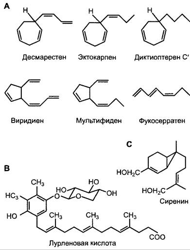
Algal gametones, particularly those of brown algae, have been studied in exceptional detail (Fig. 8.2). These unsaturated hydrocarbons frequently exert biological effects at concentrations as low as 10-11 • mol • l-1 and are synthesized from polyunsaturated Fatty acids. Some of these compounds also synchronize gamete release. The majority of gametes in bro
wn algae secrete a diverse spectrum of these hydrocarbons, yet only a single compound acts as a species-specific gametone due to a high degree of stereospecificity. The remaining hydrocarbons may serve as decoys for sperm of other species; while unable to fertilize foreign gametes, these sperm thereby forfeit their capacity to fertilize their own gametes. Upon exposure to these chemoattractants, heterokont male gametes (possessing flagella of unequal length and Morphology) exhibit accelerated flagellar beat frequencies, ultimately anchoring themselves via their longer flagella to the female gametes.
Fig. 8.2. Examples of gametones: A — from brown algae; B — from the unicellular green alga Chlamydomonas allensworthii; C — from the aquatic fungus Allomyces (Blastocladiales). Brown algal gametones are derived from polyunsaturated fatty acids (see 6.11.1). Sirenin is a sesquiterpene (see 6.16.2). Lurleic acid is presumed to originate from chloroplast plastoquinone (Fig. 6.56); the sugar is β-D-xylose
Recent investigations have also focused on negative bacterial chemotaxis and the positive chemotaxis observed in amoeboid Stages of the myxomycete Dictyostelium discoideum.
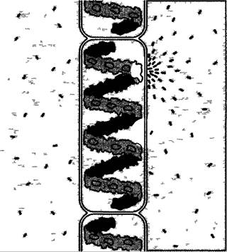
Bacterial locomotion results from an alternation between a "run" phase lasting approximately 1 s and a "tumble" phase lasting about 0.1 s (see 8.2.1) in a homogeneous environment. In the presence of a concentration gradient of a chemotactically active substance, the run/tumble frequency is modulated (Fig. 8.3): moving toward an increasing concentration of an attractant leads to prolonged runs because the frequency of tumbling decreases; conversely, in the presence of a repellent, the exact opposite occurs. Consequently, guided by the concentration gradient of the attractant, the majority of cells ultimately accumulate at the site of maximum concentration, whereas in the case of a repellent, they gather at the minimum concentration. The positive aerotactic behavior of many bacteria (swimming toward an oxygen source) formed the basis for demonstrating photosynthetic O2 evolution in Engelmann's classical bacterial experiments (Fig. 8.4). The molecular mechanisms underlying flagellar motor function and regulation are detailed in standard microbiology textbooks.
Fig. 8.3. Schematic representation of positive chemophobotaxis in a bacterium and, for comparison, positive chemotopotaxis within a concentration gradient of a chemotactically active substance (effector). For further details, see the text
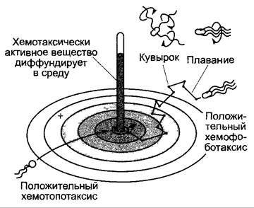
Fig. 8.4. Spirogyra cells with an accumulation of positive aerotactic bacteria (highlighted by black dashes) along the illuminated region of the ribbon-shaped chloroplast (site of photosynthetic O2 evolution). Illumination of the inter-chloroplast spaces does not induce O2 production, and consequently, bacteria fail to congregate in those zones
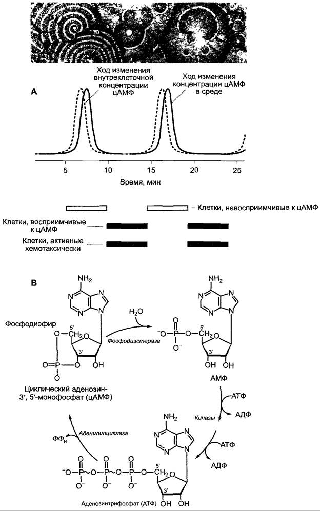
When supplied with adequate nutrients, the myxomycete Dictyostelium discoideum exists as a unicellular organism moving via amoeboid locomotion. Upon nutrient depletion, starving myxamoebae synthesize cyclic adenosine-3',5'-monophosphate (cAMP, Fig. 8.5) and secrete it into their environment. This attractant is perceived by surrounding cAMP-receptive myxamoebae, triggering positive chemotaxis that causes converging amoebae to form an aggregation center where differentiation processes subsequently take place (partially regulated by cAMP; for The life cycle of Dictyostelium, see 11.2, Fig. 5.3). Cellular synthesis and release of cAMP, as well as cellular responsiveness to this signal, oscillate cyclically with repeating maxima and minima (with a phase duration of approximately 6–10 min). This gives rise to a rhythmic topotaxis that is readily observable in experiments with myxamoebae, such as those conducted in Petri dishes (see Fig. 8.5). The cAMP receptor is localized in The Plasma Membrane, and its molecular architecture has been elucidated. It exhibits notable Homology with Bacteriorhodopsin—the light-driven archaeal proton pump (see Fig. 6.66)—with the rhodopsin of green algal eyespots serving as the phototaxis receptor (see 8.2.1.2), with vertebrate visual pigments (rhodopsin), and with other vertebrate Plasma Membrane Receptors, including numerous Hormone Receptors as well as olfactory and gustatory receptors. A common structural hallmark of these membrane receptors is the presence of seven transmembrane α-helices, alongside a signature sequence characteristic of heterotrimeric G Proteins (where G denotes GTP-binding; see cell biology textbooks). This structural conservation strongly suggests that vertebrate chemoreceptors and visual pigments evolved from the chemosensory and phototensory receptors of halobacteria and lower eukaryotes (see 7.5).
Fig. 8.5. Chemotaxis in Dictyostelium discoideum during the aggregation phase: A — rhythmic fluctuations in cAMP Synthesis and Secretion, as well as cellular cAMP sensitivity (within a synchronized cell population). The substrate tracks the wavelike migration of individual cells (or a synchronized population) toward the aggregation center, which marks the source of initial cAMP release (multiple aggregation centers are visible in the photograph); B — intracellular formation of cAMP from ATP and its extracellular degradation mediated by phosphodiesterase
Directed locomotion toward or away from light (phototaxis) is predominantly observed in photosynthetically active organisms, enabling them to optimize their ambient light intensity. However, phototaxis also occurs in non-green flagellates and in myxomycete plasmodia (see 11.2), which initially exhibit negative responses but shift to positive phototaxis following the induction of sporangium formation. Phototaxis involves both phobic and tropic reactions. Phobic responses include shock movements triggered by a sudden decrease (step-down response) or a sudden increase (step-up response) in light intensity.
Positive photophobotaxis in the purple bacterium Chromatium is mediated by a transient cessation of flagellar motion whenever ambient light intensity drops abruptly. Because the bacterial cell possesses virtually negligible inertia, it instantly comes to a standstill, resuming movement with a generally altered direction. Conversely, an increase in light intensity exerts no effect on the direction of travel. In contrast, when Rhodospirillum experiences a reduction in light intensity, the direction of flagellar rotation reverses, resulting in a directional U-turn. When presented with two competing light sources, Rhodospirillum can distinguish between them even when the brightness of the first exceeds the second by a factor of only 1.01 to 1.03, reflecting an exceptionally high differential sensitivity. In both Chromatium and Rhodospirillum, these behavioral mechanisms ultimately herd the bacteria into illuminated zones from which they cannot readily escape ("light trap").
The action spectrum for photophobotaxis in purple bacteria matches their Photosynthesis action spectrum. A critical trigger for the phobic response appears to be a sudden shift in photosynthetic electron transport. This is undeniably crucial for the photophobotactic response of gliding cyanobacteria (which reverse their gliding direction upon a drop in light intensity), where analyses have established that the redox state of plastoquinone serves as the primary regulatory switch for the phobic reaction. Negative phototaxis in the purple sulfur bacterium Ectothiorhodospira halophila is triggered by blue light. Its photoreceptor, PYP (photoactive yellow protein), is a 14 kDa chromoprotein whose light-absorbing moiety is a para-coumaric acid residue attached via a thioether bond to a single Cysteine residue of the protein (cinnamic acids, Fig. 6.115); in the dark, the para-coumaric acid exists as a phenolate anion. Light absorption causes the chromoprotein to bleach because protonation of the phenolate anion converts it into an uncharged phenolic form that absorbs in the ultraviolet region.
In Halobacterium, step-down and step-up responses are mediated by distinct receptors: the former by bacteriorhodopsin within the purple membrane—which Functions simultaneously as an energy transducer (see Fig. 6.66) and a signal generator—and the latter by a retinalidene protein, which is presumably a biosynthetic precursor of bacteriorhodopsin.
Gliding organisms such as cyanobacteria (Phormidium) or diatoms (Navicula) exhibit a specialized form of phototaxis: they select the trajectory leading toward the light source from two possible directions. They achieve this by sensing illumination gradients across the anterior and posterior poles of the cell. In Navicula, for instance, spontaneous reversals of the chosen travel direction occur at regular intervals. However, if the anterior end is more intensely illuminated than the posterior end, these directional reversals happen less frequently than when the posterior end experiences higher light intensity.
Unicellular flagellated algae typically exhibit negative photophobic reactions at high light intensities, paired with positive phototropic responses at lower light intensities. The action spectrum of phototaxis is distinct from the photosynthesis action spectrum, with sensitivity peaking in the green to blue-green spectral range. This likely represents an evolutionary adaptation to aquatic life, given that light penetration narrows predominantly to the blue-green spectrum with increasing depth (see Fig. 6.43), while minimizing competition for light absorption from other photosynthetic organisms. Directed swimming toward a light source (positive phototaxis) or away from it (negative phototaxis) equips the organism to perceive both temporal shifts in light intensity and the spatial vector of the light source.
Directional sensitivity is achieved, in part, through highly specialized Organelles known as eyespots (stigmata). These consist of a stigma laden with light-absorbing pigments and a dedicated photoreceptor region. The Structural components of the eyespot are arranged in a precise spatial geometry relative to one another and to the flagella. Two model organisms have been investigated in great depth: Chlamydomonas (possessing an eyespot typical of all motile green algae) and Euglena (possessing an eyespot typical of Euglenophyceae). Although their locomotory mechanics differ, they share a common operational principle: as the cells swim forward, they rotate about their longitudinal axis while tracing a helical path, causing the cell's longitudinal axis to execute a wobbling motion (nutation) relative to the axis of forward progression. The photoreceptive organelles, or at least the stigma, are situated peripherally; consequently, under oblique illumination relative to the direction of travel, the spatial orientation of the stigma shifts periodically with respect to the incident light vector.
In Euglena, the eyespot is composed of a loose accumulation of cytoplasmic lipid droplets rich in astaxanthin, a carotenoid also prevalent in the animal kingdom. The photoreceptor is localized within the paraflagellar body of the flagellar pocket (see 2.2.2.3; Fig. 1). Although its biochemical nature is not yet fully elucidated, action spectra indicate that its chromophore group incorporates a flavin (for methodologies on determining action spectra, see 7.7.2.4). Due to a significant spectral overlap between carotenoid and flavin absorption bands under lateral illumination, the paraflagellar body undergoes periodic, brief shading by the stigma (Fig. 8.6). This transient shading (likely mediated by Ca2+ ions) induces brief alterations in flagellar beat patterns, thereby course-correcting the cell until it swims directly toward the light source and the stigma no longer shadows the paraflagellar body (see Fig. 8.6).
Fig. 8.6. Positive phototaxis in Euglena (after W. Haupt). Rotation about the longitudinal axis causes the photoreceptor at the Base of the flagellum (the paraflagellar body) to be periodically shaded by the stigma, as illustrated in the right-hand diagram; consequently, the cell steers toward the stigma (i.e., toward the light source, situated to the left). More recent models also consider the orientation of photoreceptors relative to the plane of light polarization as a factor in light-incidence-dependent activation

In Chlamydomonas (and probably in flagellate Chlorophyceae in general), the "eyespot" or stigma in the chloroplast, located near the cell surface, consists of several (up to 8, and 4 in Chlamydomonas reinhardtii) layers of carotenoid-enriched lipid globules oriented parallel to the cell surface. The globules are of uniform size; they are located on the thylakoid membranes and held at strictly defined distances by intermediate layers devoid of lipid droplets (Fig. 8.7). Photoreceptors are located in the Plasmalemma directly above the eyespot.
Fig. 8.7. Positive phototaxis in Chlamydomonas (A — original by L.A. Staehelin, courtesy of L.A. Staehelin; B — after K.W. Foster and R.D. Smyth, with modifications): A — transmission electron micrograph of an ultrathin section through the eyespot, shown in the same orientation as the depicted cell (left side facing outward). The lipid globules are in contact with the thylakoid, with the stroma located in the intervals. The eyespot acts as a reflector (B) and enhances the light intensity at the site of the photoreceptors localized in the cytoplasm. Periodic activation of the photoreceptors under unilateral illumination occurs due to cell rotation during swimming and leads to a transient change in the movement of the flagellum adjacent to the eyespot. As a result, the cell turns toward the light source

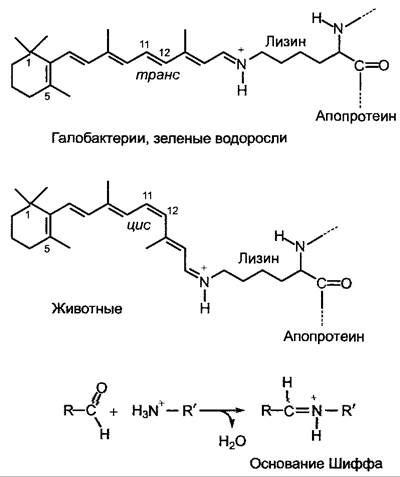
The investigation of the photoreceptor's nature using molecular biology Methods yielded a surprising result: it is a chromoprotein related to animal visual pigment, rhodopsin. Its chromophore group is represented by an isomer of retinal found in animals (Fig. 8.8), which, presumably like retinal, is synthesized from carotene and absorbs light particularly intensely in the blue-green to green region of the spectrum. Although the apoproteins of green algal sensory rhodopsin (chlamydopsin, named after Chlamydomonas) and animal rhodopsin differ in that the green algal rhodopsin proteins are shorter at both ends (C and N termini) than animal rhodopsin, they exhibit clear homology in the corresponding regions (see 8.2.1.1). While the eyespots of euglenoids serve to shade the underlying photoreceptor, the highly structured eyespots of flagellated green algae have the opposite function. They function as reflectors that increase the intensity of light falling laterally onto the site of the photoreceptors: Interference phenomena between light incident from the cell surface and reflected light play an important role, such that maximal interference Amplification of light incident perpendicularly to the orientation of the eyespot's carotenoid layers and the correspondingly reflected light occurs at wavelengths in the blue-green and green regions. Conversely, light passing through the cell is significantly attenuated due to the cancellation of interference at the site of sensory rhodopsin. Periodic excitation of the photoreceptors leads (in these cells also with the participation of Ca2+ ions) to a transient change in flagellar beating (in Chlamydomonas, this is the flagellum adjacent to the eyespot) and, consequently, to course correction as long as periodic activation of sensory rhodopsin occurs by lateral light and light reflected by the eyespot (see Fig. 8.8). It is hypothesized that the chlamydopsin photoreceptor is identical to a Ca2+-activated channel, or in other words, sensory rhodopsin represents a directly light-regulated Ca2+ channel.
Fig. 8.8. Structure of protein-bound retinal, the chromophore group of rhodopsin in halobacteria, green algae, and animals in the dark.
Retinal in rhodopsin is covalently linked via its aldehyde group to the ε-amino group of a Lysine residue of the apoprotein in the form of a Schiff base (on grey Background: formation of a Schiff base from an aldehyde and an amine). The retinal of halobacteria and green algae is present in the all-trans form in the dark and isomerizes from the 13-trans to the 13-cis form upon illumination; in animals, it is present in the 11-cis form and isomerizes to the all-trans form upon illumination. Consequently, the conformation of the apoprotein must change accordingly. It is suggested that in Chlamydomonas, the activated sensory rhodopsin (chlamydopsin) even forms a Ca2+ channel, and the influx of Ca2+ into the cell leads to the inhibition of flagellar beating on the side facing the light
In addition to chemical and light stimuli, some free-moving organisms also respond to changes in humidity (hydrotaxis), Touch stimuli (thigmotaxis), gravity (gravitaxis), or Temperature changes (thermotaxis). Plasmodia of Dictyostelium are capable of perceiving temperature gradients even of the order of 0.05 °C cm-1. Phase transitions (liquid-crystalline) of Membrane Lipids may serve as the basis for this extremely sensitive "biothermometer". If an organism perceives various environmental stimuli and responds with a taxis, cells must integrate all external signals. Thus, in Escherichia coli, negative thermotaxis induced by low temperatures is counterbalanced by chemotactic substances or even overcompensated, whereas positive thermotaxis induced by high temperatures, conversely, is counteracted by repellents.
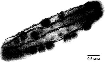
A notable feature of certain bacteria inhabiting the mud of fresh or saline waters is The ability to orient themselves in a magnetic field (magnetotaxis). In the Earth's magnetic field, this leads to downward movement into the mud, since the vertical component of the field is generally stronger than the horizontal one. The magnetic field sensor is a chain of up to 100 membrane-enclosed magnetite (Fe3O4) crystals with edge lengths up to 50 nm, which functions like a compass needle (Fig. 8.9).
Fig. 8.9. The magnetotactic bacterium Magnetobacterium bavaricum from Lake Chiemsee. The rod-shaped bacterium with flagella (not visible in the preparation) contains up to 1,000 bundled rod-shaped magnetosomes composed of magnetite (Fe3O4). The globules consist of elemental sulfur and are irrelevant to magnetotactic behavior. Magnetosomes ensure cell orientation and movement along the magnetic field vector. However, it is also hypothesized that repulsive forces between the magnetosome strands are important for stabilizing the cell body
8.2.2. Intracellular Movements
Within the cell, the positions of the cytoplasm, Nucleus, and organelles frequently change. These intracellular movements are in many respects similar to the free locomotion of unicellular organisms.
Cytoplasmic streaming (cyclosis), observed in many cells, is often triggered solely by external stimuli (e.g., light, temperature, cell damage, chemical irritation), depends on active METABOLISM (in other words, on ATP), and reaches speeds of 0.2–0.6 µm per minute (up to 6 µm per minute in internodal cells of Nitella at high temperatures). The outermost layer of cytoplasm adjacent to the plasmalemma (ectoplasm) does not participate in the movement. Since cell polarity is not altered by cytoplasmic streaming, it must be anchored in the ectoplasm or also in the plasmalemma. Structural proteins that are ATP-dependent and anchored to one another—similar to flagellar movement, plasmodial locomotion, or Muscle contraction—are responsible for cytoplasmic streaming. It remains unknown whether cytoplasmic streaming (which is by no means observed in all cells) has a physiological significance or is merely necessary for metabolism within the cell or between adjacent cells.
Cell nuclei are capable of changing their position within cells. They migrate predominantly to Regions of the most intense cell growth or exceptionally high local metabolic activity. Thus, in cells with pronounced tip growth (ROOT hairs, pollen tubes), cell nuclei are located near the growing tips; in damaged cells, they are frequently positioned near The Cell wall facing the wound; during fungal infection (see 9.3.4), they move to the sites of fungal hyphae penetration, where particularly intense cellular defense reactions take place. In the immediate vicinity of meristemoids (e.g., next to stomatal initials), cell nuclei migrate toward meristematic cells, presumably in connection with a gradient of specific substances (see 7.4.2).
Characteristically, light-dependent chloroplast movements (e.g., in algal thalli, moss leaves, fern prothalli, and among higher plants, particularly pronounced in aquatic plants) ensure an optimal position for these organelles or their relocation to sites with optimal illumination. Under low-light positioning, Chloroplasts locate near the illuminated front and back cell walls, turning their largest surfaces toward the light (maximizing light quantum absorption capabilities), whereas under high-intensity illumination, they move to the lateral walls and present the smallest possible cross-sectional surfaces to the light (high-light positioning) (Fig. 8.10), presumably to avoid light-induced damage (see 6.4.8).
Fig. 8.10. Chloroplast movements within the cell: A — arrangement of chloroplasts in a moss leaf under high- and low-light conditions, respectively (direction of light is perpendicular to the plane of the drawing); B — position of a plate-like chloroplast in a Mougeotia scalaris cell. Upon illumination of a previously dark-adapted cell (1) with short (1 min) flashes of red light (R), the chloroplast adopts the low-light position (2). The photo-reversibility of the reaction upon alternating R and FR (far-red light) illumination indicates the involvement of the Phytochrome system; light direction is perpendicular to the plane of the drawing. Chloroplast rotation takes about 30 min
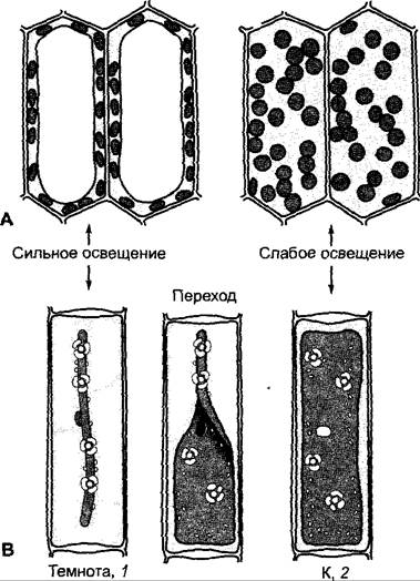
Action spectra recorded in mosses show that a flavin or flavoprotein serves as the photoreceptor in both high- and low-light responses. Light responses of plate-like chloroplasts in filamentous thalli of the alga Mougeotia (see 11.2) have been studied particularly well; it has been revealed that phytochrome participates in low-light responses, whereas high-light responses involve another blue-light receptor alongside phytochrome. According to these findings, the low-light response can be induced by red light and subsequently reversed by immediately applying far-red light; thus, we are dealing with a Class II reaction (see 7.7.2.4). In angiosperms, chloroplast positioning is governed by blue-light receptors of the phototropin group: phototropin 1 mediates the low-light response, and phototropin 2 mediates the high-light response (phototropin, see 7.7.2.4; 8.3.1.1; 8.3.2.5; Box 7.5).
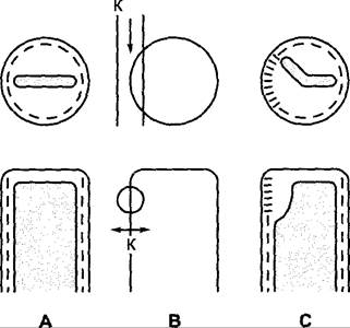
Illumination experiments using linearly polarized light demonstrated pronounced absorption dichroism of phytochrome in Mougeotia. Dichroism refers to the dependence of light absorption by a molecule on the direction of the electrical vector of the light wave. Dichroism becomes clearly noticeable when such molecules are arranged in a highly ordered manner and the preparation is irradiated with monochromatic, linearly polarized light with varying directions of oscillation, while measuring absorption and correspondingly biological activity. Furthermore, micro-irradiation of the plasmalemma and underlying ectoplasm with red light affects the local response of the chloroplast, which "turns away" from the irradiation site and partially shifts into the high-light position (Fig. 8.11). The dichroism of phytochrome indicates a highly ordered arrangement of photoreceptors within this cell; the local effectiveness of peripheral cytoplasm illumination, which is also observed in most other systems with rotating chloroplasts, implies that the photoreceptors are localized in the ectoplasm.
Fig. 8.11. Part of a Mougeotia cell in cross section (top) and in top view (below): A — before, B — during, and C — after illumination with polarized red (R) light (direction of light indicated by a single-headed arrow, polarization plane by a double-headed arrow). THE POSITION OF the chloroplast and the activity state of phytochrome (bars) are shown in A and C. The bars parallel to the upper cell surface correspond to Pr, and the transverse bars to Pfr (active phytochrome). The arrangement of phytochrome depicting dichroic properties is schematized and does not reflect the true, as yet unknown arrangement of the chromophore molecules
At the site of active phytochrome (Pfr) formation in Mougeotia, the intracellular Ca2+ concentration increases due to enhanced cellular uptake of Ca2+ and increased release from vesicles near the outer chloroplast membrane. This leads to the contraction of microfilaments adjacent to the outer chloroplast membrane (possibly involving Actin) and, consequently, to movement.
Last update: 07/08/2026
Editorial and Educational Adaptation: This material has been compiled based on the primary/original source text. The project team performed an editorial review, corrected technical inaccuracies, structured sections, and adapted the content for an educational format.
What was processed:
- elimination of formatting defects (OCR errors, structural breaks, corrupted characters);
- editorial organization of content;
- standardization of terminology in accordance with academic sources;
- verification of factual statements against the original source text.
All mentions of the author, publication year, and origin of the primary text have been preserved in accordance with the source.