PLANT MORPHOLOGY - T. A. Sautkina - 2012
CHAPTER 5. VEGETATIVE ORGANS OF PLANTS
5.4. The Leaf as a Vegetative Organ
Unlike the ROOT and stem, the leaf is a lateral vegetative organ. Because leaves exhibit extreme structural diversity, defining a leaf based purely on morphological features is practically impossible; therefore, METABOLISM/2.html">THE CONCEPT OF a "leaf" is defined by its physiological Functions. The leaf is a vegetative organ that performs the functions of Photosynthesis, Transpiration, and gas exchange. These three functions are the core characteristics of a typical leaf. At the same time, the leaves of certain plants are capable of performing other, additional functions, which leads to their modification.
As noted, during the evolution of higher plants, leaves could have originated either as enations—lateral outgrowths of axial structures—or from axial structures As a result of the latter's cladification.
5.4.1. Ontogenetic Leaf Development
The first insights into ontogenetic leaf development were provided in the works of the Italian biologist M. Malpighi (1628–1694) and K. F. Wolff (1733–1794), a German naturalist and academician of the St. Petersburg Academy of Sciences. The leaf is initiated exogenously on the stem apex. Leaf formation begins with The Emergence of a meristematic tissue protuberance just beneath the epidermis (Fig. 101). Initially, this protuberance expands uniformly in three dimensions: length, width, and thickness. However, growth in thickness soon ceases; consequently, in most plants, leaves are flat and bifacial (from Lat. bi—consisting of two parts, and facies—surface, form), meaning they have clearly differentiated upper and lower sides. Some plants develop unifacial leaves (from Lat. unifacialis—lacking distinct upper and lower sides), which are circular in cross-section, as in onions, or equifacial (isolateral) leaves (from Lat. equifacialis, isolateralis—equilateral), i.e., flattened leaves with identical upper and lower appearances, as in irises. The lateral expansion of the meristematic protuberance soon stops as well. The developing leaf primordium, having reached its maximum width within the bud, was termed the primordial leaf or primordium by the German botanist A. Eichler (1839–1887) (from Lat. primordium—beginning, origin, genesis). Longitudinal growth of the leaf primordium continues the longest. Gradually, driven by marginal Meristems, the Base of the primordium broadens and differentiates into two parts: the basal (lower) and the apical (upper). Through further Differentiation of the leaf primordium, the basal part gives rise to the leaf base and stipules (predominantly in dicotyledonous plants), while the apical part forms the leaf blade and petiole (if present in the given plant species).
Class="center">Fig. 101. Diagram of leaf formation in ontogenesis: A—initiation of the meristematic leaf protuberance; Б—enlarged meristematic protuberance (primordial leaf); В—differentiation of the primordial leaf; Г—Е—successive stages of leaf part formation; Ж—fully formed leaf: 1—apical part; 2—basal part; 3—leaf base; 4—stipules; 5а—leaf blade primordium; 5—developing and fully formed leaf blade; 6—procambium; 7—midrib; 8—lateral Veins; 9—petiole primordium; 10—petiole

Thus, a typical leaf consists of four parts, of which the leaf base and the leaf blade are mandatory. Stipules and petioles may be absent.
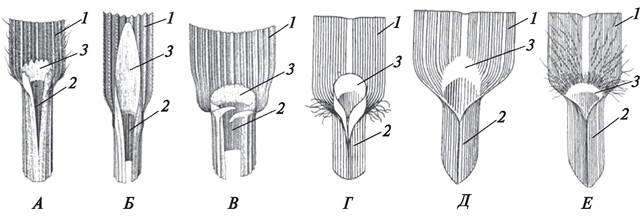
The leaf base is the portion through which the leaf attaches to the stem. Morphologically, the base is not always sharply defined. The lower boundary of the base is often located within the stem Tissues, while the upper boundary lies at the base of the stipules or petiole. In some plants, the leaf base expands significantly in width due to marginal meristem activity and wraps around the stem to varying degrees, sometimes forming a leaf sheath, as seen in grasses, sedges, and certain orchids (Fig. 102). The leaf base can grow in length through intercalary meristem activity, in which case it comes to resemble a petiole (as in Viola hirta). The base is the longest-lasting part of the leaf. In many bulbous plants (onions, tulips, hyacinths, etc.), the leaf base is represented by fleshy scales that form the bulk of the bulb and perform a storage function.
Fig. 102. Structure OF THE leaf sheath in various grass species: A—Bromus secalinus; Б—Deschampsia caespitosa; В—Lolium multiflorum; Г—Digitaria ischaemum; Д—Echinochloa crusgalli; Е—Setaria glauca:
1—leaf blade fragment; 2—leaf sheath; 3—ligule
Following the leaf base, stipules become distinct. They are most developed in representatives of primitive families within the class Dicotyledones (Magnoliopsida)—such as Fabaceae and Rosaceae. In monocots, stipules are rare (e.g., Potamogeton, Hydrocharis morsus-ranae). Stipules form as lateral outgrowths driven by marginal meristem activity. Depending on their arrangement, Three types of stipules are distinguished: lateral, interpetiolar, and median (Fig. 103).
Fig. 103. Types of stipules: A—lateral; Б—median; В—interpetiolar: 1—stipules; 2—leaves
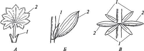
Lateral stipules are paired and located on both sides of the petiole (Rosa rugosa, Comarum palustre, Pisum sativum, etc.).
Interpetiolar stipules share the same stem node with decussate leaves and are positioned between them. Morphologically, they are virtually indistinguishable from sessile leaves. Distinguishing which structures are stipules and which are leaves is possible at the Cytology/cytology/16.html">Early stages of stem branching: lateral shoots emerge exclusively from leaf axils. Interpetiolar stipules are characteristic of Galium, Asperula, and certain other members of the Rubiaceae family.
Median stipules are single (unpaired). They are located in the leaf axil (between the stem and the leaf), are found in some monocots (Calla palustris, Potamogeton lucens, etc.), and are very rare in dicotyledonous plants (Menyanthes trifoliata).
In the early Selection/3.html">Stages of development, stipules perform a protective function for the forming leaf blade. Once the leaf emerges from the bud, the stipules either fall off (Tilia cordata) or persist and perform the core functions alongside the leaf (pea, rose). Sometimes they metamorphose into spines (Robinia pseudoacacia), thereby serving a protective function by safeguarding the plant against animal damage.
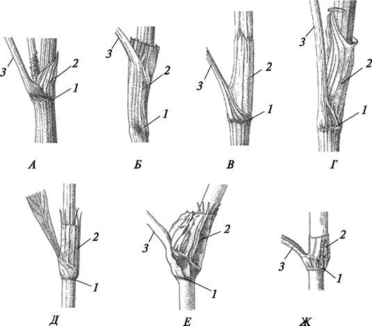
In members of the buckwheat family (Polygonaceae), membranous stipules fuse to form an ocrea (sheath), which wraps around the stem for a greater (Bistorta major) or lesser (Rumex acetosa) length. The ocrea serves a protective function. The presence of an ocrea is an important morphological feature of Polygonaceae representatives (Fig. 104).
Fig. 104. Types of ocreae in Representatives of the Polygonaceae family: A—Fagopyrum tataricum; Б—Persicaria amphibia; В—Rumex crispus; Г—Rumex acetosa; Д—Persicaria mitis; Е—Persicaria hydropiper; Ж—Persicaria lapathifolia: 1—stem node; 2—ocrea; 3—leaf petiole fragment
The leaf blade develops in length within the bud thanks to apical and intercalary meristems, which promote the enlargement of the leaf primordium and form its axial part—the midrib region. The leaf blade expands in width through The activity of the marginal meristem, which develops along the axis of the leaf. The leaf blade of a primordial leaf can form acropetally (pea, Water hemlock, begonia), basipetally (rose, maple, lily), divergently (cornflower, dandelion, yarrow), or in a parallel manner (bird cherry, palms).
In acropetal development, the leaf blade forms from the base to the apex, with older Cells located at the base. In basipetal development, The formation of the leaf blade proceeds from the apex to the base, making the leaf apex the oldest part; in divergent development, the central part is the oldest, as the leaf blade develops in diametrically opposite directions—toward the apex and the base. In parallel development, a series of marginal meristem zones appears along the axial part, and the leaf blade forms from its central part toward the margins. If the Cells of the marginal meristem divide uniformly, leaves with entire blades are formed. If zones of active meristematic Cell Division alternate with zones where cell division is slowed down, leaves with dissected blades are formed.
During the formation of a compound leaf, second-order meristematic protuberances arise on the axis of the leaf primordium, which develop into the leaflets of the compound leaf.
Upon the emergence of the leaf from the bud, the growth pattern of the leaf blade changes dramatically. In most dicotyledons, the growth of the leaf blade proceeds strictly uniformly across its entire area, primarily through an increase in the volume of the cells of the primordial leaf. However, the expansion of the leaf blade can also occur due to de novo Cell Formation, as was proven by I. G. Serebryakov for bird cherry (Padus racemosa) and linden (Tilia cordata).
5.4.2. Development of the Leaf Vascular System
Simultaneously with The Development of the leaf blade, its vascular system is formed. The leaf vascular system is represented by collateral vascular bundles that form the so-called veins, which are especially prominent on the underside of the leaf.
Vascular bundles originate from the vascular meristem (procambium), which differentiates at the base of the primordial leaf protuberance, and consist of primary xylem and phloem. Thus, the leaf blade develops its own vascular system consisting of one or more large (main) veins and numerous smaller veins permeating the entire leaf blade. The number and structure of vascular bundles vary in the main and lateral veins. Lateral veins typically contain a single bundle. The vascular bundles in the leaves of most angiosperms are closed and collateral. However, a cambium may form in the large veins of certain leaves, though it either functions poorly—producing only a small amount of secondary Vascular Tissues—or does not function at all. The smaller the veins, the more reduced their vascular bundles become. The endings of the finest veins may sometimes lack phloem. The xylem at the endings of small veins usually consists of short tracheids, while the phloem consists of short, narrow sieve elements and large companion cells. In the collateral bundles of leaves, the xylem faces the adaxial (upper) side of the leaf, and the phloem faces the abaxial (lower) side.
Vascular bundles are typically surrounded by several layers of sclerenchyma or parenchyma that form bundle sheaths. These sheaths extend all the way to the bundle endings. Minor veins play a primary role in The transport of water and photosynthesis products. They serve as the initial collection points for photosynthetic products and the terminal points for the transpiration stream flowing through the leaf mesophyll. A characteristic feature of minor veins is the presence of abundant parenchyma, especially in the phloem, which facilitates their transport functions.
5.4.3. Connection of the Leaf Vascular System with the Stem Vascular System
The development of the main vein (or veins, if there are several) proceeds in two directions—toward the apex of the leaf blade and down into the stem toward the central cylinder. The vascular bundle that passes from the leaf into the stem is called a leaf trace. A leaf trace enters the stem through a leaf gap, which is a region of parenchyma in the stem node. A leaf trace may consist of one, two, three, or multiple bundles. Depending on the number of leaf gaps formed at the stem node, nodes are classified as unilacunar, trilacunar, or multilacunar (Fig. 105). One or more vascular bundles may enter a single gap. Stem nodes are classified based on the number of leaf gaps and the number of bundles entering each gap (e.g., unilacunar one-trace, trilacunar one-trace, unilacunar three-trace, etc.). The structure of the stem node is an important taxonomic feature. The Study of stem node structural features is pursued by a specialized branch of Morphology known as nodal anatomy (from the Latin nodus, meaning node).
Fig. 105. Structure of stem nodes and diagram of nodal evolution (after Takhtajan, 1964, with additions): 1—trilacunar one-to-two-trace node; 2—trilacunar one-trace node; 3—unilacunar one-trace node; 4—multilacunar one-trace node; 5—unilacunar two-trace node; 6—unilacunar three-trace node; 7—unilacunar one-trace node

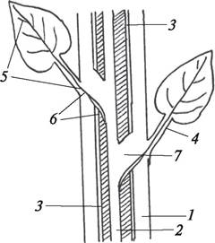
After passing through the leaf gap of the stem node, the leaf traces enter the parenchyma of the primary cortex, travel downward through it toward the underlying
node, and then bend, directed inward toward the stele to merge with the stem vascular system. Following the Integration of the leaf trace into the stem vascular system, a parenchymatous zone known as a leaf gap or leaf trace lacuna is formed in the central cylinder just above the point of entry (Fig. 106). The continuity of the stem vascular system is not disrupted by this process. Instead, it becomes complex, or "synthetic," as it incorporates both stem and leaf conducting elements. Consequently, with the formation of new leaves, new leaf traces continuously enter the stem, progressively increasing The complexity of the stem vascular system.
Fig. 106. Connection of the leaf vascular system with the stem vascular system: 1—primary cortex of the stem; 2—pith of the stem; 3—stem vascular system; 4—petiole with the midrib; 5—leaf vascular system; 6—leaf trace; 7—leaf gap
5.4.4. Types of Leaf Venation
The arrangement and interconnection of all leaf veins create various types of venation. The first Classification of venation types was proposed in 1870 by Anton de Bary, who distinguished two main types: open and closed venation.
In open venation, veins do not anastomose with one another; they extend toward the margin of the leaf blade and terminate near the edge or run directly into it. A classic example of open venation is dichotomous venation, which is characteristic of the leaves of certain ferns (such as maidenhair fern—Adianthum, staghorn fern—Platycerium) and maidenhair tree (Ginkgo biloba) from the division Gymnospermae. In dichotomous venation, the veins diverge at a very acute angle and lie closely parallel to one another.
In closed venation, veins anastomose with each other to varying degrees. Depending on the number of prominent (large, main) veins, closed venation is subdivided into the following types: pinnate, palmate, parallel, and campylodromous (arcuate) (Fig. 107).
Fig. 107. Types of leaf venation: 1—dichotomous (maidenhair tree—Ginkgo biloba); 2—pinnate-craspedodromous (black alder—Alnus glutinosa); 3—palmate-craspedodromous (Norway maple—Acer platanoides); 4—pinnate-reticulate (spotted dead-nettle—Lamium maculatum); 5—palmate-reticulate (wild cucumber—Sicyos angulatus); 6—pinnate-arcuate (bog arum—Calla palustris); 7—pinnate-camptodromous (Tatar buckwheat—Fagopyrum tataricum); 8—arcuate (Solomon's seal—Polygonatum multiflorum)

In pinnate venation, There is a single primary (midrib) vein from which secondary veins branch off on both sides (silver birch—Betula verrucosa, common lilac—Syringa vulgaris). In turn, these veins branch out to form a network of finer veins of various higher orders.
In palmate venation, several large veins radiate outward from the base of the leaf blade (field maple—Acer campestre, Guelder rose—Viburnum opulus). Just like in pinnate venation, palmate veins give rise to lateral branches that form higher-order veins (2nd, 3rd, 4th, etc.).
Depending on how the veins are arranged near the margin of the leaf blade, the pattern formed by the minor veins, and the extent of their anastomoses, venation types are distinguished as pinnate-craspedodromous and palmate-craspedodromous, pinnate-reticulate and palmate-reticulate, pinnate-camptodromous and palmate-camptodromous, as well as pinnate-arcuate and palmate-arcuate.
In craspedodromous venation, secondary veins are well developed, extending all the way to the margin of the leaf blade where they terminate (hazel—Corylus avellana, Norway maple—Acer platanoides).
In reticulate venation, fine veinlets form multiple anastomoses, dividing the entire leaf into small closed areas known as areoles (goat willow — Salix caprea, black mullein — Verbascum nigrum). In pinnate-campylodromous and palmate-campylodromous venation, lateral veins bend before reaching the leaf margin, forming loops that connect to the upward-curving veins above (variegated croton — Codiaeum variegatum). In pinnate-acrodromous and palmate-acrodromous venation, secondary veins curve gracefully toward the leaf apex (bog arum — Calla palustris, crystal anthurium — Anthurium crystallinum, common arrowhead — Sagittaria sagittifolia). In parallel venation, numerous veins extend from the leaf base parallel to the margin and converge at the apex (leaves of grasses, sedges, irises). Minor veinlets are weakly developed. In camptodromous-reticulate (arcuate) venation (lily of the valley — Convallaria majalis, Solomon's seal — Polygonatum odoratum), the veins run nearly parallel to the margin of the curved leaf blade, converging at both the apex and the base.
The evolution of leaf venation types remains a subject of considerable debate. Undoubtedly, dichotomous venation is the most ancient and primitive type. Perspectives among various authors regarding THE ORIGIN OF other venation types are contradictory and occasionally diametrically opposed. For instance, some researchers suggest that dichotomous venation gave rise to palmate venation. This transition likely occurred through a reduction in the number of primary veins and an increase in the divergence angle of lateral veins up to 90° (European wild ginger — Asarum europaeum), whereas the transition from palmate to pinnate venation may have resulted from the degeneration of major marginal veins.
Simultaneously, specialists hypothesize that pinnate venation is phylogenetically older than palmate venation. This Conclusion is supported by the fact that extant representatives of primitive families (such as Magnoliaceae and Degeneriaceae) possess simple leaves with pinnate venation.
5.4.5. Morphological Classification of Leaves
The morphological classification of leaves is not yet fully standardized. The conventional division categorizes leaves into simple and compound.
A simple leaf develops a single blade that may be entire or dissected to varying degrees. A compound leaf features multiple blades, known as leaflets, borne on a common axis (rachis). These leaflets attach to the rachis either via their own petiolules (least yellow trefoil — Trifolium campestre, garden pea — Pisum sativum) or by a sharply narrowed leaf base if sessile (lupine — Lupinus polyphyllus, woodland strawberry — Fragaria vesca).
The classification of simple leaves primarily takes into account the margin of the leaf blade and the degree of its dissection. Simple leaves are subdivided into those with entire blades and those with dissected blades.
In simple leaves with an entire blade, the margin is either entirely smooth or incised by less than 1/4 of the blade's width. If the margin is incised by 1/4 or more of the width, the leaf is classified as a simple leaf with a dissected blade.
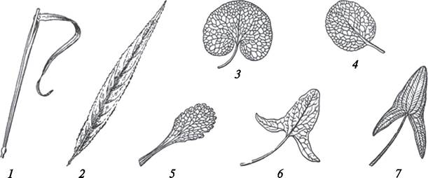
Simple leaves with entire blades can be further categorized based on blade shape, margin characteristics, and the method of attachment to the stem. Determining the shape involves analyzing The ratio of length to width, as well as the structure of the base and apex. The Morphological diversity of simple leaves with entire blades is immense. Their descriptive names reflect these specific features or their resemblance to various objects (ovate, sagittate, hastate, cordate, etc.) or geometric figures (rhombic, oval, orbicular, etc.) (Fig. 108).
Fig. 108. Simple leaves with entire blades: 1—linear leaf of grasses; 2—lanceolate (willow — Salix acutifolia); 3—reniform (European wild ginger — Asarum europaeum); 4—orbicular (shinleaf — Pyrola rotundifolia); 5—spathulate (creeping bugleweed — Ajuga reptans); 6—hastate (field bindweed — Convolvulus arvensis); 7—sagittate (common arrowhead — Sagittaria sagittifolia)
The leaf margin may be entire (greater plantain — Plantago major, spearwort — Ranunculus flammula) or toothed/lobed. Depending on The Nature of the marginal incisions, several types are distinguished: dentate (sharp Teeth and sinuses, with teeth perpendicular to the margin — Tatar saltbush — Atriplex tatarica), serrate (sharp teeth pointing forward toward the apex — stinging nettle — Urtica dioica), crenate (rounded teeth and sharp sinuses — ground ivy — Glechoma hederacea), and sinuate (blunt teeth and rounded sinuses — aspen — Populus tremula). Some plants develop more complexly incised margins (Fig. 109).
Fig. 109. Leaf margin shapes: 1—sinuate (aspen — Populus tremula); 2—dentate (halberd-leaf orache — Atriplex sagittata); 3—entire (lesser wintergreen — Pyrola minor); 4—doubly serrate (silver birch — Betula pendula); 5—doubly dentate (common hazel — Corylus avellana); 6—crenate (medicinal betony — Betonica officinalis); 7—serrate (stinging nettle — Urtica dioica)

Based on their attachment to the stem, simple leaves are divided into petiolate and sessile. Petioles can be short or long, resulting in short-petiolate (brooklime — Veronica beccabunga) and long-petiolate (white poplar — Populus alba) leaves, respectively.
Sessile leaves lack petioles and attach directly to the stem by their base. Depending on the shape and developmental Features of the base, sessile leaves may be semi-amplexicaul (opium poppy — Papaver somniferum), amplexicaul (chicory — Cichorium intybus), perfoliate (wild teasel — Dipsacus lanata), decurrent (great mullein — Verbascum thapsus), or sheathing (grasses, sedges, marsh orchids) (Fig. 110).
Fig. 110. Modes of leaf attachment to the stem: A—long-petiolate leaf of zonal geranium (Pelargonium zonale); B—short-petiolate leaf of oval-leaved privet (Ligustrum ovalifolium); C—sessile leaf of Virginia spiderwort (Tradescantia virginica); D—perfoliate leaf of golden bupleurum (Bupleurum aureum);
E—connate-perfoliate leaves of fragrant honeysuckle (Lonicera fragrantissima); F—decurrent leaf of Arabian thistle (Carduus arabicus)

Simple leaves with dissected blades are classified According to the depth of dissection and the venation pattern, which dictates the arrangement of the segments. Accordingly, botanists distinguish pinnatifid and palmatifid, pinnatipartid and palmatipartid, and pinnatisect and palmatisect leaves (Fig. 111).
Fig. 111. Simple leaves with dissected blades: 1—palmatifid leaf (Norway maple — Acer platanoides); 2—palmatipartid leaf (motherwort — Leonurus quinquelobatus); 3—palmatisect leaf (creeping buttercup — Ranunculus acris); 4—pinnatifid leaf (pedunculate oak — Quercus robur); 5—pinnatipartid leaf (fall dandelion — Leontodon autumnalis); 6—pinnatisect leaf (tansy — Tanacetum vulgare);
7—lyrate leaf (wood avens — Geum urbanum); runcinate leaf (dandelion — Taraxacum officinale)

In lobed leaves, the blade is dissected up to 1/4 of its width, with lobes positioned either on both sides of the midrib in pinnately veined leaves (common oak — Quercus robur) or radiating from a common point in palmately veined leaves (guelder rose — Viburnum opulus).
In pinnatipartid and palmatipartid leaves, the blade is dissected to about 1/2 of its width or slightly more, with the segments arranged in a manner similar to that described above (fall dandelion — Leontodon autumnalis, wood geranium — Geranium sylvaticum).
Pinnately and palmately dissected leaves possess a leaf blade divided into segments almost down to the midrib (yarrow — Achillea millefolium, tall buttercup — Ranunculus polyanthemos).
Among pinnately lobed and pinnately dissected leaves, lyrate and runcinate leaves are distinguished as special types. Lyrate leaves develop a large, whole terminal lobe or segment at the apex (remote-leaf avens — Geum rivale, yellow cress — Barbarea vulgaris). Runcinate leaves are characterized by the triangular outlines of the dissected blade sections (common dandelion — Taraxacum officinale).
Sometimes the leaf blade is dissected so extensively that it can be quite difficult to determine definitively whether the leaf is simple or compound.
The classification of compound leaves is based on the number of leaflets and their specific arrangement along the leaf rachis. Depending on this arrangement, leaves are subdivided into pinnately compound and palmately compound (Fig. 112).
Fig. 112. Compound leaves: 1—ternately compound (wild strawberry — Fragaria vesca); 2—palmately compound (horse chestnut — Aesculus hippocastanum); 3—even-pinnately compound (spring vetchling — Orobus vernus); 4—even-pinnately compound (tufted vetch — Vicia cracca); 5—odd-pinnately compound (dog rose — Rosa canina); 6—interruptedly pinnately compound (diagram); 7—bipinnately compound (diagram); 8—tripinnately compound (diagram)

Pinnately compound leaves are characterized by a bilateral arrangement of leaflets along the rachis (meadow vetchling — Lathyrus pratensis, bush vetch — Vicia sepium). Pinnately compound leaves may have an even number of leaflets, in which case the common petiole terminates in a tendril (narrow-leaved vetch — Vicia angustifolia, forest pea — Lathyrus sylvestris) or a sharp point (spring vetchling — Lathyrus vernus). Such leaves are termed even-pinnately compound. If the rachis terminates in a leaflet, the compound leaf is called odd-pinnately compound (sweetvetch milkvetch — Astragalus glycyphyllus, Siberian peashrub — Caragana arborescens).
In palmately compound leaves, the leaflets are positioned at the apex of the rachis and radiate outwards (narrow-leaved lupine — Lupinus angustifolius, yellow lupine clover — Trifolium lupinaster). A specific case of palmately compound leaves is the ternately compound leaf, which bears three leaflets on the rachis (wild strawberry — Fragaria vesca, rabbit-FOOT clover — Trifolium arvense, black medick — Medicago lupulina).
The rachis may branch, forming secondary and higher-order rachises. When the rachis branches once, bipinnately (yellow meadow-rue — Thalictrum lucidum) and biternately (bishop's weed — Aegopodium podagraria) compound leaves are formed. Lateral rachises may branch further, resulting in tripinnately (caraway-leaved parsley — Selinum carvifolia) or triternately (wild angelica — Angelica sylvestris) compound leaves.
5.4.6. Three Categories of Leaves and Heterophylly
As early as 1830, the German botanist A. Schimper (1856–1901) noted that leaves located at different levels on a plant vary in shape, structure, and function. This gave rise to the concept of three leaf categories: lower, middle, and upper (Fig. 113).
Fig. 113. Three categories of leaves: A—lily of the valley (Convallaria majalis); B—wintergreen (Pyrola rotundifolia): 1—lower leaves; 2—middle leaves; 3—upper leaves (bracts)

Lower leaves are those whose development has been arrested, effectively representing bud scales. During the early stages of SHOOT development, they perform a protective function. As the shoot matures, lower leaves either drop off, which is typical for woody plants (small-leaved linden — Tilia cordata), or persist, as seen in certain herbaceous plants (lily of the valley — Convallaria majalis, wintergreen — Pyrola rotundifolia).
Middle leaves are typical foliage leaves characteristic of each plant species, performing the functions of photosynthesis, transpiration, and gas exchange.
Upper leaves are situated at the base of flowers or inflorescences and function as bracts. In the early stages of development, bracts protect the developing flowers or inflorescences from adverse conditions. In mature plants, their STRUCTURE AND FUNCTIONS vary. In the lily of the valley, upper leaves appear as small membranous scales located near the flower pedicels and functionally inactive at this stage. In many representatives of the Araceae and Bromeliaceae families, bracts form brightly colored spathes—red (flamingos — Anthurium scherzerianum), pink (urn plant — Billbergia nutans), cream (split-leaf philodendron — Monstera deliciosa), or white (bog arum — Calla palustris, arum lily — Zantedeschia aethiopica)—which, aside from protection, serve to attract pollinating insects.
Sometimes bracts combine the function of attracting pollinators with photosynthesis, as is characteristic of cow-wheat (Melampyrum nemorosum).
Under the Influence of Environmental conditions or as a reflection of evolutionary history, the middle leaves of certain plants may exhibit varying shapes. This phenomenon is termed heterophylly (from the Greek heteros meaning different and phyllon meaning leaf), or diversity of leaf form. Heterophylly may be ecological or phylogenetic in nature.
Ecological heterophylly is induced by differing environmental conditions experienced by middle leaves located at various PARTS OF THE stem. It is especially pronounced in aquatic or semi-aquatic plants (great yellowcress — Sagittaria sagittifolia, water crowfoot — Batrachium aquatile, water parsnip — Sium latifolium), where some leaves develop underwater or on the water's surface, while others grow in the air. Occasionally, leaf dimorphism also occurs in terrestrial plants (Kashubian buttercup — Ranunculus cassubicus, harebell — Campanula rotundifolia, burnet saxifrage — Pimpinella saxifraga), which may be linked to varying light conditions at the base versus the central part of the stem (Fig. 114).
Fig. 114. Ecological heterophylly: A—flowering water crowfoot (Batrachium floribundum); B—broadleaf arrowhead (Sagittaria sagittifolia); C—staghorn fern (Platycerium alcicorne); D—aquarium cabomba (Cabomba aquatica); E—harebell (Campanula rotundifolia): 1—emergent leaves; 2—submerged leaves; 3—floating leaves; 4—dissected sporophylls; 5—entire sterile leaf; 6—basal leaves; 7—stem leaves (w. l. — water level)

Phylogenetic heterophylly manifests as differences in leaf shape depending on the age of the leaf. For instance, in certain eucalyptus species, leaves developing on young shoots are rounded, whereas those on older shoots are falcate (Fig. 115).
Fig. 115. Phylogenetic heterophylly: 1—shoot of white mulberry (Morus alba) with entire (young) and lobed (older) leaves; 2—mealy stringybark (Eucalyptus cinerea): a—young shoot with opposite sessile elliptic leaves; b—old shoot with alternate falcate drooping leaves

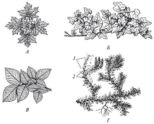
A special type of leaf variation is anisophylly (from the Greek anisos meaning unequal and phyllon meaning leaf). In anisophylly, the geometric similarity of the leaves is preserved, but they differ in size. Anisophylly occurs in certain woody plants (Norway maple — Acer platanoides, horse chestnut — Aesculus hippocastanum, English ivy — Hedera helix) during the formation of leaf mosaics at shoot tips; here, variations in petiole length and blade size prevent leaves from shading one another, thereby creating optimal conditions for photosynthesis. Anisophylly is also found in some lycophytes (species of the genera Selaginella and Diphasiastrum) (Fig. 116). W. Hofmeister believed that anisophylly develops due to unilateral illumination of the leaves. The prominent Russian botanist V. R. Zalensky (1875–1923) shared the same view.
Fig. 116. Anisophylly: A–C — leaf mosaic: A — Norway maple (Acer platanoides); B — English ivy (Hedera helix); C — common hornbeam (Carpinus betulus); D — two-ranked arrangement of leaves of different sizes in spike-moss (Selaginella sp.): 1 — marginal leaves; 2 — central leaves
5.4.7. Internal Structure of the Leaf
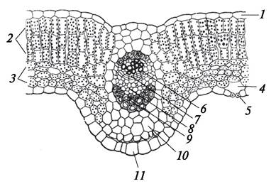
The internal structure of a leaf ensures the performance of its characteristic functions. Regardless of how much the internal structure of leaves may vary among different plants, they consist of four MAIN TYPES OF tissues: epidermis, chlorophyll-bearing parenchyma (chlorenchyma) and its other variants, mechanical tissues, and vascular tissues (Fig. 117).
Fig. 117. Internal structure of a radish leaf (Raphanus sativus): 1 — upper epidermis; 2 — palisade chlorenchyma; 3 — spongy chlorenchyma; 4 — air space; 5 — stoma; 6 — xylem; 7 — cambium; 8 — phloem; 9 — sclerenchyma; 10 — ground parenchyma; 11 — lower epidermis
The epidermis is located on the leaf surface and has a typical structure, although various plant species exhibit specific features (silica deposition in cell walls, presence of a cuticle, covering and glandular hairs, stomatal arrangement, stomatal complex types, etc.).
Based on the distribution of stomates, leaves are classified as epistomatic (from Greek epi — upon, above, and stoma — Mouth, opening), hypostomatic (from Greek hypo — beneath), and amphistomatic (from Greek amphi — on both sides).
Bifacial leaves can be either epistomatic or hypostomatic. Epistomatic leaves are typical of aquatic plants with floating leaves (white water lily — Nymphaea alba, yellow water-lily — Nuphar lutea), where Stomata are located on the upper surface of the leaf. Hypostomatic leaves are usually arranged more or less horizontally. In such leaves, stomata form on the lower surface, which serves as an adaptation to reduce water evaporation from the tissues located beneath the epidermis.
Unifacial leaves are amphistomatic, with stomata distributed across their entire surface. Equifacial leaves are also amphistomatic.
The density of stomata per unit area varies widely, ranging from 40 to 300 stomata per 1 mm². Stomatal frequency depends on environmental factors, primarily humidity and light intensity. Aquatic plants have significantly fewer stomata than terrestrial ones, and sun leaves develop more stomata than shade leaves.
Beneath the epidermis lies the mesophyll — the soft tissue of the leaf — which is represented by various types of parenchyma. The bulk of the mesophyll consists of chlorenchyma (assimilatory tissue or parenchyma). In hypostomatic and epistomatic leaves, the chlorenchyma is differentiated into palisade and spongy layers.
Palisade chlorenchyma cells are tabular in shape and typically arranged in several rows with no intercellular spaces between them. They contain a high concentration of Chloroplasts, ensuring that photosynthesis proceeds intensely. The arrangement of the palisade layer within the leaf depends on the Structural Features of the leaf blade.
In bifacial leaves, the palisade chlorenchyma lies adjacent to the upper epidermis (see Fig. 117). In equifacial (isolateral) leaves, it is found on both sides of the leaf blade (Fig. 118), whereas in unifacial leaves, the palisade tissue wraps around the entire circumference of the leaf.
Fig. 118. Internal structure of isolateral leaves: A — Oxytropis chiliohylla; B — Acantholimon diapensioides: 1 — upper epidermis; 2 — lower epidermis; 3 — palisade chlorenchyma; 4 — central vascular bundle; 5 — stoma; 6 — bundle sheath (mestome); 7 — accessory vascular bundles
Beneath the palisade layer lies the spongy chlorenchyma, where photosynthesis is less active, but gas exchange and transpiration proceed intensively. Large intercellular spaces within the spongy tissue connect with the substomatal air cavities. The differentiation of chlorenchyma into palisade and spongy tissues is related to light conditions. If a leaf is illuminated equally from all sides, this differentiation is absent, and all chlorenchyma cells are uniform (e.g., iris, gladiolus).
The leaf mesophyll contains the vascular system, formed by collateral bundles. This network of vascular bundles not only facilitates the transport of water and dissolved minerals throughout the leaf but also enables the outflow of assimilates produced during photosynthesis. In addition, vascular bundles provide mechanical support, reinforcing the leaf blade since typical strengthening tissues are scarce.
The most common mechanical tissue in the leaf blade is sclerenchyma, represented by fibers and sclereids. Fibers form a bundle sheath, which is particularly characteristic of grasses, or occur as strands above the xylem (iris) and sometimes the phloem (sunflower). Sclereids are typical of coriaceous leaves (rubber plant — Ficus elastica, Japanese camellia — Camellia japonica) and occur either singly or in groups within the mesophyll. Leaf petioles most frequently contain angular (begonia — Begonia) or lacunar (burdock — Arctium) collenchyma.
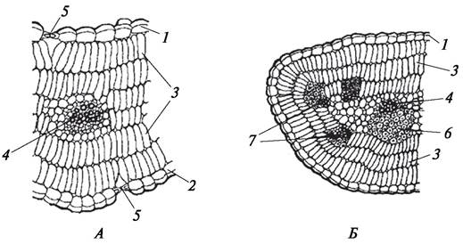
Although all angiosperms share a similar basic set of tissues, the internal anatomy of different taxa exhibits specific features. For instance, instead of a sclerenchymatous sheath, a parenchymatous bundle sheath may develop (pumpkin — Cucurbita pepo). A distinctive type is the Kranz sheath ("wreath-like"). Kranz cells are cube-shaped, living, chlorophyll-containing cells with thickened cell walls that surround or lie adjacent to the vascular bundle. These cells were first described and named by G. Haberlandt in 1884. This physiologically active tissue is characteristic of plants utilizing C4 photosynthesis and has adaptive significance (Fig. 119).
Fig. 119. Internal structure of leaves with Kranz anatomy around vascular bundles: A — Kochia prostrata; B — Atriplex dimorphostegia: 1 — epidermis; 2 — palisade chlorenchyma; 3 — vascular bundles; 4 — chlorophyll-containing Kranz cells; 5 — water-storage parenchyma cells; 6 — sclerenchyma; 7 — calcium oxalate druses

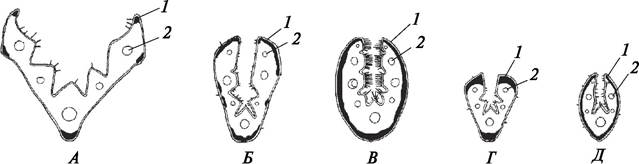
In grasses, specialized bulliform water-storage cells (formerly called motor cells) are interspersed among typical epidermal cells. They facilitate and regulate leaf rolling, a phenomenon observed in certain species during dry weather (Fig. 119, A; 120). Grasses feature a highly developed mechanical tissue system. In fescues, sclerenchyma is frequently found not only around the bundles but also along the margin of the leaf blade. The distribution pattern of sclerenchyma in narrow-leaved fescues is an important diagnostic trait used in species identification (Fig. 121). In plants adapted to waterlogged habitats, aerenchyma is well developed in the leaves, as well as in other plant Organs (bog arum — Calla palustris, floating mannagrass — Glyceria fluitans).
Fig. 120. Internal structure of a barnyard grass leaf (Echinochloa crus-galli): 1 — upper epidermis; 2 — bulliform cells; 3 — parenchymatous bundle sheath; 4 — chlorenchyma; 5 — lower epidermis
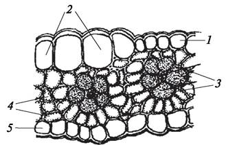
Fig. 121. Arrangement of sclerenchyma in the leaves of certain fescures: A—red fescue (Festuca rubra); B—rough-leaved fescue (Festuca trachyphylla); C—Becker's fescue (Festuca beckeri); D—valian fescue (Festuca valesiaca); E—sheep fescue (Festuca ovina): 1—sclerenchyma; 2—vascular bundles
Regardless of whether they develop in deciduous trees and consequently persist for a single growing season, or form in evergreen plants and function continuously, leaves retain their Primary Anatomical Structure throughout their entire lifespan.
5.4.8. Phyllotaxis
The arrangement of leaves on a stem (phyllotaxis) (from Greek phyllon — leaf and taxis — arrangement, order) is subject to specific regularities. Leaves are arranged along orthostichies (from Greek orthos — straight and stychos — line).
An orthostichy is an imaginary straight line connecting leaves located directly above one another. Leaves are positioned relative to each other at a specific angle—the divergence angle—which is expressed either in degrees or as a fraction of a circumference.
If one starts with a given leaf and successively connects the lower-lying leaves situated on different orthostichies until reaching a leaf located on the same orthostichy as the starting leaf, a spiral line is formed, known as the genetic spiral (Fig. 122).
Fig. 122. Patterns of leaf arrangement on the stem: 1–3 — orthostichies; a–e — genetic spiral (leaves a–d belong to a single leaf cycle)
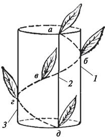
The number of leaves on the genetic spiral minus one leaf constitutes a leaf cycle.
The genetic spiral and the leaf cycle are important indicators that characterize the features of phyllotaxis. Three main types of leaf arrangement are distinguished: alternate (spiral), opposite, and whorled.
Spiral phyllotaxis is the most common type, in which a single leaf is attached at each node. Spiral leaf arrangement can be described mathematically as fractions representing the Fibonacci sequence (1/2, 1/3, 2/5, 3/8, 5/13), which reflect the density of leaf distribution on the stem. To obtain the phyllotactic formulas, the number of turns of the spiral in a single leaf cycle must be divided by the number of leaves in that cycle. The formula is a fraction whose value corresponds to the divergence angle expressed not in degrees, but as a fraction of a circle. If the genetic spiral makes only one revolution and there are only two leaves in the leaf cycle, the phyllotactic formula is 1/2, meaning that the leaves are arranged in two rows. This type of leaf arrangement is characteristic of grasses. In distichous (two-rowed) phyllotaxis, the divergence angle is 180°. If the genetic spiral makes one revolution and there are three leaves in the leaf cycle, the phyllotactic formula is 1/3. In this case, three orthostichies are formed on the stem, and the leaves are arranged in three rows. Tristichous (three-rowed) leaf arrangement is typical of sedges. With a tristichous phyllotaxis, the divergence angle is 120°. Even with A large number of leaves, the divergence angle ensures that the leaves do not shade one another, which is essential for photosynthesis. (If the phyllotactic formula is 2/5, the divergence angle in degrees can be determined by multiplying the full circle of 360° by the number of turns of the genetic spiral and dividing by the number of leaves in the leaf cycle: 360 × 2 ÷ 5 = 144°). Spiral phyllotaxis with the 2/5 formula is particularly common. It is characteristic of various representatives of both dicots (willow, briar, tobacco) and monocots (aloe).
The optimal divergence angle is considered to be 137° 30' 28". An approximately similar divergence angle is characteristic of spruce (Picea) and houseleek (Sempervivum), for which the 5/13 phyllotactic formula has been established.
In opposite phyllotaxis, There are two leaves per node. In each successive node, the direction of leaf arrangement changes, resulting in a decussate (crosswise) pattern. In whorled arrangement, there are three or more leaves per node. Phyllotaxis is an important taxonomic feature; however, in rare cases, the leaf arrangement in different individuals of the same species may vary. For example, in yellow loosestrife (Lysimachia vulgaris), the leaf arrangement is typically whorled, with four leaves per whorl. Nevertheless, some plants have three-leaved whorls. In certain loosestrifes, the leaves are arranged oppositely. The causes of variations in leaf arrangement types in loosestrife remain unclear.
The regularities of phyllotaxis have not been sufficiently studied. It is a complex morphogenic problem that remains to be solved. Apparently, the apical meristem of the stem apex, which forms leaf primordia in a specific sequence, represents a complex biological self-regulating system under Genetic control.
5.4.9. Leaf Fall (Abscission)
In most plants, the lifespan of leaves is short; afterwards, they fall off in woody plants or die off along with the entire plant in herbaceous ones. Plants that annually shed their leaves before the onset of an unfavorable growing period (cold in the temperate zone or dry in the tropics) are called deciduous. In addition to deciduous plants, there are evergreens (lingonberry, heather, spruce, pine). In these plants, leaves persist for several years and are then shed and replaced by new ones. In evergreens, leaf replacement occurs imperceptibly, as some leaves gradually die off while new ones form to replace them. During leaf fall, all of a plant's leaves are shed within a short period.
Leaf shedding—abscission—is of great biological significance. Leaf fall is a normal physiological process primarily associated with leaf Aging. In seasonal climates, leaf shedding has an adaptive character. It protects plants from excessive water loss and dehydration, as well as from Damage caused by heavy snowfalls. Furthermore, Metabolic waste products (such as sulfur, chlorine, and calcium compounds, silica, etc.) are eliminated from the plant along with the falling leaves. Leaf abscission also has major physiological significance. Before leaves fall, assimilation products formed within them through photosynthesis migrate to the overwintering buds, thereby preparing them not only for winter dormancy but also for development in the coming growing season.
Preparation for leaf fall involves physiological and morphological Changes in the leaves. In autumn, leaf color changes from green to red, yellow, or orange. This color change is associated with the disruption of the submicroscopic Structure of Chloroplasts and The breakdown of chlorophyll. Autumn leaf coloration is provided by carotenoids (carotene, xanthophyll), which, alongside chlorophyll, are contained in chloroplasts and are more stable pigments than chlorophyll.
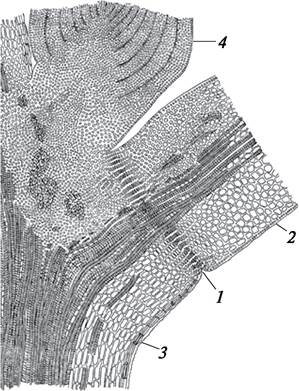
Preparation for leaf fall begins long before the leaves are actually shed. Prior to abscission, an Separation (abscission) layer forms at the base of the leaf (Fig. 123), consisting of small, easily detached parenchyma cells. The parenchyma cells of the abscission layer have thin walls capable of mucilaginous degeneration. These mucilaginous cells easily separate, leaving the leaf hanging solely by its vascular bundles (leaf veins). Under The Influence of wind, rain, and the weight of the leaf itself, the leaf tears away from the stem along the abscission layer. After the leaf falls, a leaf scar remains on the stem—an outline of the petiole base showing scars left by the vascular bundles (sometimes referred to as leaf traces) (Fig. 124). Beneath the abscission layer, a protective cork layer forms at the site of the future leaf scar. Thus, the plant's connection with the external environment is severed, and the processes of transpiration and gas exchange are restricted. The abscission layer is formed exclusively in woody plants.
Fig. 123. Abscission layer formed during leaf fall: 1—abscission layer; 2—fragment of leaf petiole; 3—fragment of plant stem; 4—axillary bud
Fallen leaves serve as reliable cover for subterranean organs (rhizomes, tubers, bulbs) of various plants, protecting them from damage during the winter period. Decaying fallen leaves enrich the soil with nutrients, which helps improve soil structure. Fallen leaves also provide a unique shelter for insects and small vertebrates.
Fig. 124. Leaf scars with leaf trace scars: A—red elderberry (Sambucus racemosa); B—white walnut / butternut (Juglans cinerea); C—horse chestnut (Aesculus hippocastanum): 1—leaf scars; 2—leaf trace scars; 3—axillary buds

Thus, leaf fall also has ecological significance, contributing to the preservation of the biological diversity of flora and fauna.
It is evident that in the course of the historical Evolution of the plant kingdom, The ability to shed leaves simultaneously arose as an adaptation to unfavorable climatic changes.
5.4.10. Metamorphoses of the Leaf and Its Parts
Like the root and stem, the leaf is capable of metamorphism, which is associated with a modification of its typical functions in response to adaptation to habitat conditions. The shoot as a whole and The Leaf as its lateral organ are most susceptible to modifications due to The Diversity of environmental factors affecting them. Often, the metamorphosis of a leaf is linked to the metamorphosis of the stem. In other cases, only the leaf or its individual parts are modified. A good illustration of the metamorphoses of various parts of the leaf is provided by leaf-derived spines (Fig. 125). In barberry (Berberis vulgaris), the entire leaf transforms into a spine. Leaf-derived spines are located at the stem node. In the axil of such spines, there is a bud from which a shortened leafy shoot develops. In very young shoots, leaf spines are absent. In barberry, all Stages of the transition of a leaf into a spine can be observed.
Fig. 125. Leaf-derived spines: A—common barberry (Berberis vulgaris); B—black locust (Robiniapseudoacacia); C—creeping thistle (Cirsium arvense): 1—spine formed from a leaf; 2—stipular spines; 3—spines representing the endings of leaf veins

Stipules can also transform into spines, as, for example, in black locust (Robiniapseudoacacia), Siberian peashrub (Caragana arborescens), and spiny cocklebur (Xanthium spinosum). In some milkvetches, the petioles of pinnately compound leaves undergo secondary thickening after the leaflets fall off, turning into long needle-like spines. The ends of leaf veins can transform into spines, as in thistles (Cirsium), or other small parts of the leaf, as in musk thistles (Carduus) and cotton thistles (Onopordum).
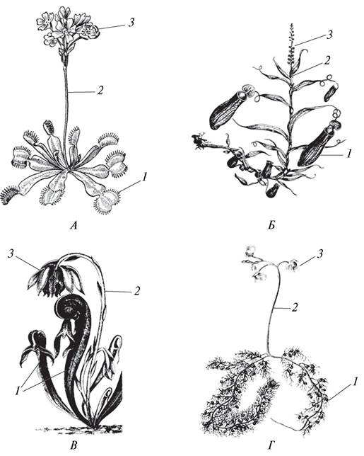
The most interesting metamorphosis is the formation of peculiar trapping organs. Leaves shaped as various trapping structures are formed in plants experiencing a deficiency of mineral nutrients, especially nitrogenous compounds, in their Nutrition. Consequently, these plants have developed A number of adaptations for capturing and digesting small invertebrates that land on their leaves. Such plants are called carnivorous. There are more than 400 such species worldwide (Fig. 126). In Belarus, carnivorous plants are represented by 10 species from four genera (sundew — Drosera, butterwort — Pinguicula, bladderwort — Utricularia, and waterwheel plant — Aldrovanda). The most widespread is the round-leaved sundew (Drosera rotundifolia), which grows almost everywhere in sphagnum bogs. Its leaves are covered along the edges and on the upper surface with glandular hairs topped with red heads that secrete droplets of sticky mucus, to which insects adhere. Substances produced by the plant paralyze the insect. When an insect lands on the leaf, the leaf margin curls inward, covering the prey, which is digested by digestive Enzymes secreted by the plant. Only indigestible chitinous remains are left on the leaf. Digestion lasts for several days, after which the leaf unfolds, and droplets of mucus reappear on the glandular hairs.
Fig. 126. Carnivorous plants: A—Venus flytrap (Dionaea muscipula); B—pitcher plant (Nepenthes sp.); C—California pitcher plant (Darlingtonia californica);
D—greater bladderwort (Utricularia vulgaris): 1—trapping leaves; 2—flowering shoot; 3—flowers
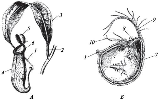
The trapping apparatus of bladderworts (Utricularia vulgaris, U. intermedia) has a more complex structure. In this aquatic plant, alongside dissected photosynthetic leaves, spherical trapping bladders up to 2–5 mm in diameter develop. The entrance opening of the bladder is surrounded by hairs that actively oscillate toward the entrance, followed by a valve that snaps shut as soon as any small aquatic invertebrate gets inside. Digestion of the prey takes place inside the bladder (Fig. 127).
Fig. 127. Trapping organs (metamorphosed leaves): A—pitcher plant (Nepenthes sp.); B—greater bladderwort (Utricularia vulgaris): 1—leaf petiole; 2—sheath-like expanded base of the leaf petiole; 3—phyllode (a leaf-like flattened portion of the leaf petiole); 4—pitcher (metamorphosed leaf blade); 5—"lid", upper part of the leaf blade; 6—rim of the pitcher; 7—trapping bladder; 8—entrance opening of the bladder; 9—structure closing the entrance to the bladder; 10—"tentacles"
In pitcher plants (Sarracenia), which are perennial rhizomatous herbaceous plants native to North America, rosettes are formed at the apex of the rhizome
consisting of ascidia—long (75–100 cm), narrow (5–8 cm in diameter) pitcher-shaped leaves with reddish veins. The inner surface of the leaves is lined with downward-pointing rigid hairs. Secretory fluid accumulates at the bottom of the pitcher leaves, where trapped insects drown and are subsequently digested. The prey is lured by a sweetish nectar produced by the trap leaves. This is a passive carnivorous plant, as it does not snap its traps shut. Presumably for this reason, certain wasp species have adapted to exploit these pitchers to their advantage by laying their eggs inside them in the sugary liquid. Sarracenia plants grow predominantly in swampy forests and sphagnum bogs. Some species of Sarracenia have been introduced into cultivation as "curiosity" houseplants.
A very distinctive Modification of the leaf occurs in species of the genus Nepenthes, whose representatives are distributed in tropical Asia, Madagascar, the Seychelles and Philippine Islands, the Malay Archipelago, and tropical Australia. The genus includes about 70 species, among which are shrubs, subshrubs, and lianas, with some species being epiphytes.
In Nepenthes, alongside ordinary photosynthetic leaves, trapping leaves also develop (see Fig. 127). Their long petioles transform at the base into a leaf-like, wide, flat phyllode (a leaf-like flattened petiole), while the middle section bears a modified leaf blade—the pitcher. In many species, the pitcher is covered by a lid formed by the upper part of the leaf blade. The pitchers are brightly colored: crimson, light green, with red spots and stripes, etc. The margins of the pitcher secrete nectar; its inner walls are very slippery in the upper part, while the lower part, as well as the bottom, is studded with secretory glands. Insects attracted by the nectar land on the rim of the pitcher, slip to the bottom, and fall into the digestive enzyme fluid.
Leaf-like petioles (phyllodes) are also characteristic of Australian phyllodinous acacias (Acacia longifolia, A. armata). In these plants, true pinnately compound leaf blades do not develop, and the function of photosynthesis is performed by the phyllodes. In a hot climate, the formation of phyllodes is an adaptation to reduce transpiration. Furthermore, since phyllodes are oriented with their narrow edge (edge-on) toward the sun, this protects the plant from overheating (Fig. 128).
Fig. 128. Metamorphosis of the leaf petiole in blackwood acacia (Acacia melanoxylon): 1—typical bipinnately compound leaf; 1a—common leaf petiole; 2—Formation of the phyllode; 3—remaining fragment of the compound leaf on the phyllode; 4—phyllode, with the leaf blade of the compound leaf completely reduced

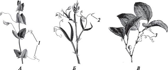
Leaves can be fully or partially transformed into tendrils. For example, in plants of the Fabaceae family, tendrils are usually formed only from the upper parts of a pinnately compound leaf (pea — Pisum, vetchling — Lathyrus). At the same time, in some legume species, the entire leaf blade transforms into a tendril (yellow vetchling — Lathyrus aphaca). Stipules can also be modified into tendrils (smilax — Smilax excelsa) (Fig. 129).
Fig. 129. Leaf-derived tendrils: A—yellow vetchling (Lathyrus aphaca); B—hairy vetchling (Lathyrus hirsutus); C—rough bindweed (Smilax exelsa): 1—tendrils formed by the common leaf petiole (rachis); 2—tendrils formed from the leaflets of a compound leaf; 3—stipular tendrils
In succulent plants, leaves perform a water-storing function (stonecrop — Sedum). The fleshy scales of bulbs also perform a storage function, accumulating primarily CARBOHYDRATES. During the formation of rhizomes, leaves are transformed into scales.
Thus, certain leaf metamorphoses are analogous to stem metamorphoses.
Last update: 07/08/2026
Editorial and Educational Adaptation: This material has been compiled based on the primary/original source text. The project team performed an editorial review, corrected technical inaccuracies, structured sections, and adapted the content for an educational format.
What was processed:
- elimination of formatting defects (OCR errors, structural breaks, corrupted characters);
- editorial organization of content;
- standardization of terminology in accordance with academic sources;
- verification of factual statements against the original source text.
All mentions of the author, publication year, and origin of the primary text have been preserved in accordance with the source.