BOTANY WITH BASICS OF HYDROBOTANY (AQUATIC PLANTS OF UKRAINE) - B.Ye. Yakubenko - 2011
VIII. BASICS OF PLANT MORPHOLOGY AND ANATOMY
Morphology and Anatomy of the Root
Primary Anatomical Structure of the Root
The Introduction/19.html">Primary Structure of the ROOT is formed by the complex of primary Meristems of the root apical meristem (growing cone). It consists of three main components: the epiblema (rhizodermis), the primary cortex, and the central vascular cylinder (stele).
The primary structure can be observed in a young root a few millimeters away from the tip. It is characteristic of all vascular plants; however, in monocots, it persists throughout their entire life, whereas in dicots and gymnosperms, it is restricted only to the root tips and is replaced by Secondary structure as the root grows.
Externally, the root is covered by the epiblema, a primary protective tissue that performs an absorptive function. Its characteristic feature is the presence of numerous fine root hairs, which increase the root's absorption surface area tens of times and supply the entire plant with Water and mineral nutrients. Epiblema Cells are living and thin-walled. Under different conditions, root hairs develop unevenly even on the roots of the same plant—they form more intensively in dry soil than in wet soil. In marsh and aquatic plants, root hairs are either absent or develop only when the plants transition to more xeric growing conditions.
Beneath the epiblema lies the well-developed layer of the primary cortex. The primary cortex is differentiated into three zones: the exodermis, the mesodermis (middle cortex), and the endodermis. Directly beneath the epiblema is the exodermis. It is single- or multi-layered, formed by tightly compressed cells whose walls eventually become suberized. As the root tip pushes deeper into the soil, the epiblema cells rapidly break down, and the exodermis cells take over the protective function.
The mesodermis is the most massive tissue. It is formed by rounded, thin-walled, living parenchyma cells with large intercellular spaces. The loose arrangement of the bulk parenchyma provides optimal aeration. In aquatic and marsh plants, large air cavities develop, some plants form various storage cavities, and mechanical elements occasionally occur. The function of mesodermis cells is the conduction of nutrients from the root hairs through the central cylinder to The Vascular System, as well as the synthesis and storage of reserve nutrients.
The endodermis is the innermost layer of the primary cortex, consisting of a single layer of tightly adjoining cells. It comprises two Cell types: some have horseshoe-shaped Cell wall thickenings (Casparian strips) and eventually die off, while others are thin-walled with dense Cytoplasm—these are passage cells that conduct water and nutrients from the mesodermis to the central cylinder and are located opposite the protoxylem ridges. The endodermis performs a mechanical function and also regulates the radial movement of nutrient solutions from the periphery (the mesodermis) toward the central cylinder, where the conductive elements of the xylem are located.
The entire central part of the root is occupied by the central axial cylinder (stele). It begins with the pericycle, the primary lateral meristem of the root consisting of living, thin-walled cells; in most plants, it is single-layered, though in some it is multi-layered. Inside the pericycle are procambial cells that eventually transform into primary conducting Tissues—primary xylem and primary phloem. The primary xylem conducts water and minerals from the root to other plant Organs, ensuring the upward flow. It appears as a multi-rayed star and consists of protoxylem and metaxylem vessels. The number of xylem rays can vary among plants. Areas of primary phloem are located between the xylem rays. The primary phloem is represented by polygonal, living sieve tubes with companion cells, providing the downward flow of organic assimilates. The conducting tissues are arranged along different radii of the stele, forming a radial closed vascular bundle. Sometimes the central part of the root may be occupied by sclerenchyma or thin-walled parenchyma.
The roots of hygrophytes exhibit certain structural features. In some fully submerged plants, the roots are reduced. Often, they lie in the surface layers of the soil and show weak branching. In certain plant species, thick and sturdy rhizomes develop, which act as anchors and store reserve nutrients.
The root of the slender sedge (Carex acuta L.) (Fig. 59), which exhibits exclusively primary structure, displays specific adaptations to waterlogged conditions. The root is covered by an epiblema characterized by the complete or partial absence of root hairs. Beneath the epiblema lie several layers of the exodermis, followed by a specialized tissue—aerenchyma (air-storage parenchyma). It is distinguished by large intercellular spaces filled with air, and its cells contain starch grains. Below this are one or two layers of typical mesodermis. The endodermis is well-defined due to The Development of Casparian strips. The central cylinder begins with the pericycle, with sclerenchyma cells occasionally wedge-formed between its cells. The vascular bundle is closed and radial, consisting of a triarch primary xylem. Sclerenchyma is situated between the large metaxylem vessels. Small patches of primary phloem are located between the xylem rays. The central part of the root is filled with parenchymal cells.
The root of the common reed (Phragmites communis Trin.) also possesses specific features associated with growth in waterlogged environments. Externally, the root is covered by an epiblema whose cells have thickened, cuticle-covered walls. Further inward lies the subepiblemal parenchyma and a sclerenchymatous ring. The exodermis is absent. The mesodermis is represented by a system of air cavities—a massive aerenchyma. Its cells radially transport aqueous mineral solutions to the central cylinder. The innermost layer of the primary cortex, the endodermis, features weakly expressed Casparian strips. The central cylinder begins with the pericycle, followed by a closed radial vascular bundle.
Class="center">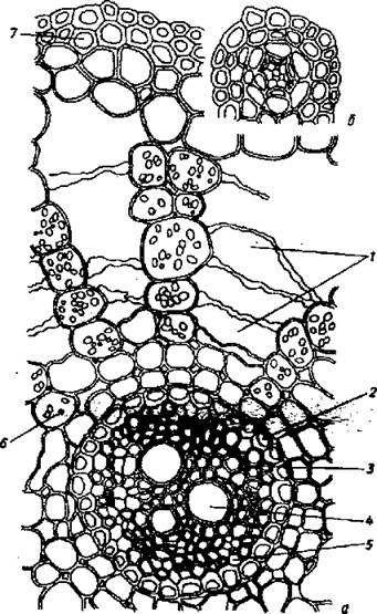
Fig. 59. Cross-section through a thick (a) and thin (b) root of the slender sedge:
1 - well-developed cortical parenchyma (aerenchyma), 2 - endodermis, 3 - phloem,
4 - vascular xylem, 5 - pericycle, 6 - starch grains, 7 — exodermis
The root of lesser duckweed (Lemna minor L.). Duckweed is a small plant growing on the water surface in ponds and other stagnant water bodies. Its body consists of modified stems functioning as leaves. Straight, unbranched rootlets are located on the lower side. Externally, the root is covered by an epiblema, whose large rectangular cells perform the absorptive function. The primary cortex is thin, through which the central axial cylinder is visible. In addition to the cortex and axial cylinder, the root tip features a distinct, prominent root cap. The root cap cells contain METABOLISM/14.html">Chloroplasts, and some contain raphides—needle-like crystals of calcium oxalate. A cavity is located between the root cap and the epiblema. The root cap is formed from epiblema cells.
Last update: 07/08/2026
Editorial and Educational Adaptation: This material has been compiled based on the primary/original source text. The project team performed an editorial review, corrected technical inaccuracies, structured sections, and adapted the content for an educational format.
What was processed:
- elimination of formatting defects (OCR errors, structural breaks, corrupted characters);
- editorial organization of content;
- standardization of terminology in accordance with academic sources;
- verification of factual statements against the original source text.
All mentions of the author, publication year, and origin of the primary text have been preserved in accordance with the source.