BIOLOGY Volume 2 - A Guide to General Biology - 2004
19. HOMEOSTASIS
19.6. The Liver
19.6.2. Functions of the Liver
It is estimated that the Liver performs several hundred Functions, involving thousands of different Chemical Reactions. A massive volume of Blood continuously flows through it (approximately 20% of the total blood volume). The Liver and Kidneys are the primary Organs responsible for maintaining the chemical constancy of the blood. All nutrients absorbed in the digestive tract pass directly into the liver, where they are either stored or converted into other substances required by the body at any given time.
The Role of Insulin. Sugars (primarily glucose) enter the liver from the digestive tract via the HEPATIC PORTAL VEIN—the only blood vessel in which sugar levels fluctuate significantly. This helps explain the liver's role in carbohydrate metabolism as the organ that maintains blood glucose concentration at around 90 mg% (90 mg per 100 mL), regardless of when or how much we have eaten. A drop in this level (hypoglycemia) is particularly dangerous for the body because certain Tissues, such as the Brain, are unable to store glucose, which serves as a vital respiratory substrate. In the liver, all hexoses (six-carbon sugars), including FRUCTOSE AND GALACTOSE, are converted into glucose, which then undergoes Condensation to form an insoluble storage polymer—Glycogen. The amount of glycogen stored in the liver can reach up to 100 g, although skeletal Muscles may store even larger quantities. The process of converting glucose into glycogen is called Glycogenesis and is stimulated by insulin. This hormone is produced by The Pancreas in response to high blood sugar levels (Section 17.6):
Class="center">
THE ROLE OF Glucagon. By breaking down into glucose, glycogen prevents blood glucose levels from falling below 60 mg%. This breakdown process is known as Glycogenolysis and involves the activation of the enzyme phosphorylase by the hormone glucagon. Glucagon is also produced by the pancreas and released in response to low blood sugar levels (Section 17.6.6). In moments of danger, stress, or cold exposure, phosphorylase is additionally activated by adrenaline, released by The adrenal medulla, and noradrenaline, released by both the adrenal medulla and sympathetic nerve endings (Section 17.6.5):

LACTIC ACID AS A PRODUCT OF Anaerobic Respiration. In muscles, glycogen cannot be converted directly into glucose via the pathway described above, because muscles do not release this sugar into the bloodstream to supply other organs, unlike the liver. Glycogen is broken down in muscles only when glucose is needed for their own respiratory demands. In this case, glycogen is converted into glucose-phosphate, which is then broken down to pyruvic acid (Glycolysis), used to synthesize ATP through aerobic respiration or anaerobic glycolysis. Lactic acid produced in skeletal muscles during glycolysis can subsequently be converted into glucose, which is then transported to the liver and converted into glycogen:

Gluconeogenesis. When hepatic glycogen reserves are depleted due to glucose utilization, glucose can be synthesized from any non-carbohydrate precursor. This process is called gluconeogenesis and occurs when liver glycogen stores are exhausted. Low blood glucose levels (hypoglycemia) stimulate the sympathetic Nervous system to trigger the release of adrenaline, which, as noted above, helps meet the body's immediate demand for glucose. Low blood glucose also stimulates the Hypothalamus to release corticotropin-releasing hormone (Section 17.6.5), which induces the anterior Pituitary Gland to secrete adrenocorticotropic hormone (ACTH). Under The Influence of ACTH, the synthesis and release of glucocorticoid Hormones (primarily cortisol, also known as hydrocortisone) are enhanced. These hormones stimulate the mobilization of Amino Acids, glycerol, and Fatty acids from tissues into the bloodstream, as well as the synthesis in the liver of Enzymes that catalyze The conversion of Amino Acids and glycerol into glucose—effectively carrying out gluconeogenesis. Fatty acids are broken down to yield acetyl-coenzyme A and are subsequently oxidized in the Krebs cycle.
FAT FORMATION. CARBOHYDRATES that cannot be utilized by the body or stored as glycogen are converted into fats and stored in this form, predominantly subcutaneously.
An Overview of carbohydrate metabolism involving the liver, muscles, and other tissues is shown in Fig. 19.22.
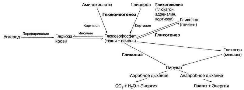
Fig. 19.22. General scheme of carbohydrate metabolism.
The liver plays a crucial role in protein metabolism, which includes:
1) deamination;
2) urea formation;
3) Transamination;
4) plasma Protein Synthesis.
DEAMINATION. The body is unable to store amino acids, even when dietary intake is abundant; therefore, any amino acids that cannot be immediately utilized in protein synthesis or gluconeogenesis are deaminated in the liver. This process is described in Section 20.4.
TRANSAMINATION. This is the process of synthesizing amino acids through the enzymatic transfer of an amino group from an amino acid to a oxo acid, resulting in The formation of a new Amino Acid and a new oxo acid. The general reaction is represented as follows:

For instance, glutamic acid (an amino acid) can be produced through the following transamination reaction:

Transamination is one of the pathways for synthesizing amino acids that are deficient in the diet, meaning it serves as yet another homeostatic function of the liver. However, Essential Amino Acids (sec. 8.7.8) cannot be synthesized in the liver via transamination and must be obtained from food.
PLASMA PROTEIN SYNTHESIS. Proteins are vital components of plasma, and the majority of these proteins are synthesized from amino acids in the liver.
1. The predominant plasma protein is albumin, which has a normal serum concentration of about 4 g per 100 ml (4 g%). It plays a crucial role in maintaining the plasma osmotic potential, which balances the hydrostatic pressure in Blood Vessels. Due to the mutual antagonism of these two factors, fluid balance between the blood vessels and the extracellular space is maintained (sec. 14.6). In addition, albumin acts as a transport vehicle in the Circulatory system, serving as a carrier for calcium, Bile components, various salts, and certain Steroid Hormones, for example.
2. Globulins are large-molecule proteins with a blood concentration of about 3.4 g%. α- and β-Globulins bind and transport hormones (including thyroxine and insulin), Cholesterol, Lipids, iron, and Vitamins B12, A, D, and K. Gamma-globulins are Antibodies and are produced not by the liver, but by lymphocytes and other Cells of The Immune System. They participate in the Immune Response (sec. 14.9). Other important Plasma Proteins include blood clotting factors, such as prothrombin and fibrinogen, the functions of which are described in sec. 14.8.5.
Under normal conditions, the liver does not store lipids, but merely participates in their metabolism and transport. In this regard, hepatocytes perform the following functions:
1) convert excess carbohydrates into fat;
2) extract and break down cholesterol from the blood, or synthesize it when necessary;
3) hydrolyze fats into glycerol and fatty acids during glucose deficiency, which are then used in cellular respiration. Fatty acids are broken down into acetyl groups that bind with coenzyme A to form acetyl-CoA. In this form, they enter the Krebs cycle for further oxidation (sec. 9.3.5). Furthermore, fatty acids can be converted into other substances and "exported" from the liver. Glycerol, as mentioned above, is used to produce glucose (gluconeogenesis).
Vitamin Storage
The liver primarily stores Fat-soluble vitamins—A, D, E, and K. The liver of certain fish, particularly cod and halibut, is especially rich in vitamins A and D (which is why fish oil is beneficial). Vitamin K is essential for The Biosynthesis of blood clotting factors.
The liver also stores certain Water-Soluble Vitamins, particularly B-complex vitamins (nicotinic acid, folic acid, and vitamin B12), as well as Vitamin C. Vitamin B12 and folic acid are required by the Bone Marrow to produce red Blood Cells, and a deficiency of these substances leads to various types of anemia.
Mineral Storage
Along with iron and potassium (see "Hemoglobin Breakdown" below), the liver stores elements required by the body in trace amounts. These are known as Trace Elements, such as copper, zinc, cobalt, and molybdenum. Approximately one thousandth of the dry mass of human liver tissue consists of iron. The bulk of this reserve is held temporarily; iron is released when old red blood cells are destroyed in the liver and is stored until needed for The production of new red blood cells in the bone marrow.
Blood Reservoir Function
Blood vessels draining the Spleen and intestines converge to form the hepatic portal vein. Together with the hepatic artery, it supplies a massive volume of blood to the organ, acting as a blood reservoir. However, the blood does not stagnate here, but flows slowly yet continuously through numerous sinusoids. Impulses from sympathetic Neurons and adrenaline secreted by the adrenal medulla cause many hepatic blood vessels to constrict, releasing more blood into the general Circulation. If the total blood volume increases (e.g., during a transfusion), the hepatic and other Veins dilate to accommodate the excess blood.
Erythrocyte Production
In the fetus, red blood cells are produced in the liver, but this function is gradually taken over by the bone marrow (sec. 14.3.2). Once this process is complete, the liver assumes the exact opposite role, becoming one of the sites where red blood cells and hemoglobin are broken down.
Hemoglobin Breakdown
The lifespan of a human red blood Cell is approximately 120 days. Toward the end of this period, they age and are subsequently destroyed by phagocytic macrophages in the liver, spleen, and bone marrow. The hemoglobin they contain is released into the Blood Plasma. Specialized macrophages extract it from the plasma and deliver it to the liver, spleen, and Lymph Nodes. The macrophages of the liver are called Kupffer cells. Inside the macrophages, hemoglobin is broken down into heme and globin. Globin is the protein portion of the hemoglobin molecule and is subsequently hydrolyzed into amino acids, which are utilized in various ways depending on the body's needs. Iron is split off from the heme, leaving behind a green pigment called biliverdin, which is then converted into a yellow pigment—bilirubin, a component of bile. The accumulation of bilirubin in the blood leads to a characteristic symptom of certain liver disorders known as jaundice, i.e., the yellowish discoloration of the Skin and the whites of the eyes.
Iron forms a complex with the protein transferrin in the blood plasma and is transported through the body in this form, to be reused by bone marrow cells for hemoglobin synthesis. Excess iron is stored in hepatocytes in the form of the complex protein ferritin.
Bile Formation
Bile is a viscous, greenish-yellow fluid secreted by hepatocytes and stored in a concentrated form in the Gallbladder. A human produces 500–1000 mL of bile daily. It consists of approximately 98% water, 0.8% bile salts, 0.2% bile pigments, 0.7% inorganic salts, and 0.6% cholesterol.
From the liver, bile enters the duodenum, where it plays a key role in the Digestion AND ABSORPTION of fats. Furthermore, it serves as a pathway for The excretion of Metabolic waste products from the body (specifically, bile pigments). The release of bile into the digestive tract (causing the gallbladder to contract) is triggered by cholecystokinin—also known as pancreozymin—a hormone produced by the intestinal mucosa (sec. 8.4.3).
Bile salts are derivatives of the steroid cholesterol, which is synthesized by hepatocytes. The most common of these salts are sodium glycocholate and sodium taurocholate. They are secreted along with cholesterol and Phospholipids in the form of large spherical particles called micelles. Bile salt molecules behave like detergents, featuring a hydrophilic end and a hydrophobic end. Cholesterol and phospholipids hold the polar bile salt molecules together so that all their hydrophobic ends point in the same direction. In the intestine, these hydrophobic ends attach to dietary lipid droplets, while the hydrophilic ends bind to water. This reduces the surface tension of the lipid droplets, causing them to break down into a fine emulsion. As a result, the surface area available for interaction with lipase—the enzyme that breaks down lipids into glycerol and fatty acids for intestinal absorption—is dramatically increased. Thus, bile salts effectively activate lipase, although their action is purely physical. If bile lacks sufficient bile salts, the concentration of cholesterol rises, which can cause it to precipitate on the walls of the bile ducts or gallbladder, forming so-called gallstones. These stones can block the bile duct, leading to highly painful symptoms.
Bile pigments serve no physiological function; they are simply waste products slated for elimination from the body.
Cholesterol Production
Cholesterol produced in the liver acts as a precursor for the Synthesis of Other Steroids. The primary source of cholesterol is diet. For instance, many dairy products are rich not only in cholesterol but also in fatty acids from which it can be synthesized. Thyroxine enhances both hepatic cholesterol synthesis and its excretion via bile. Excess blood cholesterol can lead to its deposition in arterial walls, reducing their elasticity (arteriosclerosis) and narrowing their lumen (atherosclerosis). The resulting atherosclerotic plaques damage the endothelium, triggering intravascular blood clotting, or thrombosis. Blood clots (thrombi) can obstruct blood vessels (thromboembolism), disrupting tissue Blood supply and causing tissue death (necrosis). Thromboembolism is particularly dangerous in the coronary Arteries of the Heart, leading to myocardial infarction, and in the cerebral arteries, causing a stroke. Despite these risks, a certain amount of dietary cholesterol is essential for the reasons outlined at the beginning of this section.
Hormone Breakdown
Almost all hormones are broken down in the liver to some extent. Testosterone and aldosterone are degraded fairly rapidly, whereas insulin, glucagon, digestive and Adrenal hormones, Female Sex Hormones, vasopressin (ADH), and thyroxine are broken down more slowly. By removing these BIOLOGICALLY ACTIVE SUBSTANCES from the blood, the liver neutralizes their effects, essentially acting as a homeostatic antagonist to the Endocrine System.
Detoxification
Detoxification refers to the inactivation of toxins (poisons). Even common metabolites and medications can become toxic if their concentration in the blood exceeds normal levels for any reason. Detoxification is one of the many homeostatic Functions of the liver that maintain the chemical constancy of the blood. Bacteria and other pathogens are cleared from the blood in the sinusoids by Kupffer cells, while the toxins they release are neutralized in hepatocytes through various biochemical reactions: oxidation, reduction, methylation (attachment of a –CH3 group), or condensation, which involves joining with another organic or inorganic molecule. Following detoxification, these substances are excreted from the body by the kidneys as harmless products. The most toxic substance found in the blood is ammonia, the metabolism of which is discussed in sec. 20.4.
Substances such as alcohol and nicotine are also detoxified in the liver. Prolonged consumption of large amounts of alcoholic beverages can cause liver damage, such as cirrhosis. Alcohol is oxidized in the liver by the enzyme Alcohol dehydrogenase.
Certain metabolic activities in the liver can pose potential hazards to the body. For example, growing evidence suggests that certain food additives can be converted by the liver into toxic or carcinogenic substances. Even the popular pain reliever paracetamol (acetaminophen), when consumed in excess, is converted into a substance that interferes with enzyme systems and can damage the liver and other tissues.
Heat Production
It is widely believed that the High Metabolic Rate of the liver makes it one of the body's Main sources of heat; however, recent evidence suggests this notion is likely incorrect. Many metabolic processes in the liver are endothermic, meaning they absorb rather than release energy. During severe body cooling, the hypothalamus can ramp up exothermic processes in the liver by stimulating the release of adrenaline and thyroxine. Yet, at a "normal" Temperature, the liver can be considered "thermally neutral": its temperature is only 1–2 °C higher than that of other Internal Organs.
The overall functions of the liver are summarized schematically in Fig. 19.23.

Fig. 19.23. General Overview of liver functions.
Last update: 06/08/2026
Editorial and Educational Adaptation: This material has been compiled based on the primary/original source text. The project team performed an editorial review, corrected technical inaccuracies, structured sections, and adapted the content for an educational format.
What was processed:
- elimination of formatting defects (OCR errors, structural breaks, corrupted characters);
- editorial organization of content;
- standardization of terminology in accordance with academic sources;
- verification of factual statements against the original source text.
All mentions of the author, publication year, and origin of the primary text have been preserved in accordance with the source.