BIOLOGY Volume 2 - A Guide to General Biology - 2004
19. HOMEOSTASIS
19.6. The Liver
The Liver is the largest internal organ in the body, accounting for 3–5% of its total mass. It is located directly beneath the Diaphragm in the Abdominal cavity. Its primary function is to maintain Homeostasis, and it hosts numerous metabolic processes essential for keeping The chemical composition of the Blood constant. Many of its Functions are related to Processing substances absorbed from the digestive tract. Due to its extensive vascularization, the liver can store a large volume of blood (acting as a blood reservoir), thereby regulating circulating blood volume (Fig. 19.19). Despite the vast array of metabolic functions performed by the liver, its histological Structure is relatively simple and uniform.
Class="center">
Fig. 19.19. A. Diagram of the liver's Blood supply and its connection to the digestive tract. B. The same shown in a more schematic form. ПА — hepatic artery; ПВ — hepatic vein; ВВП — HEPATIC PORTAL VEIN.
19.6.1. Structure of the Liver
The liver consists of several lobes, the shape and size of which vary depending on The amount of blood contained within them. Externally, the liver is enclosed by a two-layered envelope: the outer layer is formed by the smooth Peritoneum, and the inner layer by a fibrous capsule known as Glisson's capsule.
Liver Cells are called hepatocytes. Aside from these, the liver contains only nerve elements and cells associated with Blood and Lymphatic vessels. Hepatocytes feature large nuclei, a well-developed Golgi apparatus, numerous Cell/35.html">Mitochondria and Lysosomes, as well as abundant Glycogen granules and lipid droplets. The hepatocytes fit closely together, and at sites of contact with blood capillaries, they form microvilli through which substance exchange occurs between the hepatocytes and the blood.
The structure of the liver is most easily studied using the pig's liver, because the functional units of this organ—the lobules—are more clearly defined here than in other mammals. A liver lobule resembles a prism with a cross-section of about 1 mm (Fig. 19.20). The diagram of its structure is shown in Fig. 19.21. Branches of the hepatic artery, hepatic portal vein, and Bile duct (interlobular ducts) run between the lobules. The portal vein delivers nutrients absorbed in the digestive tract to the organ, while the hepatic artery supplies oxygen.
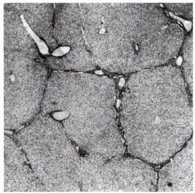
Fig. 19.20. Cross-section of a pig's liver. A branch of the hepatic vein (central vein) runs through the center of each lobule. Between the lobules (at the corners) are interlobular bile ducts, along with branches of the hepatic artery and hepatic portal vein. The lobules consist of rows of glandular cells—hepatocytes—separated by blood sinusoids and branches of the bile canaliculi.

Fig. 19.21. A. Diagram of a cross-section through a liver lobule. B. Simplified diagram of a portion of a liver lobule. The arrows indicate the direction of Blood flow through the sinusoids and bile flow through the bile canaliculi.
At the center of each lobule runs a branch of the hepatic vein, called the central vein. It connects to the hepatic artery and portal vein not through capillary networks, but via functionally equivalent expanded channels known as sinusoids, which are lined with thin endothelial cells containing pores up to 10 nm in diameter. Adjacent sinusoids are separated from one another by so-called hepatic trabeculae (or cords) formed by at least two rows of hepatocytes. These trabeculae radiate outwards from the center of the lobule like the spokes of a wheel. Blood from the periphery of the lobule (from the artery and portal vein) slowly flows through the sinusoids toward its center (the hepatic vein), washing over the hepatocytes and exchanging substances with them.
Within the hepatic trabecula, between the rows of hepatocytes, lies a bile canaliculus (or capillary) into which the bile produced by the hepatocytes is secreted. The bile flows toward the periphery of the lobule (i.e., in the opposite direction to the blood in the sinusoids), where it empties into the interlobular duct. These ducts unite with one another, ultimately forming the common bile duct that transports bile out of the liver.
The liver contains another type of cell—Kupffer cells (stellate reticuloendothelial cells). Kupffer cells are anchored to the walls of the sinusoids by cytoplasmic extensions. As is now understood, these cells are macrophages (section 14.3.2). They are capable of phagocytosis and play a role in breaking down Aging red Blood Cells and destroying pathogenic organisms.
Last update: 06/08/2026
Editorial and Educational Adaptation: This material has been compiled based on the primary/original source text. The project team performed an editorial review, corrected technical inaccuracies, structured sections, and adapted the content for an educational format.
What was processed:
- elimination of formatting defects (OCR errors, structural breaks, corrupted characters);
- editorial organization of content;
- standardization of terminology in accordance with academic sources;
- verification of factual statements against the original source text.
All mentions of the author, publication year, and origin of the primary text have been preserved in accordance with the source.