LEHNINGER PRINCIPLES OF BIOCHEMISTRY - VOL 3. INFORMATION PATHWAYS - 2017
PART III. INFORMATION PATHWAYS
26. RNA METABOLISM
26.2. RNA Processing
Many bacterial RNA molecules and virtually all eukaryotic RNA molecules undergo some degree of Processing following synthesis. In RNA METABOLISM, some of the most fascinating transformations occur precisely during this post-synthetic processing. Surprisingly, certain Enzymes that catalyze these reactions are made of RNA rather than protein. The discovery of such catalytic RNAs, called ribozymes, revolutionized our understanding of RNA function and the origins of life.
A newly synthesized RNA molecule is called a primary transcript. Primary transcripts of eukaryotic mRNAs and bacterial and eukaryotic tRNAs probably undergo the most extensive processing. RNA molecules with specialized Functions also undergo processing.
Eukaryotic mRNA primary transcripts typically contain a single Gene, but the polypeptide-coding sequences may be interrupted by other segments. These noncoding regions that interrupt the coding sequences are called introns, and the coding regions are called exons (for introns and exons in DNA, see Chapter 24). During splicing, introns are removed from the primary transcript, and the exons are joined to form a continuous sequence that corresponds to the functional polypeptide. In addition, the ends of eukaryotic mRNA are modified. A modified residue, known as the 5' cap, is attached to the 5' end. The 3' end is cleaved, and a sequence of 80 to 250 A residues is added in its place, forming a poly(A) tail. Sometimes, the complex protein machineries involved in each of these three mRNA Processing reactions do not act independently; instead, they associate with one another and with the phosphorylated CTD sequence of Pol II, with each complex influencing the Functions of the others. Other Proteins involved in mRNA transport to the Cytoplasm bind to the mRNA while still in The Nucleus, coupling transcript processing with its transport. In essence, a synthesized eukaryotic mRNA exists as a complex, multicomponent assembly consisting of dozens of proteins. The composition of this complex changes throughout primary transcript processing, cytoplasmic transport, and mRNA release on the ribosome for Translation. These Structure/178.html">Protein Complexes modulate mRNA activity and trafficking. These processes are illustrated in Figure 26-12 and described in more detail below.
Class="center">Figure 26-12. Formation of the primary transcript and its processing during mRNA maturation in Eukaryotic Cells. Before Synthesis of the primary transcript is complete, a cap appears at the 5' end (shown in red). The noncoding sequence (intron) located beyond the last exon is highlighted in orange. Splicing can occur either before or after the Cleavage and polyadenylation steps. All processes depicted here take place in the nucleus.

Prokaryotic and eukaryotic tRNA primary transcripts lose terminal sequences through processing (cleavage) and sometimes introns (splicing). Many bases and sugars in tRNAs are modified; mature tRNA contains many unusual bases not found in other Nucleic Acids (see Figure 26-23). Many specialized RNAs also undergo complex processing, often involving the removal of fragments from one or both ends of the sequence.
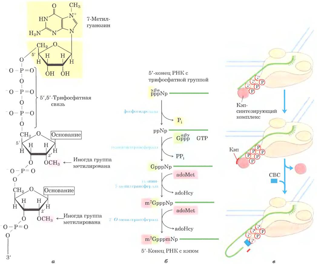
Figure 26-13. The 5' cap on mRNA. (a) In nearly all eukaryotic mRNA molecules, a 7-methylguanosine (m7G) residue is attached to the 5' end via an unusual 5',5'-triphosphate linkage. Methyl groups are also frequently found at the 2' position of the First and Second NUCLEOTIDES (shown in pink). Yeast RNA molecules lack 2'-methyl groups. In the second nucleotide, a 2'-methyl group is generally found only in vertebrate RNA. (b) The formation of the 5' cap occurs in 4 to 5 distinct steps (adoHcy, S-adenosylhomocysteine). (c) Cap synthesis is carried out by enzymes associated with the C-terminal domain (CTD) of polymerase II. The cap is linked to the CTD via a cap-binding complex (CBC).
Thus, all RNA molecules are subject to complete, regulated degradation. The turnover rate of RNA molecules determines their steady-state levels within The Cell and the speed with which cells can shut down the expression of a gene whose product is no longer needed. For example, during The Development of Multicellular Organisms, certain proteins are expressed only at a specific developmental stage, and the mRNA molecules encoding these proteins are synthesized and degraded at precisely timed intervals.
A cap is added to the 5' end of eukaryotic mRNA
In eukaryotes, most mRNA molecules possess a 5' cap—a 7-methylguanosine residue linked to the 5'-terminal residue of the mRNA via an unusual 5',5'-triphosphate bridge (Figure 26-13). The function of the 5' cap is to protect the mRNA from ribonucleases. In addition, the cap binds to a specific cap-binding protein complex and participates in the interaction of mRNA with the ribosome to initiate translation (Chapter 27).
The 5' cap is formed by the Condensation of a GTP molecule with the triphosphate at the 5' end of the transcript. The guanine is then methylated at the N-7 position; additionally, the 2'-hydroxyl groups of the first two nucleotides adjacent to the cap are frequently methylated (Figure 26-13a). S-adenosylmethionine provides the methyl groups. These reactions take place very early in Transcription, after The addition of the first 20 to 30 nucleotides of the transcript. Until the cap is synthesized, all three copying enzymes—and through them, the 5' end of the transcript itself—remain associated with the C-terminal domain of RNA polymerase II. The capped 5' end is subsequently released from the enzymes and binds to the cap-binding complex (Figure 26-13c).
Both introns and exons are transcribed from DNA into RNA
In Bacteria, a polypeptide chain is typically collinear with its encoding DNA sequence, which forms a continuous segment of the DNA template required for the accurate reproduction of the polypeptide. However, the notion that all genes are continuous was overturned as early as 1977, when Phillip Sharp and Richard Roberts independently established that Many eukaryotic genes have a discontinuous structure containing noncoding sequences (introns).
The vast majority of vertebrate genes contain introns; histone genes are among the few exceptions. The situation varies in other eukaryotes. Many genes in the yeast Saccharomyces cerevisiae lack introns, but introns are quite common in other yeast species. Introns have also been discovered in several bacterial and archaeal genes. Introns in DNA are transcribed by RNA polymerases along with the rest of the gene. Subsequently, introns are excised from the primary transcript during splicing, and the exons are joined to form the mature, functional RNA. In eukaryotic mRNAs, most exons are no longer than 1,000 nucleotides, with many spanning only 100 to 200 nucleotides; these exons encode segments of 30 to 60 Amino Acids within a longer polypeptide. Introns range in size from 50 to 20,000 nucleotides. Genes of higher eukaryotes, including humans, typically contain far more DNA within introns than within exons. Many genes contain introns, and some harbor dozens of them.
RNA catalyzes intron splicing
There are four groups of introns. Group I and Group II introns differ in the details of their splicing mechanism, but they share one remarkable property: they undergo self-splicing, meaning they splice in the absence of protein enzymes. Group I introns are present in certain nuclear, mitochondrial, and chloroplast genes encoding rRNA, mRNA, and tRNA. Group II introns are typically found in Mitochondrial and Chloroplast mRNA primary transcripts of Fungi, Algae, and plants. Group I and Group II introns have also been found in some bacteria. These introns do not require high-energy Cofactors (such as ATP) for splicing. The splicing mechanism in groups I and II involves two transesterification steps (Figure 26-14), during which 2'- or 3'-hydroxyl groups of ribose perform a nucleophilic attack on a phosphorus atom, replacing an old phosphodiester bond with a new one while maintaining Energy balance. These reactions closely resemble the DNA breakage and reunion Reactions Catalyzed by topoisomerases (see Figure 24-21) and site-specific recombinases (see Figure 25-40).
Figure 26-14. Transesterification reaction. The first step of Group I intron splicing. In this example, the 3'-OH group of a guanosine molecule acts as a nucleophile, attacking the phosphodiester bond between the U and A residues at the exon-intron junction in the mRNA molecule (see Figure 26-15).

The splicing reaction of Group I introns proceeds with the participation of a guanosine nucleoside or nucleotide cofactor, which is not used as an energy source: the 3'-hydroxyl group of guanosine acts as a nucleophile in the first step of splicing. The 3'-hydroxyl group of guanosine forms a standard 3',5'-phosphodiester bond with the 5' end of the intron (Figure 26-15). Next, the 3'-hydroxyl group of the exon—which is displaced at this stage—acts as a nucleophile in a similar reaction at the 3' end of the intron. As a result, the intron is precisely excised, and the exon ends are joined.
Figure 26-15. Mechanism of Group I intron splicing. The nucleophile in the first step can be guanosine, GMP, GDP, or GTP. The excised intron is completely degraded.

Group II intron splicing follows the same general pathway, except that the nucleophile in the first step is the 2'-hydroxyl group of an internal A residue within the intron (Fig. 26-16). This reaction proceeds through a branched, lariat-shaped intermediate.
Fig. 26-16. Mechanism of group II intron splicing. The chemical principles are identical to those of group II self-splicing, with the exception of the nucleophile involved in the initial step and the formation of a lariat intermediate, in which one of the branches contains a 2', 5'-phosphodiester bond.

Self-splicing introns were first discovered by Thomas Cech and his colleagues in 1982 while investigating the splicing mechanism of a group II intron in the rRNA of the ciliated protozoan Tetrahymena thermophila. The researchers transcribed intron-containing Tetrahymena DNA in vitro using purified bacterial RNA polymerase. Remarkably, RNA splicing occurred with high precision in the absence of any Tetrahymena proteins. The discovery of RNA molecules with catalytic activity marked a major milestone in our understanding of biological systems.

Most introns do not undergo autosplicing and are not designated by group numbers. The third and largest group of introns is found in the primary transcripts of nuclear mRNA. They are called spliceosomal introns because their removal takes place within and is catalyzed by a large protein complex called the spliceosome. Within the spliceosome, introns are excised by the same mechanism as group II introns (via lariat formation). Spliceosomes consist of specialized RNA-protein complexes known as small nuclear ribonucleoproteins (snRNPs). Each snRNP contains a single eukaryotic RNA molecule 100–200 nucleotides long, termed small nuclear RNA (snRNA). Five snRNAs (U1, U2, U4, U5, and U6) participate in splicing reactions and are typically abundant in eukaryotic nuclei. Proteins and RNAs in snRNPs are highly conserved across all eukaryotes (from yeast to humans).
Spliceosomal introns typically have a GU sequence at the 5' end and an AG sequence at the 3' end. These sequences mark the splice sites. The U1 snRNA molecule contains a sequence complementary to the sequences near the 5' splice site of nuclear mRNA introns (Fig. 26-17a), and the U1 snRNP binds to this region in the primary transcript. The recruitment of U2, U4, U5, and U6 leads to the assembly of the spliceosome (Fig. 26-17c). Through its snRNPs, the spliceosome incorporates five RNA molecules and approximately 50 proteins, making its supramolecular structure almost as complex as that of the ribosome (see Chapter 27). About 50 additional proteins associate with the spliceosome at various stages of splicing; many of these proteins are involved in multiple processes such as splicing, mRNA export to the cytoplasm, translation, and mRNA decay. ATP is required for spliceosome assembly, but the RNA cleavage-ligation reactions themselves appear to be ATP-independent. Some introns in mRNA are removed by a less common spliceosome in which U1 and U2 are replaced by U11 and U12. Spliceosomes containing U1 and U2 remove introns with (5') GU and AG (3') terminal sequences (see Fig. 26-16), whereas spliceosomes containing U11 and
U12 excise rare group introns with (5') AU and AC (3') terminal sequences. Spliceosomes involved in nuclear RNA splicing may have evolved from more ancient group II introns, with snRNAs replacing the catalytic domains of their self-splicing ancestors.
Some Components of the splicing apparatus appear to be linked to the CTD of RNA polymerase II, which represents a fascinating reaction mechanism (Fig. 26-17c). As the first splice site is synthesized, it associates with the CTD 'tail' of the spliceosome. The second synthesized splice site is then captured by this complex, which facilitates the approximation of the intron ends and its excision. Following splicing, the intron remains in the nucleus and is eventually degraded.
Fig. 26-17. Mechanism of splicing in primary mRNA transcripts. a — RNA base-pairing during the formation of spliceosomal complexes. Near the 5' end of U1 snRNA, There is a sequence complementary to the 5' splice site of the intron. Base-pairing of U1 with this region of the primary transcript helps define the 5' splice site during spliceosome assembly (Ψ — pseudouridine; see Fig. 26-24). U2 then binds to the intron region containing the A residue (pink), which carries out the nucleophilic attack in the splicing reaction. The binding of U2 snRNA creates a loop that shifts and helps activate adenylate, whose 2'-OH group can form a lariat-like structure via a 2',5'-phosphodiester bond. b — Spliceosome assembly. First, the U1 and U2 snRNPs associate, followed by the binding of the remaining snRNPs (the U4/U6 and U5 complexes) to form an inactive spliceosome. Following internal rearrangements, this complex is converted into an active spliceosome, from which U1 and U4 are excluded, while U6 associates simultaneously with the 5' splice site and with U2. This is followed by catalytic steps analogous to those of group II intron splicing (see Fig. 26-15). c — Coordination of splicing with transcription provides a mechanism for bringing two splice sites together. For details, see text.

Group IV introns, found in certain tRNAs, differ from group I and II introns in requiring ATP and an endonuclease for their splicing. The splicing endonuclease cleaves the phosphodiester bonds at both ends of the intron, and the two exons are joined via a mechanism reminiscent of the DNA ligase reaction (see Fig. 25-17).
Spliceosomal introns appear to be unique to eukaryotes, whereas introns of other groups are widespread. Genes containing group I and II introns have now been discovered in both bacteria and bacterial Viruses. Bacteriophage T4, for example, contains several genes with group I introns. Introns may be more common in archaea than in bacteria.
The 3' end of mRNA molecules features characteristic structures
In eukaryotes, most mRNA molecules have a sequence of 80 to 250 adenine residues at the 3' end, forming the so-called poly(A) tail (polyadenylation sequence). This tail serves for the binding of one or more specific proteins. The poly(A) tail and its associated proteins likely help protect the mRNA from enzymatic degradation. Many prokaryotic mRNA molecules also possess poly(A) tails, but these tails tend to stimulate mRNA decay rather than protect it against degradation.
The addition of the poly(A) tail is a multi-step process. The transcript is synthesized past the boundary of the region where the tail is to be added, and is then cleaved at this site by the endonuclease activity of a large enzyme complex associated with the CTD of RNA polymerase II (Fig. 26-18). The mRNA region where cleavage occurs contains two characteristic elements: a conserved sequence (5') AAUAAA (3') located 10 to 30 nucleotides upstream of the cleavage site (closer to the 5' end), and a less well-characterized G- and U-rich sequence located 20 to 40 nucleotides downstream of the cleavage site. Cleavage generates a free 3'-hydroxyl group at the end of the mRNA, to which polyadenylate polymerase immediately adds A residues via the reaction
RNA + n ATP → RNA-(AMP)n + n PPi
where n = 80–250. This enzyme does not require a template, but instead uses the cleaved mRNA as a primer.
Fig. 26-18. Addition of the poly(A) tail to the primary RNA transcript in eukaryotes. Pol II synthesizes RNA, proceeding beyond the signal sequences that specify the cleavage site, which include the conserved sequence (5') AAUAAA located upstream of the cleavage site. &① The signal sequence binds to an enzyme complex comprising an endonuclease, polyadenylate polymerase, and several other multisubunit proteins involved in sequence recognition, cleavage facilitation, and poly(A) tail length regulation. &② The RNA downstream (toward the 3' end) of the AAUAAA sequence is cleaved by the endonuclease 10 to 30 nucleotides away. &③ Polyadenylate polymerase synthesizes a poly(A) tail 80 to 250 nucleotides in length, starting from the transcript cleavage site.

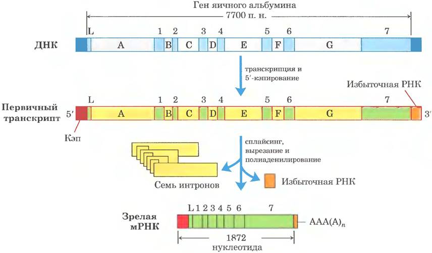
The complete set of processing reactions for a typical eukaryotic mRNA is shown in Fig. 26-19. In some cases, the coding region of the mRNA also undergoes "editing" (for details, see Section 27.1). This may involve reactions such as the insertion or deletion of bases within the coding regions of primary transcripts, or sequence alterations (e.g., the enzymatic deamination of a C residue to form a U residue). One striking example of processing occurs in parasitic Protozoa such as trypanosomes: long stretches of their mRNA are synthesized completely devoid ofuridylate, and U residues are subsequently inserted during editing.
Fig. 26-19. Schematic Overview of eukaryotic mRNA processing. The Ovalbumin gene contains introns A through G and exons 1 through 7, as well as sequence L (L encodes the signal peptide that directs the protein out of the cell; see Fig. 27-34). During processing, approximately three-quarters of the RNA is removed. Before transcription termination, Pol II manages to synthesize the primary transcript well past the cleavage and polyadenylation site (excess RNA). Termination signals for Pol II have not yet been identified.
Alternative RNA Processing gives rise to multiple products from a single gene
One of the paradoxes of modern Genomics is that organismal complexity does not correlate with the number of protein-coding genes, or even with The amount of genomic DNA (p. 460, pt. 1). However, the traditional approach, which focuses primarily on protein-coding genes, overlooks The complexity of an Organism's transcriptome. As our knowledge of RNA functions in Genome Organization expands, new and intricate elements are continually being discovered.
Some eukaryotic mRNA transcripts yield only a single mature mRNA and a corresponding polypeptide, whereas others can undergo processing in multiple ways to produce diverse mRNAs and distinct Polypeptides. The primary transcript contains molecular signals for all alternative processing pathways, and the pathway preferred by a given cell is determined by processing factors—RNA-binding proteins that trigger one specific route.
Complex transcripts may feature more than one cleavage and polyadenylation site, Alternative Splicing sites, or both. When two or more cleavage and polyadenylation sites are present, the closer the site is chosen to the 5' end, the larger the portion of the primary transcript sequence that is removed (Fig. 26-20a). This mechanism, termed poly(A) site Selection, generates diversity in the variable domains of immunoglobulin heavy chains (see Fig. 25-46). Through alternative splicing at different Developmental Stages of the fruit fly (Fig. 26-20b), three distinct forms of the Myosin heavy chain are generated from a common primary transcript. Alternative RNA processing options can operate simultaneously to synthesize two different Hormones: the calcium-regulating hormone Calcitonin in the rat Thyroid Gland and the calcitonin gene-related peptide (CGRP) in the rat Brain (Fig. 26-21). Numerous other variations of alternative splicing exist (Fig. 26-22). Many genes in the mammalian genome (perhaps the majority) undergo alternative splicing, which vastly increases the repertoire of proteins encoded by genes. This process plays a far less prominent role in lower eukaryotes; for instance, only a few genes undergo alternative splicing in yeast.
Fig. 26-20 Two mechanisms of alternative processing of complex transcripts in eukaryotes. (a) Alternative cleavage and polyadenylation variants. Two polyadenylation sites, A1 and A2, are shown. (b) Alternative splicing pathways. Two different 3'-terminal cleavage sites are shown. Both mechanisms allow The production of distinct mature mRNAs from the same transcript.

Fig. 26-21. Alternative Processing of the calcitonin gene transcript in rats. The primary transcript contains two polyadenylation sites; one is used more frequently in brain cells, and the other in The Thyroid Gland. In the brain, splicing eliminates the calcitonin exon (exon 4); in the thyroid, this exon is retained. The resulting Peptides are further processed to yield the final hormones: the calcitonin gene-related peptide (CGRP) in the rat brain and calcitonin in the thyroid gland.

Fig. 26-22. General scheme of alternative splicing. Exons are depicted as light- and dark-green rectangles, whereas introns and untranslated regions are shown as yellow lines. Asterisks indicate the positions where polyadenylation should occur. Black lines on the left side of the diagram represent possible exon-joining pathways leading to the corresponding mRNAs (on the right side of the diagram). Splicing pathways shown above and below the transcript produce the upper and lower products, respectively. Red and orange rectangles denote 5' caps and 3' untranslated regions, respectively.

Fig. 26-23. Selected modified bases in rRNA and tRNA generated via post-transcriptional reactions. Abbreviated base names are given in parentheses. Note the unusual ribose attachment site in pseudouridine. Only a few Examples are shown out of the 96 known nucleotide modifications discovered in various RNAs (currently, 81 types of modifications are known in tRNA and 30 types in rRNA). A complete list of modified bases can be found in the RNA Modification Database (http://mods.rna.albany.edu/mods/).

rRNA and tRNA molecules also undergo processing
mRNA is not the only transcript subjected to post-transcriptional processing. Ribosomal RNAs in prokaryotic, archaeal, and Eukaryotic cells are generated from longer precursors termed preribosomal RNAs or pre-rRNAs. Transfer RNAs likewise originate from longer precursors. These RNA molecules may also contain various modified nucleosides; some examples are illustrated in Fig. 26-23.
Fig. 26-24. Processing of pre-RNA transcripts in bacteria. ① Prior to cleavage, the 30S RNA precursor is methylated at specific bases (red dashes), and certain uridine residues are converted into pseudouridine (blue dashes) or dihydrouridine (black dashes). Methylation sometimes targets bases or 2'-hydroxyl groups. ② Cleavage releases rRNA and tRNA precursors. Cleavage at sites 1, 2, and 3 is carried out by the enzymes RNase III, RNase P, and RNase E, respectively. As discussed below, RNase P is a ribozyme. ③ The final 16S, 23S, and 5S rRNA products are generated through the action of various specific Nucleases. The seven copies of the pre-rRNA gene in the *E. coli* chromosome differ in the number, arrangement, and type of tRNAs embedded within the primary transcript. Some gene copies carry additional tRNA gene segments between the 16S and 23S rRNA segments, as well as at the 3' end of the primary transcript.

Ribosomal RNAs.
In bacteria, the 16S, 23S, and 5S rRNAs (along with some tRNAs, although most tRNAs are encoded elsewhere) are produced from a single precursor—the 30S RNA, which is approximately 6,500 nucleotides long. During processing, terminal fragments and the spacer regions between rRNA sequences are removed from the 30S RNA (Fig. 26-24). The 16S and 23S rRNAs contain modified nucleosides. *E. coli* harbors 11 modifications in its 16S rRNA sequence: one pseudouridine and 10 nucleosides methylated on the base and/or the 2'-hydroxyl group. The 23S rRNA contains 10 pseudouridines, one dihydrouridine, and 12 methylated nucleosides. In bacteria, each modification is typically catalyzed by a dedicated enzyme. The methylation reaction utilizes S-adenosylmethionine as a cofactor. Pseudouridine formation requires no cofactors.
The *E. coli* genome encodes seven pre-rRNA molecules. All these genes share similar rRNA-coding regions, but their spacer regions differ. The region between the 16S and 23S rRNA genes typically encodes one or two tRNA molecules, with different pre-rRNA transcripts yielding distinct tRNAs. Sequences encoding tRNA molecules have also been identified at the 3' end of the 5S rRNA in certain primary transcripts.
In eukaryotes, the situation is more complex. RNA polymerase I synthesizes a 45S pre-rRNA transcript, which is cleaved in the nucleolus to yield the 18S, 28S, and 5.8S rRNAs characteristic of eukaryotic Ribosomes (Fig. 26-25). As in bacteria, processing involves endo- or exonucleolytic cleavage reactions as well as nucleoside modifications. Some pre-rRNAs contain introns that must be excised. The overall process is initiated in the nucleolus within large complexes that assemble on the rRNA precursor as it is synthesized by Pol I. There is a close coupling between rRNA transcription, rRNA maturation, and ribosome assembly in the nucleolus. Each complex contains ribonucleases that cleave pre-rRNAs, enzymes that modify specific bases, A large number of small nucleolar RNAs (snoRNAs) that guide nucleoside modifications and certain cleavage reactions, and ribosomal proteins. In yeast, the process involves pre-rRNAs, over 170 non-ribosomal proteins, snoRNAs for modifying every nucleoside (~70 in total, as some mediate Two Types of modifications), and 78 ribosomal proteins. Human cells possess an even greater number of modified nucleosides—around 200—and consequently a larger set of corresponding snoRNAs. During ribosome assembly, the COMPOSITION OF THE complexes changes, and many intermediate complexes rival the ribosome and snRNA themselves in complexity. In most eukaryotes, 5S rRNA is produced by a different polymerase (Pol III) as an independent transcript.
Fig. 26-25. Processing of pre-RNA transcripts in vertebrates. During transcription, the 45S primary transcript is incorporated into a 90S nucleolar preribosomal complex, where rRNA Processing and ribosome assembly are coupled. ① The 45S precursor is methylated at more than 100 out of 14,000 nucleotides (on bases or 2'-OH groups), several uridine residues are converted to pseudouridine, and various other modifications occur. ② A series of enzymatic cleavages of the 45S transcript yields 18S, 5.8S, and 28S rRNAs, and ribosomal subunits gradually begin to form through the addition of ribosomal proteins. Small nucleolar RNAs (snoRNAs) integrated into protein complexes (snoRNPs) within the nucleolus, resembling spliceosomes, participate in all cleavage and modification reactions. 5S rRNA molecules are produced independently.

Nucleoside modifications in eukaryotic rRNA most frequently involve The conversion of uridine to pseudouridine and adoMet-dependent methylation (typically at the 2'-hydroxyl group). These reactions are mediated by snoRNP complexes—protein-RNA complexes consisting of a snoRNA and four or five proteins, including the modifying enzyme. There are two classes of such complexes, distinguished by key conserved sequence elements (boxes). H/ACA box complexes participate in pseudouridine formation, whereas C/D box complexes are involved in 2'-OH methylation. Unlike in bacteria, a single eukaryotic enzyme may participate in modifications at different sites under the guidance of appropriate snoRNAs.
These snoRNAs consist of 60 to 300 nucleotides. Many are encoded within the introns of other genes and are co-transcribed with those genes. Each snoRNA contains a 10-to-21-nucleotide sequence that is fully complementary to a specific region of the rRNA. Conserved sequences in the remainder of the snoRNA form structures that bind to corresponding proteins within the snoRNP (Fig. 26-26).
Fig. 26-26. Role of snoRNAs in rRNA modification. (a) To direct methylation reactions, the RNA base-pairs with a C/D box snoRNA. The methylation sites in the target rRNA (dark green) are located within the region paired with the C/D box snoRNA. Highly conserved sequences of the C and D boxes (as well as C' and D') serve as binding sites for the proteins that make up the larger snoRNP complex. (b) The RNA base-pairs with an H/ACA box snoRNA to convert uridine into pseudouridine. The uridine-to-pseudouridine conversion sites in the target rRNA (green) also lie within the regions that pair with the snoRNA, and the conserved H/ACA box sequences act as protein-binding sites.

Transfer RNAs.
Most cells synthesize between 40 and 50 distinct tRNAs, with eukaryotic cells containing multiple copies of many tRNA genes. Transfer RNA molecules are generated from longer precursors through the enzymatic removal of nucleotides from both the 5' and 3' ends (Fig. 26-27). In eukaryotes, the primary transcripts of several tRNAs contain introns that must be removed. When a single primary transcript houses two or more different tRNAs, they are separated via enzymatic cleavage. Endonuclease RNase P, found in all organisms, removes the RNA segment from the 5' end of tRNA molecules. This enzyme consists of both Protein and RNA components. The RNA component is essential for catalytic activity; in bacterial cells, the enzyme can even perform processing without the protein component. Thus, RNase P serves as yet another example of catalytic RNA, the actions of which are explored in greater detail below. The 3' end of tRNA molecules is modified by one or more nucleases, including the exonuclease RNase D.
Fig. 26-27. tRNA Processing in bacteria and eukaryotes. Key processing steps are illustrated using yeast tRNATyr (a Tyrosine-specific tRNA; see Chapter 27). The nucleotide sequence highlighted in yellow is cleaved from the primary transcript. End processing occurs first—targeting the 5' end initially, followed by the 3' end. Next, the CCA sequence is added to the 3' end; this step is obligatory in the processing of eukaryotic tRNAs and those bacterial tRNAs that lack this sequence in their primary transcript. Concurrently with end processing, specific bases in the remaining portion of the transcript are modified (Fig. 26-23). For the eukaryotic tRNA shown here, the final step involves the splicing of a 14-nucleotide intron. Introns appear in certain eukaryotic tRNAs but are absent from bacterial tRNAs.

tRNA precursors can undergo further post-transcriptional processing. The 3'-terminal trinucleotide CCA (3'), to which an amino acid attaches during Protein Synthesis (Chapter 27), is missing in some bacterial and all eukaryotic tRNA precursors and is therefore added during processing (Fig. 26-27). This function is carried out by tRNA nucleotidyltransferase—an unusual enzyme that binds three ribonucleoside triphosphates at separate active sites and catalyzes the formation of phosphodiester bonds to build the CCA (3') sequence. Consequently, the synthesis of this sequence requires no DNA or RNA template; the enzyme's binding site itself serves as the template.
The final type of tRNA processing involves the modification of specific bases via methylation, deamination, or reduction reactions (Fig. 26-23). In pseudouridine formation, the base (uracil) is excised and reattached to the sugar moiety at the C-5 position. Several of these modified bases occupy conserved positions across all tRNA molecules (Fig. 26-27).
RNAs with specialized functions undergo diverse processing pathways
The number of identified specialized RNA classes is growing rapidly, along with our expanding understanding of their functions. Many of these RNA molecules undergo processing.
Small nuclear and small nucleolar RNAs not only facilitate RNA processing but are themselves synthesized as larger precursors before undergoing processing. Many snRNAs are encoded within the introns of other genes. As introns are excised from pre-mRNAs, snRNP complex proteins bind to the snRNA sequences, while ribonucleases trim excess RNA segments from the 5' and 3' ends. The snRNAs destined for spliceosomes are synthesized as pre-snRNAs by RNA polymerase II, with ribonucleases removing surplus RNA fragments from both ends. Specific nucleosides within snRNAs can undergo 11 distinct modifications, most commonly 2'-O-methylation and the conversion of uridine to pseudouridine.
MicroRNAs constitute a specialized class of RNAs involved in the Introduction/30.html">Regulation of Gene Expression. The non-coding sequences of these RNAs, which are roughly 22 nucleotides long, are complementary to specific regions of mRNAs. They regulate mRNA function by either inducing its cleavage or repressing its translation. MicroRNAs have been discovered in numerous multicellular eukaryotes, ranging from worms and fruit flies to plants and mammals. It is estimated that up to 1% of The Human Genome encodes microRNAs, and these molecules may interact with as many as one-third of human mRNAs. Their role in gene regulation is discussed in Chapter 28.
MicroRNAs are synthesized from much larger precursors through a multi-step pathway (Fig. 26-28). The primary transcripts for microRNAs (pri-miRNAs) vary significantly in size; some are encoded within the introns of other genes and are co-expressed with them. Their role in gene regulation is also examined in detail in Chapter 28.
Fig. 26-28. Synthesis and processing of microRNAs. The primary microRNA transcript is an extended RNA sequence of variable length known as a pri-miRNA. Two endoribonucleases of the RNase III family, Drosha and Dicer, play active roles in its processing. In the nucleus, the pri-miRNA is first trimmed into a 70–80 nucleotide microRNA precursor (pre-miRNA) by a protein complex comprising Drosha and the DGCR8 protein. Subsequently, the pre-miRNA is exported to the cytoplasm, where it is acted upon by the Dicer protein, yielding a nearly mature microRNA base-paired with a short RNA fragment. This complementary RNA strand is unwound and removed by an RNA helicase, and the mature microRNA is incorporated into protein complexes such as RISC (RNA-induced silencing complex), which then bind to the target mRNA. If complementarity between the microRNA and its target is extensive, target mRNA cleavage ensues; if complementarity is only partial, the resulting complex blocks target mRNA Translation.

Catalytic RNAs drive several reactions of RNA metabolism
The investigation of post-transcriptional processing in RNA molecules has led to one of the most thrilling breakthroughs in modern biochemistry: the discovery of catalytic RNAs (ribozymes). The best-characterized ribozymes include group I self-splicing introns, RNase P, and the hammerhead ribozyme (see below). The activity of these ribozymes centers around two primary reactions—transesterification (Fig. 26-14) and the Hydrolysis of phosphodiester bonds. Often, the substrate for a ribozyme is an RNA molecule, which may even form a part of the ribozyme itself. Base pairing can occur between the RNA substrate and the RNA catalyst, optimally positioning the substrate for the reaction.
Fig. 26-29. The hammerhead ribozyme. Virus-like entities known as Viroids possess a tiny RNA genome and typically rely on a helper virus to replicate and/or package themselves. Certain viroid RNA molecules contain small segments that trigger site-specific RNA cleavage reactions coupled to Replication. These regions are called hammerhead ribozymes because their Secondary structure resembles a hammerhead, with 'a' representing the minimal sequence required for catalytic activity. Conserved nucleotides outlined in boxes are essential for catalytic function. The arrow marks the self-cleavage site. 'b' shows the three-dimensional structure (PDB ID 1MME, depicted as a space-filling CPK model in Fig. 8-25, vol. 1). The chains are colored identically to panel 'a'. Hammerhead ribozymes belong to metalloenzymes; they require Mg2+ ions for their activity. The phosphodiester bond at the self-cleavage site is indicated by the arrow. The hammerhead ribozyme

Ribozymes vary substantially in size. Group I self-splicing introns can exceed 400 nucleotides, whereas the hammerhead ribozyme consists of two RNA chains totaling just 41 nucleotides (Fig. 26-29). Much like protein enzymes, the three-dimensional structure of ribozymes is critical for their function. Ribozymes are inactivated by heating above their melting Temperature, or by exposure to Denaturing Agents or complementary oligonucleotides that disrupt normal base pairing. They can also be inactivated by modifying key nucleotides within their structure. The Secondary structure of the group I self-splicing intron from the 26S rRNA precursor in Tetrahymena is illustrated in Fig. 26-30.
Fig. 26-30. Secondary STRUCTURE OF THE self-splicing rRNA intron from Tetrahymena. Intron sequences are highlighted in yellow, and exon sequences in green. Yellow lines indicate bonds between adjacent nucleotides within the sequence (a necessary compromise to represent a volumetric molecule in two dimensions; the long blue line between residues C and G denotes normal base pairing); all sequence nucleotides are shown. The catalytic core responsible for self-splicing activity is enclosed in a gray box. Certain base-paired regions are numbered (P1, P3, P2.1, P5a, etc.) According to the established conventions for this RNA molecule. The region P1, which contains the internal guide sequence (boxed), houses the 5' end of the intron (red arrow). A portion of this sequence pairs with the 3' end of the exon, bringing the 3' and 5' splice sites into direct contact (red and blue arrows). The three-dimensional structure of a large fragment of this intron is presented in Fig. 8-25, vol. 1.

Enzymatic properties of group I introns
Beyond their ability to accelerate reaction rates, group I self-splicing introns share other features with classical enzymes, including kinetic behavior and Specificity. The binding of the guanosine cofactor (Fig. 26-14) to the group I intron from Tetrahymena rRNA (Fig. 26-26) reaches saturation (Km ≈ 30 µmol/L) and is subject to competitive inhibition by 3'-deoxyguanosine. The intron executes the cleavage reaction with remarkable precision, largely owing to the so-called internal guide sequence, which can base-pair with exon sequences near the 5' splice site (Fig. 26-30). This pairing facilitates the proper alignment of the specific bonds destined to undergo cleavage and rejoining.
Because the intron undergoes chemical alteration during the splicing reaction (its ends are cleaved), it might seem to lack a defining characteristic of an enzyme: The ability to mediate multiple rounds of catalysis. However, detailed studies have revealed that the 414-nucleotide intron excised from Tetrahymena rRNA can function (in vitro) as a true enzyme (in vivo, it is rapidly degraded). A series of intramolecular cyclization and cleavage reactions within the excised intron leads to the removal of 19 nucleotides from its 5' end. The linear RNA comprising the remaining 395 nucleotides (designated L-19 IVS, for intervening sequence lacking 19 nucleotides) promotes nucleotide transfer reactions in which certain oligonucleotides are elongated at the expense of others (Fig. 26-31). The most effective substrates for this RNA are oligonucleotides such as the synthetic oligomer (C)5, which can base-pair with the same guanine-rich internal guide sequence that previously aligned the 5' exon end for self-splicing.
Fig. 26-31. Catalytic activity of L-19 IVS in vitro. 'a' — L-19 IVS is generated by the autocatalytic removal of 19 nucleotides from the 5' end of the excised Tetrahymena intron. The cleavage site within the internal guide sequence (boxed) is indicated by the arrow. The G residue (on a pink Background) attached During the first step of the splicing reaction (see Fig. 26-15) becomes part of the excised sequence. A portion of the internal guide sequence remains at the 5' end of L-19 IVS. 'b' — L-19 IVS elongates certain RNA oligonucleotides at the expense of others through a cycle of transesterification reactions (steps ①–④). A pivotal role in this cycle is played by the 3'-OH group on the terminal G residue at the 3' end of L-19 IVS (noting that this is distinct from the G residue added during splicing). One of the ribozyme's optimal substrates is (C)5, as it can bind to the guide sequence retained within the intron. Although this catalytic activity is not utilized within the cell, it holds profound significance for models of modern evolutionary hypotheses discussed at the end of this chapter.

The enzymatic activity of the L-19 IVS ribozyme is manifested in a cycle of transesterification reactions whose mechanism resembles self-splicing. Each ribozyme molecule can process approximately 100 substrate molecules per hour without being altered in the course of the reaction; thus, the intron acts as a catalyst. Its action conforms to Michaelis-Menten kinetics, is specific for oligonucleotide RNAs as substrates, and can be completely blocked by an inhibitor. The Kcat/Km ratio (specificity constant) is 103 M-1 • s-1, which is lower than that of many enzymes, but compared to the uncatalyzed reaction, the ribozyme increases The rate of hydrolysis by a factor of approximately 1010. Such acceleration is achieved through substrate orientation, Covalent Catalysis, and metal ion catalysis—mechanisms shared by protein enzymes.
Characteristics of other ribozymes.
E. coli RNase P consists of RNA (M1 RNA, 377 nucleotides) and protein (Mr = 17,500). In 1983, Sidney Altman and Norman Pace, along with their colleagues, discovered that under certain conditions, M1 RNA is capable of independent catalysis, cleaving tRNA precursors at a precisely defined site. The protein component likely stabilizes the RNA or facilitates its functioning in vivo. The RNase P ribozyme recognizes the three-dimensional shape of the substrate (pre-tRNA) as well as the CCA sequence, thereby being able to cleave 5'-terminal sequences from various tRNA molecules (Fig. 26-27).
The list of known ribozymes continues to grow. Certain viroids—small RNA molecules associated with plant RNA viruses—contain a structure that triggers a self-cleavage reaction. This class of catalytic molecules includes the hammerhead ribozyme shown in Fig. 26-29, which catalyzes the hydrolysis of an internal phosphodiester bond. The splicing reaction in the spliceosome appears to be carried out in a catalytic center formed by the U2, U5, and U6 snRNAs (Fig. 26-17). And perhaps most importantly, the RNA component of ribosomes catalyzes protein synthesis (see Chap. 27).
The discovery of catalytic RNA molecules has broadened our understanding of catalytic function in general and played a crucial role in shaping our concepts regarding the ORIGIN AND EVOLUTION of life on our planet (see Sec. 26.3).
mRNAs in the cell are degraded at different rates
Gene Expression is regulated at multiple levels. A decisive factor governing gene expression is the concentration of the corresponding mRNA within the cell. The concentration of any molecule depends on two factors: the rate of synthesis and the rate of degradation. When the rates of mRNA Synthesis and degradation become equal, the mRNA is at a steady-state concentration. A change in either the synthesis rate or the degradation rate leads to the accumulation or depletion of mRNA. Metabolic pathways of degradation prevent the accumulation of mRNA in the cell and the synthesis of unneeded proteins.
The degradation rates of mRNAs from different eukaryotic genes vary widely. The half-life of mRNAs for products needed only briefly may be several minutes or even seconds. Conversely, mRNAs for genes whose products are required constantly can be stable across many cell generations. The average half-life of mRNA in vertebrate cells is approximately 3 hours, and the pool of each mRNA type turns over roughly 10 times during a single cell generation. The half-life of bacterial mRNA is much shorter—only about 1.5 min; this is likely due to Metabolic Regulation.
Messenger RNA is degraded by ribonucleases present in all cells. In E. coli, the process begins with one or more endoribonuclease-mediated cuts, followed by 3' —> 5' exoribonuclease degradation. In lower eukaryotes, the primary metabolic pathway begins with the shortening of the poly(A) tail, followed by 5'-end decapping and 5' —> 3' degradation of the mRNA. The 3' —> 5' degradation pathway also exists and may be the primary pathway in higher eukaryotes. All eukaryotes possess a complex called the exosome, which contains up to 10 conserved 3' —> 5' exoribonucleases that participate in the processing of the 3' ends of rRNA and tRNA, as well as in mRNA degradation.
The hairpin-like structure of bacterial mRNA with a rho-independent terminator (Fig. 26-8) provides protection against degradation. Similar hairpin structures stabilize certain PARTS OF THE primary transcript, which accounts for the nonuniform degradation of transcripts. In eukaryotic cells, the 3'-poly(A) tail and 5'-cap are vital for the stability of many mRNA molecules.
Polynucleotide phosphorylase generates random RNA-like polymers
In 1955, Marianne Grunberg-Manago and Severo Ochoa discovered the bacterial enzyme polynucleotide phosphorylase, which catalyzes the following reaction in vitro:


Polynucleotide phosphorylase was the first discovered of all nucleic acid-synthesizing enzymes (shortly thereafter, Arthur Kornberg discovered DNA polymerase). The reaction catalyzed by polynucleotide phosphorylase differs fundamentally from all the polymerase reactions mentioned above in that it is independent
of a template. This enzyme utilizes ribonucleoside 5'-diphosphates as substrates and does not act on homologous 5'-triphosphates or deoxyribonucleoside 5'-diphosphates. The RNA polymer synthesized by polynucleotide phosphorylase contains conventional 3', 5'-phosphodiester bonds, which are cleaved by Ribonuclease. This reaction is readily reversible, and the equilibrium shifts toward polyribonucleotide degradation upon an increase in phosphate concentration. The presumed role of the enzyme in the cell is The breakdown of mRNA molecules into nucleoside diphosphates.
Because polynucleotide phosphorylation does not require a template, the resulting polymer does not contain specific base sequences. The reaction proceeds equally well with any one of the four nucleotide diphosphates or with all four combined, and the composition of the resulting polymer depends solely on the relative concentrations of the 5'-diphosphate substrates in the medium.
Polynucleotide phosphorylase can be used in the laboratory to synthesize RNA polymers with A wide variety of base sequences. Synthetic RNA polymers of this kind played a critical role in deciphering The Genetic Code for amino acids (Chap. 27).
Summary of Section 26.2 RNA Processing
■ Eukaryotic mRNA molecules are modified by the addition of a 7-methylguanosine residue at the 5' end, as well as by the cleavage and polyadenylation of the 3' end to form a long poly(A) tail.
■ Many primary mRNA transcripts contain introns (noncoding regions) that are removed by splicing. The excision of group I introns, found in certain rRNA molecules, requires a guanosine cofactor. Some group I and group II introns are capable of self-splicing without the participation of protein enzymes. Nuclear mRNA precursors contain group III introns (the most abundant type), which are excised with the aid of RNA-protein complexes called snRNPs organized into spliceosomes. Group IV introns undergo protein enzyme-mediated splicing; these introns are found in certain tRNA molecules.
■ The function of many eukaryotic mRNAs is regulated by complementary microRNAs. MicroRNA molecules are generated from longer precursors through a series of processing reactions.
■ Ribosomal and transfer RNA molecules are derived from longer precursor molecules cleaved by nucleases. During maturation, certain bases are modified by enzymes. Some nucleoside modifications are guided by snoRNAs within snoRNP protein complexes.
■ Self-splicing introns and the RNA component of RNase P (which cleaves the 5' end of tRNA precursors) are examples of ribozymes. These biological catalysts possess The properties of true enzymes. Typically, they catalyze hydrolytic cleavage and transesterification using RNA as a substrate. A combination of such reactions is facilitated by the excised group I intron from Tetrahymena rRNA, generated during the RNA polymerization reaction.
■ Polynucleotide phosphorylase synthesizes RNA-like polymers from ribonucleoside 5'-diphosphates in a reversible reaction, adding or removing ribonucleotides at the 3'-hydroxyl end of the polymer. The enzyme degrades RNA in vivo.
Last update: 06/08/2026
Editorial and Educational Adaptation: This material has been compiled based on the primary/original source text. The project team performed an editorial review, corrected technical inaccuracies, structured sections, and adapted the content for an educational format.
What was processed:
- elimination of formatting defects (OCR errors, structural breaks, corrupted characters);
- editorial organization of content;
- standardization of terminology in accordance with academic sources;
- verification of factual statements against the original source text.
All mentions of the author, publication year, and origin of the primary text have been preserved in accordance with the source.