Fundamentals of Biochemistry - A. A. Anisimov 1986
Biological Oxidation and Bioenergetics
Oxidative Phosphorylation
7.3.1. The Respiratory Chain. Protons and electrons are split off from substrates during The Tricarboxylic Acid Cycle. They enter the Coenzymes NAD+ and FAD+, which transfer them to the respiratory chain formed by oxidation-reduction Enzymes located in The inner mitochondrial membrane. Moving from one electron carrier to another, electrons descend to progressively lower energy levels, releasing their energy in portions. In the final link of the chain, they reduce molecular oxygen. The energy released during The transfer of electrons along the respiratory chain is conserved in the phosphate bonds of ATP.
The synthesis of ATP from ADP and phosphoric acid, which occurs utilizing the energy released during The oxidation of substances in living Cells and is coupled with the transfer of electrons along the respiratory chain, is called Oxidative Phosphorylation. It was discovered in the early 1930s by V. A. Engelhardt.
The respiratory chain is frequently referred to as the redox chain, which stands for reduction-oxidation chain, since oxidation-reduction processes occur repeatedly within it. It is also called the Electron Transport Chain, electron-transporting chain, or electron transfer chain. These concepts are broader than the "respiratory chain," as similar systems function in the membranes of METABOLISM/14.html">Chloroplasts, nuclei, and microsomes.
The Components of the respiratory chain that carry out electron transfer and oxidative phosphorylation are integrated into the inner mitochondrial membrane and function as multienzyme complexes or assemblies. A single complete electron transport chain consists of A large number of molecules. For instance, in Cell/35.html">Mitochondria isolated from bovine Heart, The electron transport chain comprises approximately 80 protein molecules. Only 1/3 of them are direct electron carriers, while the remaining 2/3 are Proteins performing auxiliary Functions.
The respiratory chain includes a non-protein electron carrier—ubiquinone, sometimes referred to as coenzyme Q (abbreviated as CoQ). It is a benzoquinone derivative with an isoprenoid side chain containing from 6 to 10 five-carbon isoprene residues, depending on the species of Organism. In most mammalian Tissues, ubiquinone Q10 functions (where 10 is the number of isoprene residues in the side chain), whereas in Yeasts, it is Q6. Ubiquinone can participate in one- or two-electron transfer. Upon partial reduction (one-electron transfer), it forms a relatively stable semiquinone; upon full (two-electron) reduction, it forms hydroquinone (ubiquinol).
Thus, ubiquinone and its hydroquinone form a redox pair. They transfer hydrogen atoms—that is, electrons and protons—from one group of carriers to another. Ubiquinone can migrate within the lipid phase of the membrane, acting as a mobile substrate for enzymes embedded in the membrane.
Class="center">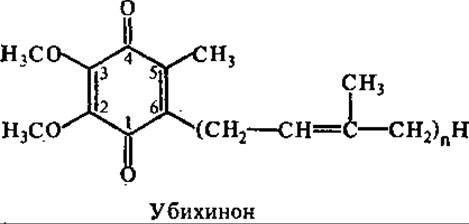
Electrons and protons are split off from substrates in the Reactions of the Krebs cycle predominantly by pyridine-dependent dehydrogenases, resulting in The formation of NADH. The latter is oxidized by a flavin-dependent enzyme, NADH dehydrogenase. It transfers electrons and protons to ubiquinone, which passes them on to the cytochrome system.
Many flavin-dependent dehydrogenases are known. They contain flavin adenine dinucleotide (FAD) or flavin mononucleotide (FMN) as prosthetic groups, which are firmly—often covalently—bound to the protein. The isoalloxazine ring of riboflavin participates in oxidation-reduction reactions: two hydrogen atoms are transferred to FAD or FMN, yielding the reduced forms FADH2 and FMNH2. Frequently, flavin enzymes and the complexes they form contain metal atoms, most commonly in the form of iron-sulfur center proteins (Fe-S proteins). In these proteins, iron is not present as a heme group, but is coordinated to sulfur atoms in the Cysteine residues of the protein. Some proteins contain labile sulfur atoms.
From certain substrates (succinic acid, glycerol phosphate, fatty acid CoA derivatives), flavin enzymes remove hydrogen directly, bypassing pyridine-dependent dehydrogenases and NADH. Flavin dehydrogenases transfer electrons and protons to ubiquinone in the respiratory chain. Examples include succinate dehydrogenase and glycerol-3-phosphate dehydrogenase. They contain iron-sulfur centers. During the Oxidation of Fatty acid CoA derivatives, the flavoprotein strips hydrogen atoms from the aliphatic chain and transfers them to a second flavoprotein, from which they pass to a third flavoprotein containing iron-sulfur centers that delivers the hydrogen atoms to ubiquinone (Fig. 7.2).
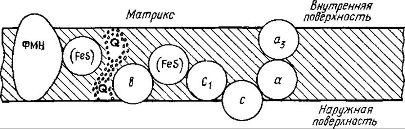
Fig. 7.2. Arrangement of the Main Components of the respiratory chain in the inner mitochondrial membrane:
a, a3, b, c, c1 — components of the cytochrome system; see text for other designations
The most important flavin-dependent dehydrogenase of the respiratory chain is NADH dehydrogenase, which mediates the oxidation of NADH. The Structure of the NADH dehydrogenase complex has not yet been fully elucidated. It contains at least 10 distinct polypeptide chains, FMN, and iron-sulfur center proteins. In a native preparation, there are 16 Fe and 16 S-2 per molecule of FMN. Electron paramagnetic Resonance (EPR) spectroscopy studies have identified four iron-sulfur centers. Proteins containing iron-sulfur centers have been found In the second major dehydrogenase of the respiratory chain—succinate dehydrogenase—and in the enzymatic complex that oxidizes reduced ubiquinone with oxidized cytochrome c.
Succinate dehydrogenase is an enzyme of the inner mitochondrial membrane; isolated from bovine heart Muscle, M = 97,000 (consisting of two subunits with M = 70,000 and 27,000). The larger component contains the substrate-binding site and FAD. Iron-sulfur centers are found in both subunits. Electrons flow from succinate to FAD, then to the iron-sulfur centers of the large subunit, and from there to the iron-sulfur centers of the small subunit.
From reduced ubiquinone, electrons pass to the cytochrome system. Cytochromes are a group of iron-containing proteins present in all aerobic cells. They contain iron-porphyrin prosthetic groups similar to those found in Hemoglobin and Myoglobin. During electron transfer, the valence of the iron within the cytochromes changes reversibly:
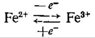
The Role of cytochromes in cellular Respiration was first investigated by D. Keilin (1925). A large number of cytochromes have now been isolated from various species of living organisms. They are divided into groups based on structural differences in their heme prosthetic moieties. Four types of heme are distinguished, designated by the letters a, b, c, and d. They differ in the side chains of their porphyrin structures. When designating cytochromes with well-established structures, a numerical index is added to the letter to indicate the subgroup to which the cytochrome belongs. For example, five different cytochromes have been identified in the mitochondria of higher animals and plants: a, a3, b, c, c1.
Cytochromes differ from one another in their protein moieties, The Nature of their porphyrin side chains, and the manner in which the heme is attached to the protein. The absorption spectrum of reduced cytochromes exhibits three main absorption bands in the visible region. In order of decreasing wavelength, they are designated as the α-, β-, and γ-bands; of these, the α-band usually appears as a sharp peak. Consequently, when designating cytochromes with insufficiently studied structures, the wavelength (in nanometers) of the α-absorption band of the reduced form is specified. For instance, the respiratory chain includes two cytochromes of the b group: b562 and b566. At the same time, cytochromes are frequently named after incidental features. For example, cytochrome P-450 is localized in The Endoplasmic reticulum, so named because its carbon monoxide (CO) complex exhibits an absorption maximum at a wavelength of 450 nm.
Only the final cytochrome of the respiratory chain—cytochrome c oxidase (cytochrome aa3)—reacts with oxygen. It is the only cytochrome possessing catalytic, i.e., enzymatic, properties; other cytochromes are not enzymes. The oxidase molecule contains two Cu atoms and two heme a-type groups: a and a3. Cytochrome c oxidase from the inner mitochondrial membrane of bovine heart Muscle consists of six different protein subunits. Cytochrome oxidase from Yeast and Neurospora contains seven subunits. Cytochrome oxidase carries out the rapid oxidation of cytochrome by molecular O2. It is the terminal oxidase of the mitochondrial respiratory chain. Cytochrome oxidase is localized in the inner mitochondrial membrane, to which it is relatively firmly bound, forming an asymmetric bridge across the Two Sides of the membrane. It is hypothesized that heme a3 is located on the inner side, while heme a—which interacts first with cytochrome c—is located on the outer side of the inner mitochondrial membrane. Oxygen appears to approach cytochrome oxidase from the matrix side, and a four-electron reduction of the O2 molecule to Water takes place.
NADH serves as a universal donor of H atoms for the respiratory chain. If NADPH is generated during substrate oxidation, it is typically utilized as a reductant in biosynthetic processes. However, a transhydrogenase reaction can occur, through which hydrogen atoms are transferred from NADPH to NAD+ and subsequently enter the respiratory chain:

A critical step in establishing the Sequence of electron transfer among the components of the respiratory chain was The Use of specific inhibitors (Fig. 7.3). For instance, rotenone (an insecticide) inhibits NADH dehydrogenase, causing all respiratory chain components to transition to an oxidized state. Amobarbital (sodium barbital) prevents the reduction of ubiquinone. The antibiotic antimycin A blocks the oxidation of cytochromes b, whereas cyanides, azide, and H2S bind to cytochrome oxidase. Based on these findings and thermodynamic calculations, the electron carriers can be arranged as shown in Fig. 7.3.
Each component of the respiratory chain in the inner mitochondrial membrane is interposed between its reductant and oxidant. This establishes conditions for a flow of electrons from the substrate, through sequentially arranged electron carriers, to O2. All proteins are embedded within the bulk of the membrane and form an integral part of it, whereas only cytochrome c is loosely attached to the outer surface of the membrane. The Active Site of NADH dehydrogenase is located on the inner side of the membrane, where it reacts with NADH. The O2-binding site of cytochrome a3 is likewise situated on the matrix-facing side of the membrane (see Fig. 7.2).
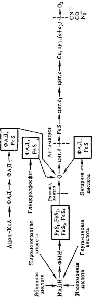
Fig. 7.3. The mitochondrial respiratory chain.
The arrows indicate the entry points of electrons from various substrates into the electron transport chain and their subsequent transport; the dashed lines denote the sites of inhibitor action.
As electrons pass along the respiratory chain, they lose a significant portion of their energy at specific segments, which is entirely sufficient for ATP generation. These are the sites of oxidative phosphorylation coupling, where proteins known as coupling factors operate. The latter form a coupling device, or the H+-ATPase complex, which synthesizes ATP from ADP and Pi. It spans the inner mitochondrial membrane, which is therefore referred to as the coupling membrane. In addition to proteins involved in Electron Transport and energy conversion, the inner mitochondrial membrane contains structural proteins and Lipids.
7.3.2. Energetics of Electron Transport. Reactions Involving the transfer of electrons from an electron donor (reductant) to an electron acceptor (oxidant) are called oxidation-reduction (redox) reactions. In some of these reactions, electron transfer is accomplished via the transfer of hydrogen atoms.
Reductants and oxidants form redox pairs:
The capacity of a reductant to donate electrons to an oxidant is typically expressed by the standard reduction potential (TE'0). It is measured at pH 7.0 and a Temperature of 25°C. The standard reduction potential is numerically equal to the electromotive force (in volts) developed in a half-cell in which the oxidant and reductant, present at concentrations of 1.0 M under standard conditions, are in equilibrium with an electrode capable of reversibly accepting electrons from the reductant.
The reduction potential of the reaction H2 ⇄ 2Н+ + 2е- is adopted as the standard. At a gaseous H2 pressure of 1.0 atm, an H+ ion concentration of 1.0 M (corresponding to pH 0), and 25°C, it is conventionally taken as 0. At pH 7.0, the standard reduction potential of the H2 ⇄ 2Н+ system is —0.42 V.
Knowledge of the standard reduction potentials of various biological systems makes it possible to determine the direction of electron flow. According to the Laws of Thermodynamics, if two redox-active components have different reduction potentials, the substance with the higher potential will act as the oxidant in the system, whereas the substance with the lower potential will serve as the reductant. In the respiratory chain, all reactions proceed down a thermodynamic gradient from the component with the most negative potential—NADH (—0.32 V)—to oxygen, which has a high positive potential (+0.82 V). Based on this
principle, all electron carriers of the respiratory chain can be arranged in a specific sequence: NAD→FAD→cytochrome b→cytochrome c→cytochrome a→O2.
When two redox pairs react with each other, the Standard Free energy change AG0' can be calculated using the formula
∆G0' = — nF∆E'0, (1)
where ∆G0' is the standard free energy change in kilojoules, n is the number of transferred electrons, F is the Faraday constant (96.5 kJ/V·mol), and ∆Е'0 is the difference between the standard reduction potentials of the electron acceptor and donor. For example, when a pair of electron equivalents is transferred from NADH (E'0 = —0.32 V) to molecular oxygen (E'0 = +0.82 V)—that is, passing through the entire respiratory chain—the free energy change is: ∆G0' = -2∙96.5∙[0.82—(—0.32)] = —220 kJ.
Thus, electron transport through the entire respiratory chain from NADH to molecular oxygen results in a large change in free energy. Under physiological conditions, the formation of ATP from ADP and inorganic phosphate requires —34.5 kJ. Using equation (1), one can calculate that a potential difference of 0.2 V is sufficient for ATP synthesis. Consequently, the transfer of a single electron pair through the entire respiratory chain releases enough energy to synthesize multiple ATP molecules.
7.3.3. Coupling Sites in the Respiratory Chain. The respiratory chain contains three sites where Electron transport is accompanied by a substantial change in potential difference (free energy). The decrease in free energy at each of these sites is large enough to drive the formation of ATP from ADP and Pi. It is precisely at these segments of the respiratory chain that energy is conserved in the phosphate bonds of ATP (Fig. 7.4).
Coupling sites were more precisely identified by measuring the oxidative phosphorylation P/O ratio. It was proposed in 1939 by the Soviet scientist V. A. Belitzer as a measure of oxidative phosphorylation efficiency. These classical experiments demonstrated that the electron transport chain performs one or more phosphorylations coupled to oxidation. The P/O ratio is the number of moles of ATP formed from ADP and Pi per gram-atom of oxygen consumed.
Fig. 7.4. Decrease in free energy driven by the transfer of an electron pair along the respiratory chain from NADH to oxygen
To date, P/O ratios have been determined for the oxidation of A number of substrates by mitochondria. For instance, for NADH and substrates oxidized by NADH dehydrogenases (malic, pyruvic, and isocitric acids), P/O = 3, whereas for the oxidation of succinic acid to fumaric acid, P/O = 2. Consequently, electrons and protons split off from NADH pass through three coupling sites of the respiratory chain, whereas those from succinic acid pass through only two (the second and third) (see Fig. 7.3).
The work of B. Chance was of paramount importance in elucidating the localization of coupling sites within the respiratory chain. He developed THE CONCEPT OF five metabolic states of the respiratory chain. State 1 is observed in the absence of oxidation and phosphorylation substrates, with the respiration rate being extremely low. In State 2, respiration is limited because the oxidizable substrate is absent, even though the phosphorylation substrate (ADP) is added in sufficient quantities. State 3 is the most active state, characterized by a high respiration rate. Both oxidation substrates and the phosphorylation substrate (ADP) are added in excess to the incubation medium. The respiration rate is limited solely by The rate of substrate penetration into the mitochondria and The activity of oxidative phosphorylation enzymes.
Once all the added ADP is converted to ATP, respiration slows down again. This is State 4, or the state of Respiratory Control. An electrochemical H+ ion potential accumulates across the coupling membrane, which cannot discharge due to the lack of ADP. The Membrane Potential impedes the movement of electrons along the respiratory chain, and substrate oxidation consequently ceases as well. The cessation of respiration is never complete, because sooner or later a "leak" of the membrane potential occurs, sustaining a low baseline respiration rate, i.e., respiratory control1. Mitochondria exhibit a very high affinity for ADP. A fresh addition of ADP restores the high respiration rate (State 3), which continues until ADP or oxygen is exhausted. The latter condition results in anaerobiosis, or State 5.
1 V. P. Skulachev introduced the concept of "proton control," which refers to the inhibition of H+-ATPase activity, as well as respiratory and photosynthetic Electron Transport Chains, mediated by the proton Electrochemical Potential gradient or one of its components. This concept is somewhat broader than respiratory control, as it applies not only to respiratory but also to photosynthetic redox chains.
Using spectrophotometric techniques, B. Chance and coworkers compared the oxidation states of electron carriers across all five states of the Respiratory Chain and determined the locations of the coupling sites within it. When the respiratory chain is inhibited due to a lack of ADP (State 4), it stalls at the coupling sites. Under these conditions, the carrier positioned upstream of the coupling site must be in the reduced form, accumulating electrons because its oxidation is hindered. Conversely, the second carrier, located downstream of the coupling site, remains in its most oxidized form because its reduction is impeded. Upon subsequent addition of ADP to the mitochondria, the respiratory chain transitions to State 3—its most active state. The respiration rate increases approximately 50-fold, and electron transfer at the coupling sites is accelerated. The first carrier (upstream of the coupling site) becomes oxidized, whereas the second carrier (downstream of the coupling site) becomes reduced.
As a result, B. Chance mapped the coupling sites in the respiratory chain: they correspond to segments where electron transfer is accompanied by a large change in potential difference: the first is between NADH and FP1, the second is between cytochrome b and cytochrome c1, and the third is located at the cytochrome c oxidase segment. Thus, the respiratory chain parcels out the energy released during electron transfer into discrete "portions." In three instances, these exceed the 34.5 kJ·mol-1 required to generate ATP from ADP and phosphate.
7.3.4. Mechanism of Energy Coupling in Mitochondria. The question concerning The Mechanism of energy coupling addresses how the energy of respiratory chain electrons is transformed into the energy of ATP phosphate bonds. Numerous schemes have been proposed to explain this phenomenon, all of which can be classified into three groups.
The first (chronologically) is the chemical concept, or the intermediate coupling factor concept. It views oxidative phosphorylation by analogy with substrate-level phosphorylation in Glycolysis. It is hypothesized that a high-energy bond is formed on the oxidation product, and this energy is subsequently transferred to ATP. Thus, a direct Conversion of the chemical energy released during the oxidation of electron carriers into the energy of an ATP precursor is proposed. Coupling between these two reactions—oxidation and ATP synthesis—occurs via common intermediates. Since inorganic phosphate is not directly involved in the electron transport chain, it is assumed that intermediate coupling factors, to which phosphate binds, mediate the energy transfer. In the diagram below, symbols A and B denote the electron carriers of the respiratory chain in their oxidized or reduced forms, X and Y represent hypothetical intermediate coupling factors, and Pi stands for inorganic phosphate.
The reduced carrier AH2 is oxidized by the subsequent carrier B, yielding a high-energy compound A~X. Subsequently, A is displaced by another coupling factor, Y. In the resulting X~Y complex, one of the intermediate factors is replaced by Pi, forming the X~P compound, which serves as the direct phosphoryl donor for ADP:
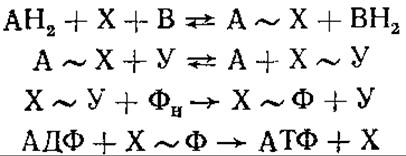
The chemical coupling hypothesis failed to account for two major facts. First, despite extensive searches, the actual existence of high-energy Intermediates of the A~X type could not be proven. Second, it remained unclear why an intact mitochondrial membrane is strictly required for oxidative phosphorylation.
These contradictions are readily resolved by the chemiosmotic (or proton-motive) concept. Formulated by the English biochemist P. Mitchell in 1961, its experimental validation was substantially advanced by V. P. Skulachev. The chemiosmotic theory posits that the chemical energy released during electron transport is converted into the electrical energy of a membrane potential, which is subsequently transformed into the chemical bond energy of ATP. According to this theory, the inner mitochondrial membrane (the coupling membrane) exhibits high electrical resistance and very low permeability to charged particles, primarily protons and hydroxide ions. The redox enzymes comprising the respiratory chain are embedded across the inner mitochondrial membrane.
It is postulated that the membrane also houses a system of enzymes—proton pumps—driven by the electron flow along the respiratory chain. Utilizing the energy released during electron transfer, these pumps extrude protons (without their counter-anions) from the matrix into the intermembrane space. Consequently, the intermembrane space becomes acidified, and the outer face of the coupling membrane acquires a positive charge. Simultaneously, an excess of OH- accumulates in the mitochondrial matrix, charging the inner side of the membrane negatively. Thus, a proton concentration gradient (ΔpH) and an electrical potential gradient (Δψ) are generated simultaneously across the coupling membrane.
The chemical energy of oxidation is thus transformed into electrical energy and stored as a membrane potential. The inner mitochondrial Membrane Functions analogously to a capacitor, with its surfaces acting as capacitor plates separated by a lipid insulator layer. The resulting electrochemical proton gradient (ΔμH+) therefore consists of a chemical component (ΔpH) and an electrical component (Δψ). The return flow of protons down their concentration gradient toward the matrix side of the membrane occurs via the ATP synthase complex. This very proton flux serves as the driving force for ATP synthesis. According to Mitchell's theory, one molecule of ATP is synthesized for every two protons passing through the membrane. Consequently, the electron transport chain must contain three proton pumps corresponding to the three phosphorylation sites.
The respiratory chain loops across the inner mitochondrial membrane three times. Each pair of electrons from respiratory substrates transported from NADH to oxygen effectively extracts three pairs of H+ ions from the inner matrix, which are then translocated outward into the intermembrane space. As a result, the transfer of two electrons leads to the synthesis of three ATP molecules (Fig. 7.5).

Fig. 7.5. Schematic representation of oxidative phosphorylation occurring in the inner mitochondrial membrane via a proton-motive mechanism
Thus, ATP synthase acts as a proton pump. It utilizes the downhill movement of protons along their gradient to synthesize ATP.
Today, the chemiosmotic theory is supported by a wealth of experimental evidence. For instance, E. Racker employed Bacteriorhodopsin—a retinal-containing chromoprotein—for this purpose. Upon absorbing light energy, bacteriorhodopsin generates a proton electrochemical gradient across the coupling membrane. H+-ATPase was isolated from bovine heart mitochondria and bacteriorhodopsin from halophilic Bacteria; these two proteins were subsequently incorporated into plant Phospholipids. This chimeric proteoliposome—a complex combining components from three distinct biological kingdoms (animals, plants, and bacteria)—demonstrated photosynthetic capability: upon Light absorption, it synthesized ATP from ADP and phosphate. Bacteriorhodopsin generated a proton current driven by light energy, which the H+-ATPase then utilized for ATP Biosynthesis.
In recent years, Yu. A. Ovchinnikov, V. P. Skulachev, and their coworkers successfully measured directly the electromotive force generated upon illumination of bacteriorhodopsin. The protein was embedded in a phospholipid film separating two compartments of a cell filled with an electrolyte solution and equipped with electrodes. Upon illuminating the bacteriorhodopsin (using a 15 ns laser flash), a potential difference of approximately 0.25 V arose across the membrane—a value sufficient to drive ATP synthesis.
The third Concept of the oxidation-phosphorylation coupling mechanism is the conformational hypothesis. It views mitochondrial function by analogy with Muscle contraction. Direct experiments demonstrate that mitochondria can undergo contraction and relaxation (detected via changes in light scattering). According to the conformational hypothesis, the chemical energy of oxidation is first utilized to perform mechanical work (altering mitochondrial conformation) and is only subsequently converted into the chemical bond energy of ATP. The energy released during electron transport in the respiratory chain is directly employed to drive the inner mitochondrial membrane into a new, high-energy conformational state. This stage can be likened to compressing a spring. Energy is then transferred to ATP, the membrane reverts to its initial conformation, and the "spring" relaxes. These tenets of the conformational scheme have not received experimental confirmation. In 1974, P. Boyer modified the conformational hypothesis, suggesting that conformational changes are driven by mitochondrial ATPase (ATP synthase).
Most researchers currently believe that The problem of Oxidative phosphorylation is best resolved within the framework of Mitchell's chemiosmotic theory. It outlines core principles and provides a universal approach to addressing specific questions and mechanisms of energy coupling. Nevertheless, the chemical and conformational hypotheses should probably not be dismissed entirely, as their utility in deciphering and refining specific aspects of Mitchell's theory remains undeniable.
The most challenging aspect of oxidative phosphorylation is elucidating the mechanism by which the electrochemical potential of H+ ions drives the ATP synthesis reaction. In explaining the utilization of electrochemical potential energy, P. Mitchell postulated a direct interaction between the proton flux and the active site of the ATPase system.
The mitochondrial ATPase complex (H+-ATPase, or ATP synthase) comprises a soluble catalytic domain (F1 factor) and membrane-embedded components (F0 complex). The F1 factor is a protein with a molecular mass (M) of 360,000–380,000. It possesses a complex quaternary structure consisting of five types of subunits: α (M = 54,000); β (M = 50,000); γ (M = 33,000); δ (M = 17,000); ε (M = 11,000).
The most probable subunit Stoichiometry of the F1 factor corresponds to α3β3γδε3. The principal catalytic Properties of the F1 factor reside in the α- and β-subunits. The active site is located near their interface, specifically on the β-subunit. A nucleotide-binding site with high affinity for substrates—ADP and Pi—is situated on the α-subunit of the enzyme (Fig. 7.6). The role of the γ- and δ-subunits is to mediate the linkage between the F1 factor and the remaining components of the ATPase complex. The ε-subunit functions as a natural inhibitor of ATPase activity.
The F0 complex contains four types of Polypeptides: the oligomycin sensitivity-conferring protein (OSCP, M = 19,000), F2 factor (M = 30,000), F6 factor (M = 8,000), and a DCCD1-binding proteolipid (M = 6,500). The F0 complex anchors the F1 factor to the membrane and participates in forming the proton-conducting pathway within the H+-ATPase complex. This pathway exhibits high Specificity, as it is permeable exclusively to protons.
According to the chemiosmotic theory of energy coupling, the ATP synthase complex couples the electrochemical proton gradient generated by the respiratory chain to ATP synthesis reactions. The specific roles of the ATPase are as follows: 1. Translocation of protons across The Lipid Bilayer from the outer side of the mitochondrial membrane to the F1 factor (translocation step). 2. Synthesis of ATP from ADP and Pi (catalytic step). 3. Ensuring the directionality of proton flow to harness electrochemical potential energy for maintaining high ATP concentrations (coupling step). The first two processes are carried out by the F0 and F1 factors, respectively, while the third requires coordinated functioning of both factors.
The DCCD-binding proteolipid of the F0 factor plays a primary role in proton translocation. It contains two hydrophobic regions and one hydrophilic region located near the middle of the molecule. It is hypothesized that the proteolipid spans the membrane in a hairpin conformation, positioning its polar segment on the outer surface of the membrane to serve as the H+ ion entry point into the channel.
1 DCCD (N,N'-dicyclohexylcarbodiimide) is a proton conductance inhibitor. Proteolipids are hydrophobic proteins containing covalently bound Fatty acids.
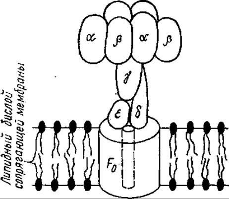
Fig. 7.6. Schematic model of the Molecular Organization of the ATP synthase complex:
α, β, γ, δ, ε are subunits of the coupling factor F1, F0 is the membrane component of the ATP synthase complex, and the dashed line indicates The pathway of protons
Proton translocation apparently involves Glu, Arg, and Tyr residues within the proteolipid. The most likely mechanism of proton transfer is a Relay-type transmission along the proton-donor and acceptor groups of these Amino Acids. Protons are driven electrophoretically down their concentration gradient.
The catalytic stage, namely the synthesis of ATP from ADP and H3PO4, can be formally viewed as a dehydration reaction: АДФ + Н3РO4 ⇄ АТФ + Н2O.
According to Peter Mitchell's initial hypotheses, the Coupling of oxidation and phosphorylation occurs through the utilization of ∆μH+ energy during the pumping of protons into the catalytic site of ATPase, which is located close to the proton-conducting pathway. The electrophoretic movement of protons along this path leads to their accumulation at the catalytic site. Two protons attack the phosphate oxygen and, upon combining with it, form a water molecule. This renders the phosphate group highly reactive and capable of binding directly to ADP. Phosphate protonation is energy-dependent; Mitchell postulated that it is precisely at this stage where ∆μН+ energy is utilized.
However, recent studies have led Paul Boyer (USA) to conclude that the rapid and reversible synthesis of ATP can occur at the ATPase catalytic site without the expenditure of ∆μН+ energy. The rate-limiting step of this reaction is the release of synthesized ATP from the enzyme's catalytic site into the matrix—that is, from the hydrophobic phase into the aqueous phase. It is precisely this process that is accelerated manifold (1,000-fold) upon membrane energization.
Energy-dependent protonation of specific groups within the ATPase complex, driven by ∆μН+ energy, can induce Conformational Changes in the F1 factor. These changes facilitate the rapid release of synthesized ATP from the catalytic site. It must be acknowledged that many specific aspects of ATP synthase function remain unresolved to date.
According to I. A. Kozlov and V. P. Skulachev (1977, 1984), alterations in The properties of the enzyme sites responsible for binding ATP, ADP, and H3PO4, as well as the nature of interaction between these sites, play a major role in the release of ATP from the ATPase catalytic site. Overall, the ATP synthase reaction proceeds along the same pathway as the ATPase reaction, via the reversal of all Hydrolysis steps.
According to A. D. Vinogradov (1984), the coupling factor F1 catalyzes either ATP synthesis or hydrolysis while existing in two alternative, mutually interconvertible states, the equilibrium between which is governed by The ratio of ATP to ADP in the mitochondria.
Proton ATPases isolated from the mitochondria of animals, higher plants, and Fungi, as well as from chloroplasts and bacterial cells, share a common structural layout and apparently operate via the same mechanism. A crucial feature is the reversibility of the reaction catalyzed by the ATPase complex. Under appropriate conditions, the F1—F0 complex can hydrolyze an ATP molecule and utilize the energy thus released to pump protons—that is, to generate a transmembrane electrochemical proton gradient.
Last update: 06/08/2026
Editorial and Educational Adaptation: This material has been compiled based on the primary/original source text. The project team performed an editorial review, corrected technical inaccuracies, structured sections, and adapted the content for an educational format.
What was processed:
- elimination of formatting defects (OCR errors, structural breaks, corrupted characters);
- editorial organization of content;
- standardization of terminology in accordance with academic sources;
- verification of factual statements against the original source text.
All mentions of the author, publication year, and origin of the primary text have been preserved in accordance with the source.