NEONATAL SURGERY - 1976
2. SPECIAL SECTIONS
8. Malformations of Individual Organs and Systems
Congenital Esophageal Malformations
Among Congenital Malformations of the Esophagus, esophageal atresia, tracheoesophageal fistulas, partial stenosis, chalasia, and achalasia are the most common. Infants with esophageal malformations generally require urgent surgical intervention. According to G. A. Bairov (1968), esophageal atresia occurs in approximately 1 out of every 3,500 newborns. In Finland, 1 in 3,000 infants is born with esophageal atresia (Louhimo, 1970). The Embryogenesis OF THE esophagus and its primary malformations are schematically illustrated in Fig. 46.
Class="center">Fig. 46. Formation of the esophagus, potential mechanisms of malformation development, and their main variants.
The esophagus and Trachea originate from the initial segments of the common primitive gut tube. The region of the future esophagus is highlighted in red (a). Next, a groove appears on the midline of the pharyngeal floor, which rapidly transforms into a tubular outgrowth—the trachea (b)—running parallel to the intestinal tube. The caudal end of the trachea begins to enlarge and bifurcate (c), forming the lung buds. Distal to the Pharynx, the digestive tube narrows noticeably to form the esophagus (b, c, d). The directions of normal growth and Separation of these structures are indicated by arrows in figure (c).
If the direction and rate of growth of the trachea and esophagus mismatch (compare Fig. e with Fig. c), esophageal atresia with a tracheoesophageal fistula may develop (Gruenwald).
The likely cause of atresia and stenosis of the esophagus, as well as the entire gastrointestinal tract, is considered to be a disruption of vacuolization processes during the solid cord stage, which the esophagus undergoes alongside other structures of the intestinal tube between the 20th and 40th days of gestation. Fig. f illustrates the potential outcomes of epithelial proliferation upon the resolution of the solid stage: normal restoration of the esophageal lumen, atresia, and stenosis. Fig. g schematically presents the main clinical variants of esophageal atresia.
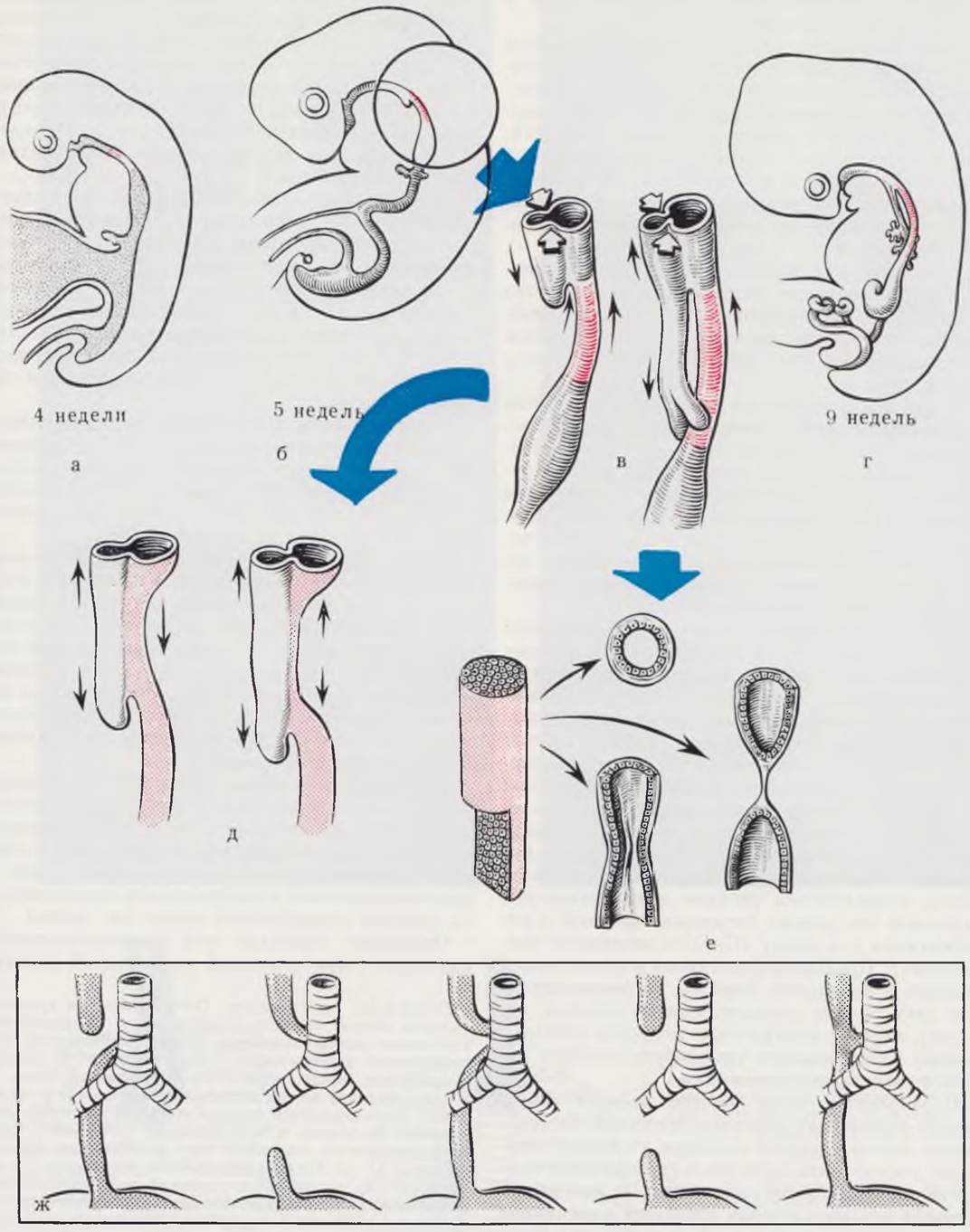
The most frequent esophageal malformation, occurring in 85–95% of all patients (Guendet, 1968; Kummer, 1970; Koop, 1971), is atresia of the upper blind-ending segment accompanied by a lower tracheoesophageal fistula.
Clinical presentation. The manifestations of esophageal atresia are distinct. It is crucial to bear in mind the possibility of this malformation. Just 2 to 3 hours after birth, the upper blind pouch of the esophagus and the nasopharynx become filled with mucus, and the infant develops profuse frothy discharge from the Mouth. Some of the mucus is aspirated, leading to episodes of cyanosis. Initially, maternity hospital staff often fail to pay proper attention to these frothy secretions; a nurse may suction the nasopharynx, and the infant's condition temporarily improves. However, the frothy oral secretions and cyanotic spells persistently recur, and signs of aspiration Pneumonia develop rapidly.
Such persistent frothy oral secretions and cyanotic episodes make it possible to suspect esophageal pathology within the first 4 to 5 hours of the newborn's life, and certainly before the first feeding. The Diagnosis is confirmed by catheterizing the esophagus using a thin, rounded-tip urethral catheter. The catheter is inserted through the Nose into the blind esophageal pouch, and mucus is aspirated with a syringe. Subsequent insufflation of air through the catheter results in a characteristic noisy escape from the nasopharynx (a positive Elefant sign). If the upper esophageal segment communicates with the trachea, respiratory disturbances will dominate the clinical picture, as the Contents of the upper esophageal pouch enter the trachea through the fistula.
In recent years, the majority of foreign authors (Romstadt, 1966; Raffensperger, 1970; Velona, 1971) have strongly recommended routine esophageal probing for all newborns without exception. Mandatory esophageal probing has been implemented in Kyiv, Riga, and Leningrad. This simple Procedure easily diagnoses esophageal malformations immediately after birth, enabling the timely transfer of the infant to a surgical department.
The presence of a lower tracheoesophageal fistula is diagnosed by physical examination and Percussion of the Abdominal cavity. When the lower esophageal segment does not communicate with the trachea, air fails to enter The Stomach and intestines; the abdomen appears scaphoid, and percussion across all Abdominal Regions elicits a dull sound. In patients with a lower tracheoesophageal fistula, the stomach and intestines are distended with air, which is readily detected upon abdominal inspection and percussion.
Thus, the diagnosis and even the Determination of the specific form of esophageal atresia present no difficulty in the majority of patients. Even without esophageal catheterization, the diagnosis of esophageal atresia can be established within the first hours of the infant's life.
Diagnosing tracheoesophageal fistulas without atresia poses significant challenges. Depending on the caliber of the fistula, the clinical picture in such patients is dominated either by respiratory disorders—especially during feeding while the infant is lying on their left side—or by recurrent pneumonias. In these cases, the diagnosis is refined through radiological examination or direct visualization of the fistula via tracheobronchoscopy.
Once the diagnosis is established, oral feedings are withheld. A thin, preferably double-lumen catheter is inserted nasally into the upper blind esophageal pouch, through which mucus is carefully aspirated every 20 to 30 minutes. The infant is placed in a HEAD-elevated position to prevent the regurgitation of acidic gastric contents into the Lungs and the subsequent development of aspiration pneumonia. The infant is transported to the surgical department in this position. Continuous suctioning of mucus from the upper esophageal pouch during transport is mandatory. Prior to dispatching the infant to the surgical department, injections of vikasol and Antibiotics are administered (see the chapter "Preoperative Preparation"). We do not recommend performing radiological examinations in the maternity hospital, as this imposes an additional radiation load and frequently leads to the contrast agent being aspirated from the esophagus into the tracheobronchial tree, precipitating pneumonia.
Upon admission to the surgical unit, a radiological examination of the infant is mandatory, aimed at confirming the diagnosis and determining the presence and severity of aspiration pneumonia. In recent years, the clinic has abandoned The Use of contrast media in the upper esophageal pouch. For most patients, a radiogram utilizing a radiopaque catheter inserted into the proximal esophageal segment is quite sufficient to clarify the diagnosis (Fig. 47). If contrast enhancement of the esophagus becomes necessary, iodolipol (no more than 1–1.5 ml) is used. Following the radiograph, the iodolipol is aspirated, and only then is the catheter removed. Leaving the contrast medium in the esophagus (Fig. 48) leads to its subsequent aspiration into the tracheobronchial tree.
Fig. 47. Chest radiograph of a newborn. Diagnosis: esophageal atresia. The catheter delineates the upper segment of the esophagus. There is no communication between the lower segment and the trachea.

Fig. 48. Chest radiograph of a newborn. Diagnosis: esophageal atresia. Iodolipol left in the esophagus has been aspirated into the trachea. Massive right-sided pneumonia.

When interpreting pulmonary changes, it should be kept in mind that modern diagnostic X-ray equipment is, in most cases, not specifically adapted for imaging newborns. The resulting lung radiographs tend to be overexposed, a factor that must be accounted for when reading the images. The actual extent of lung damage in such patients is significantly greater than what is apparent on the radiograph.
Concomitant malformations are frequent in infants with esophageal atresia, occurring in 20–50% of cases according to various authors (G. A. Bairov, 1968; Okmian, 1966; Forrester, Cohen, 1970; Toyama, 1971), which considerably worsens Treatment outcomes.
Treatment. Esophageal atresia requires surgical intervention. Infants admitted to the clinic within the first 10 – 12 hours after birth do not require special preoperative preparation. In cases of late admission accompanied by pronounced aspiration pneumonia, preoperative preparation and pneumonia treatment are carried out following standard protocols until the patient's condition improves. A gastrostomy is performed to ensure adequate Nutrition.
Most authors prefer a right-sided extrapleural or transpleural approach. In recent years, right-sided lateral thoracotomy with a vertical Skin incision along the midaxillary line (see above) has been considered the procedure of choice for the Surgical treatment of esophageal atresia.
The surgery is performed under endotracheal anesthesia with artificial pulmonary ventilation.
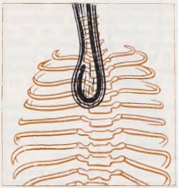
Surgical technique. After opening the thoracic cavity, the lung is retracted medially. The mediastinal Pleura is incised, and the azygos vein is mobilized, ligated, and transected. Guided by the Vagus nerve and trachea, the ends of the esophagus are located within the mediastinal Tissues. Locating and isolating the proximal segment is facilitated by the preliminary insertion of a catheter into it. A primary (direct) anastomosis is feasible when the diastase does not exceed 1.5 cm. Bridging a diastase of up to 1.5 cm is achieved primarily through the mobilization of the proximal segment. Mobilization of the distal segment may compromise its Blood supply and subsequently lead to anastomotic leakage. After esophageal mobilization, the distal segment is transfixed with a nylon ligature near its junction with the trachea, ligated, and transected. We consider end-to-end anastomosis using a single row of interrupted inverting sutures on an atraumatic needle (No. 00000 – 000000) placed circumferentially to be the method of choice (Fig. 49). If the wall of the distal segment is well-developed, a two-row Haight anastomosis may be used. It should be borne in mind, however, that the slightest tension typically causes the second row of sutures to cut through the wall of the distal segment. Complex suture techniques have been entirely abandoned.
Fig. 49. Types of esophageal sutures.
a — single-row inverting interrupted suture along the entire circumference of the anastomosis; b — two-row telescopic Haight anastomosis; c — mechanical suture.

In recent years, mechanical sutures created with various stapling devices have found application in neonatal esophageal surgery (G. A. Bairov, 1973; Yu. L. Doroshevsky, 1973; Okmian et al., 1969).
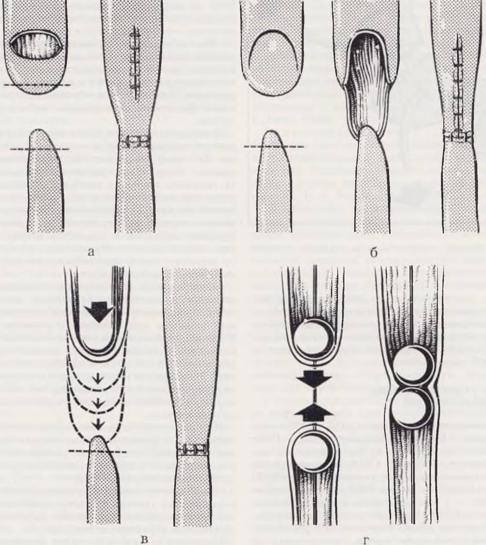
Sulamaa suggests suturing the tracheoesophageal fistula without transecting it, followed by an end-to-side anastomosis (proximal end to distal segment); this approach is used in selected patients with no diastase and a small diameter of the distal segment. The esophagus can be lengthened by 4 – 5 mm using various Methods (Fig. 50). Okmian performs a full-thickness transverse incision across half the circumference of the proximal esophageal wall, followed by longitudinal closure of the wound. In our view, this method is the simplest and we use it with success. The anterior lip of the anastomosis,
as a rule, is sutured over a catheter with an outer diameter of no more than 2 mm, which is left in the esophagus for 2 – 5 days. The catheter is used to feed the infant and, when necessary, to aspirate gastric contents.
Fig. 50. Methods of esophageal lengthening: Okmian's technique (a); Ten-Kate — Bairov technique (b); bougienage according to Howard (c); traction lengthening according to Rehbein (d).
Attempting to replace an esophageal defect using a transverse colon segment via Waterston's method is justified in newborns only when performed by a highly experienced surgeon who has mastered the technique of esophagoplasty. Soave (1972) successfully performed two such operations. He cites compiled data from Holder (1963), showing a 16.7% rate of direct anastomotic dehiscence out of 747 cases of esophageal atresia. For esophagoplasty in esophageal atresia, some authors (Vidue, Levy, 1970) use Pericardium, while others (D. E. Bablyak, 1973) propose harvesting strips from it to anchor the esophagus above and down the anastomotic suture line in order to reduce tension on the latter. These methods should be considered poorly advisable, as any failure of the anastomotic sutures and the subsequent development of purulent intrapleural complications would expose the opened pericardial sac to the risk of infection.
Following the creation of the anastomosis, hemostasis is checked. The integrity of the mediastinal pleura is restored. In questionable cases, the pleural cavity is drained for 2 – 3 days. The wound is closed tightly in layers (the drain is brought out through a separate stab wound in the chest wall).
When an anastomosis is not feasible, the operation is concluded by transfixing and ligating the tracheoesophageal fistula, alongside gastrostomy and esophagostomy. Esophageal replacement using a segment of the Large Intestine is performed at an older age.
In the postoperative period, in the absence of complications, enteral feeding via the catheter left in the esophagus is initiated as early as the 2nd day after surgery. The pleural drain is removed on the 2nd to 3rd day.
The Outcomes of surgical treatment for esophageal atresia have improved significantly. According to various authors, 43% to 85% of infants with esophageal atresia are successfully saved (Livaditis, 1968; Rickham, 1971). In recent years, we have successfully saved every second to third infant admitted to the clinic within the first 2 days of life.
The treatment outcomes for premature infants born with esophageal atresia are considerably worse. Consequently, Howard (1965) suggests abandoning radical surgery in premature infants in favor of a staged procedure.
The First stage, upon the infant's admission to the clinic, involves performing a gastrostomy (Fig. 51) or a laparotomy followed by transecting the stomach at the border of its upper and middle thirds and bringing both ends out onto the abdominal wall. A tube is inserted into the duodenum to establish enteral feeding. The infant is kept in a position with the head of the bed elevated. The contents of the proximal blind esophageal pouch are continuously aspirated through a tube. A thick, elastic rubber catheter is introduced into the proximal segment 2 – 3 times a day to gently stretch the esophagus. In this manner, the esophagus can be stretched by 2 – 2.5 cm over 2 – 3 weeks. The next stage involves a thoracotomy, division of the tracheoesophageal fistula, and anastomosis of the esophageal ends. After the anastomosis and chest wall wound have healed, the gastrostomy is closed. This approach improves treatment outcomes in premature infants. The successful application of this method has been reported by Lafer and Boley (1966).
Fig. 51. Double gastrostomy with gastric division according to Meeker.

Children who have undergone surgery for esophageal atresia require subsequent dispensary follow-up for the early detection and management of potential esophageal strictures (which develop postoperatively in every 3rd to 5th child).
Congenital Heart and great vessel defects
Heart defects occur in 0.3 – 1.3% of newborns (L. M. Bolkhovitinova, 1961; V. V. Badmaeva, 1962; B. A. Konstantinov et al., 1966; Nadas, 1963). In the USA, approximately 10,000 children die annually from Congenital heart defects, and over 6,000 in England, with more than 2/3 of them dying During the first weeks and months of life (Aberdeen, 1969). According to our observations, 70% of children born with heart defects die within the 1st year of life, one-third of whom die in the first 4 weeks of life. Among newborns, mortality from heart defects accounts for 9 to 11% of total mortality. M. L. Petrov-Maslakov and I. I. Klimets (1965) state that during the 1st week of life, heart defects rank second only to birth trauma as a cause of neonatal mortality. Lambert (1966) considers them the primary cause of lethality in this age group. Meanwhile, more than half of the children who die in the first weeks and months of life have technically correctable defects (V. I. Burakovsky, B. A. Konstantinov, 1970).
Although heart defects in newborns constitute the dominant type of cardiovascular pathology at this age (accounting for 80 – 90% of all organic heart diseases), they do not encompass the entirety of pathology within this system.
All cardiovascular diseases in newborns are divided into primary, characterized by direct damage to The Heart, and secondary, where hemodynamic disturbances are caused by functional or anatomical disorders in any other organ.
Primary lesions include: 1) congenital heart and vascular defects; 2) myocarditis; 3) cardiomyopathies; 4) arteriovenous fistulas. Secondary conditions include: 1) hyaline membrane disease; 2) circulatory failure resulting from hemolytic anemia; 3) cardiomegaly with atrioventricular valve insufficiency resulting from birth asphyxia; 4) acute "Cor Pulmonale" following aspiration complications; 5) circulatory failure due to birth-related craniocerebral trauma.
Despite the wide variety of etiologies and pathogenetic mechanisms of these conditions, they share a common feature: they are most often congenital in nature or associated with the labor and delivery process. Congenital pathologies include heart and vascular defects resulting from embryopathy or Chromosomal aberrations. Neonatal myocarditis also originates intrauterinely, as many Viruses are capable of crossing the Placenta from mother to fetus.
Cardiovascular lesions associated with the delivery process include the consequences of asphyxia, aspiration, and resuscitation, such as acute "cor pulmonale," cardiomegaly in craniocerebral trauma, etc.
The anatomical and hemodynamic characteristics of newborns are such that they hinder not only the topical diagnosis of a defect, but also the determination of the general Nature of the pathology.
The clinical picture of various heart defects is characterized by stereotypy and the predominance of general symptoms. Local signs fail to fully develop and are poorly expressed. Such a classic symptom of a Heart defect as a murmur may be absent in newborns. When a murmur is pronounced, its diagnostic value is limited, as it typically lacks the range of tones and other characteristics acquired with age. Dyspnea and cyanosis in infants during the first weeks of life, alongside heart defects, can also manifest As a result of respiratory, cerebral, metabolic, and other disorders.
Cyanosis in a newborn can be of vasomotor, pulmonary, cerebral, metabolic, or cardiac origin. A careful physical examination and simple tests help determine the underlying cause of the cyanosis.
Vasomotor cyanosis usually appears during the 2nd to 3rd week of life, less commonly within the first months. It is characterized by peripheral localization (hands, feet). The Lips and mucous membranes always remain pink. In septicopyemia, cyanosis can become generalized.
Pulmonary cyanosis is most frequently associated with Atelectasis, appears shortly after birth, and has a generalized character. Typically, this type of bluish discoloration diminishes when the infant cries and upon oxygen inhalation.
In hyaline membrane disease, the condition begins with dyspnea, and cyanosis reaches its peak within 24 — 48 h. As is well known, this pathology is most commonly observed in premature infants and full-term newborns whose mothers have Diabetes Mellitus.
Other causes of pulmonary cyanosis—such as pneumonia, diaphragmatic hernia, tracheoesophageal fistula, and pulmonary agenesis—are diagnosed using general clinical methods and X-ray Examination.
Cerebral cyanosis is observed in cerebral edema caused by an intracerebral Hemorrhage. In addition to a specific medical history, a characteristic hemicyanosis with a clear axial demarcation is often noted, which led foreign authors to term this symptom the "harlequin color change" (or "harlequin fetus").
Metabolic cyanosis occurs in neonatal tetany when serum calcium levels are below 8 mg/100 ml and hyperphosphatemia is present, as well as in methemoglobinemia. Administration of calcium gluconate or methylene blue AIDS in identifying these forms of cyanosis.
Cardiac cyanosis is shunt-related in origin, typically systemic in nature, and carries a consistently grave prognosis. Cardiac cyanosis worsens with crying, is accompanied by dyspnea, hepatomegaly, and a gallop rhythm, and is combined with varying degrees of circulatory failure. This latter syndrome and arterial hypoxemia are the primary causes of high mortality rates in newborns with congenital heart defects.
Anamnestic and physical examination of a newborn with suspected heart defect is conducted according to general guidelines. Electrocardiography is mandatory. Notably, recording standard leads alone can provide valuable diagnostic information. For instance, detecting right atrial and right ventricular hypertrophy is possible as early as 48 — 72 h after birth, while left-sided hypertrophy can be identified immediately after birth. The latter sign most frequently points to a Congenital heart defect (tricuspid atresia, pulmonary atresia with an intact ventricular septum). For more detailed information, chest leads must be recorded, with data interpretation tailored to the patient's age (M. Sandrucci, G. Bono, 1966).
X-ray examination makes it possible to assess the pulmonary pattern, the size and configuration of the cardiac silhouette, and The Nature of its contours. Interpretation of radiographs is performed taking age-related norms into account (V. I. Burakovsky, B. A. Konstantinov, 1970).
The greatest diagnostic value in recognizing heart defects and determining prognosis is provided by specialized instrumental examination methods: cardiac catheterization and radiopaque angiocardiography. Indications for their use are established when there is firm conviction that the infant has a heart defect.
The Procedures are performed under special premedication or anesthesia. A catheter for pressure recording, blood sampling, and contrast agent administration (1.5 — 2 ml/kg) is inserted via the axillary vein, the great saphenous vein of the thigh, the main femoral vein, or through the umbilical vessels.
In newborns, selective angiocardiography is safer and provides sufficient information, allowing clinicians to bypass cardiac catheterization in the vast majority of cases. Only a comprehensive examination of the sick infant, progressing from simpler to more complex methods as necessary, makes it possible to unravel cardiovascular pathology.
The majority of congenital heart defects exhibit a distinct chronological age pattern in their initial clinical manifestations. Anomalies in which hemodynamics are disrupted during the Cytology/cytology/16.html">Early stages of placental Circulation manifest within the first 4 weeks of life (first group). These include valvular atresia and hypoplasia of a particular cardiac chamber.
Defects of the second group are observed in newborns, but may also manifest at an older age. These defects cause acute circulatory disturbances only after birth—due to the onset of pulmonary Respiration and an increase in Cardiac Output (complete transposition of the great Arteries, persistent truncus arteriosus, cor biloculare, etc.).
Defects of the third group are not detected in newborns. This category includes atrial septal defects, ventricular septal defects, aortopulmonary septal defects, etc. In septal defects, The Development of the clinical picture in neonates is hindered by high pulmonary arteriolar resistance. For example, in ventricular septal defects, a blood shunt occurs as pulmonary vascular resistance decreases—i.e., starting from two months of age, which is when the first symptoms of the disease appear. In Tetralogy of Fallot, mechanisms associated with age-related physiological features operate during the 1st month, preventing
the Development of the pathological effect (postembryonic polycythemia, low basal metabolic rate, etc.).
The fourth group comprises defects in which hemodynamic disturbances can manifest at any age depending on the specific anatomical variant.
Distinguishing these four categories narrows The Scope of investigation during the topical diagnosis of congenital heart defects in newborns (Table 16).
Table 16. Classification of Congenital heart defects According to the age chronology of initial symptoms
Defects manifesting exclusively in newborns |
Defects manifesting predominantly in newborns |
Defects not manifesting in newborns |
Defects occurring in all age groups |
1. Hypoplastic left heart syndrome |
1. Complete transposition of the aorta and pulmonary artery |
1. Atrial septal defect |
1. Patent ductus arteriosus |
2. Pulmonary valve atresia with intact ventricular septum |
2. Truncus arteriosus communis |
2. Ventricular septal defect |
2. Coarctation of the aorta |
3. Infradiaphragmatic total anomalous pulmonary venous drainage |
3. Cor biloculare |
3. Tetralogy of Fallot (except cases with pulmonary atresia) 4.4. Anomalous Water/144.html">Origin of the left coronary artery from the pulmonary artery |
3. Pulmonary valve stenosis 4. Aortic stenosis 5. Tetralogy of Fallot with pulmonary atresia 6. Tricuspid atresia 7. Single ventricle 8. Ebstein's anomaly 9. Complete (supradiaphragmatic) anomalous pulmonary venous drainage 10. Atrioventricular septal defect |
The proportion and significance of individual congenital heart defects vary across different age periods. Therefore, in this chapter, we provide a clinical Description of the heart defects most commonly encountered in newborns.
Complete transposition of the aorta and pulmonary artery
Complete transposition of the aorta and pulmonary artery is one of the most common congenital heart defects in newborns. According to autopsy data, its incidence ranges from 8.4% to 37.5% of all cardiovascular diseases in neonates (B. A. Konstantinov, 1969; Donzelot, Dallaines, 1954). This defect has been described as "the most frequent cause of cyanosis and Heart Failure" in infants or as "the most common defect requiring surgery" in children during their first weeks and months of life (Cooley, 1966).
In complete transposition of the great arteries, the aorta arises from the right ventricle, lying anterior to the orifice of the pulmonary artery. The latter originates from the left ventricle and is positioned posterior to the aorta.
Gas exchange and, consequently, the patient's survival are possible only if there are additional communications between the SYSTEMIC AND PULMONARY circulations. Most commonly, these include a patent foramen ovale, a ventricular septal defect, an atrial septal defect, or a patent ductus arteriosus. Shunting of blood occurs through these pathways, and it is only in this manner that a portion of oxygenated blood can reach the aorta, while a portion of venous blood enters the lungs. The magnitude of the shunt in vessel transposition is determined by the size of these communications, vascular resistance, and other factors. The amount of oxygenated blood entering the aorta in such patients is small; therefore, immediately after birth, the infant experiences severe arterial hypoxemia of a degree unseen in any other defect. We have observed newborns in whom arterial oxygen saturation was 20 – 40% HbO2.
Shortly after birth, patients exhibit a significant increase in circulating blood volume as a compensatory mechanism for tissue Hypoxia. This inevitably leads to cardiac overload and the development of circulatory failure.
Thus, the hemodynamics in complete transposition of the great arteries are characterized by the separation of the circulations, wherein the right ventricle Functions as a pump for the systemic circulation. Gas exchange is maintained through variable blood shunts via additional communications.
Clinical presentation. A high cardiac output leads to cardiac overload and the development of circulatory failure. The defect occurs predominantly in boys. Infants are born with a normal birth weight. The initial symptom—cyanosis—appears on the first day of life and is intense, persistent, and generalized. During the 1st to 2nd week, there is no dyspnea, the heart is not enlarged, and its configuration is normal. Edema, hepatomegaly, and other signs of circulatory failure are absent at this time. From the 3rd to 4th week of life, tachypnea, feeding difficulties, tachycardia with gallop rhythm, hepatomegaly, pulmonary rales, peripheral edema, and ascites develop. The cardiac shadow rapidly increases in size, progressively acquiring an obliquely positioned oval shape or that of an "egg on its side" (as described by English authors).
A deceptive "well-being" in a cyanotic newborn during the first 2 to 3 weeks of life, followed by rapidly developing and relentlessly progressing cardiac decompensation, is a characteristic feature of complete transposition of the great arteries.
However, one should not wait for decompensation to develop in a cyanotic newborn before making a diagnosis. Considering that the average life expectancy for a child with complete transposition of the great arteries is 3 months (Hanlon, Blalock, 1948), it becomes clear how little time a physician has to establish a topographic diagnosis and how crucial it is during the first 2 to 3 weeks of life. Objective Examination, aside from profound and total cyanosis, reveals a precordial bulge and, in some patients, a systolic murmur to the left of the Sternum. Complete or near-complete aphonia is more characteristic. Symptoms of circulatory failure, as mentioned above, appear from the 3rd to 4th week of life.
Electrocardiography demonstrates right axis deviation with marked right ventricular hypertrophy. Right ventricular overload is generally not detected.
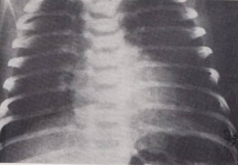
Radiological findings indicate normal or increased pulmonary blood flow (hypervolemia) and normal heart size. From the 3rd to 4th week of life, the cardiac silhouette enlarges to the left, highlighting the narrow vascular pedicle in the anteroposterior projection, and increasingly resembles an oval (Fig. 52).
Fig. 52. Chest radiograph of an infant with complete transposition of the aorta and pulmonary artery, and a patent foramen ovale. The cardiac silhouette is enlarged and oval-shaped.
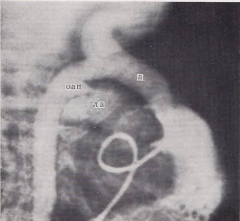
If this defect is suspected, the infant should undergo selective radiopaque angiocardiography from the right ventricular cavity with mandatory biplane imaging. A definitive sign of the defect is the visualization of the aorta arising from the right ventricle and lying anterior to the pulmonary artery. Furthermore, the aortic bulb is located unusually high and seemingly anterior to the cardiac shadow (Fig. 53).
Fig. 53. Selective angiocardiogram of an infant with complete transposition of the aorta and pulmonary artery, patent foramen ovale, and patent ductus arteriosus. The aorta (a) is opacified from the right ventricle and lies anterior to the pulmonary artery (pa), with the latter filling via the ductus arteriosus (da).
Differential diagnosis OF the defect is presented in Table 17.
Table 17. Differential diagnosis of major cardiac disorders in newborns
Diagnosis |
Onset of initial symptoms from birth |
Initial symptom |
Dyspnea |
Cyanosis |
Pulse |
ECG |
Cardiomegaly |
Circulatory failure |
Enhanced pulmonary pattern |
Note |
|
1. Hypoplastic left heart syndrome |
First day |
Sudden dyspnea |
+++ |
+ |
Not palpable |
Right axis deviation |
+ + + |
+++ |
+++ |
Occurs twice as often in boys; 75% mortality in the 1st week of life |
|
2. Pulmonary valve atresia |
From birth |
Cyanosis |
+++ |
++ |
Normal |
Left heart overload |
++ |
+++ |
— |
||
3. Infradiaphragmatic pulmonary venous drainage |
24 h – 7 days |
" |
++ |
++ |
Weak |
Right axis deviation |
— |
++ |
+++ |
||
4. Transposition of great arteries |
From birth |
Cyanosis |
+ |
+ + + |
Normal |
Right axis deviation |
++ |
++ |
— or ++ |
Cardiac silhouette resembles an "egg on its side". Occurs 2 1/2 times more frequently in boys |
|
5. Coarctation of the aorta |
Any |
Dyspnea |
++ |
— |
Bounding in upper extremities |
Right axis deviation |
++ |
++ |
— or + |
||
6. Tricuspid atresia |
From birth |
Cyanosis |
+ — |
++ |
Normal |
Left axis deviation |
+ |
— |
— or + |
||
7. Truncus arteriosus |
1 – 4 weeks |
Dyspnea |
++ |
+ — |
Bounding, full |
Combined hypertrophy |
+ |
+ — |
+ + or — |
||
8. Tetralogy of Fallot with pulmonary atresia |
1 – 4 weeks |
Cyanosis |
+ — |
Normal |
Right axis deviation |
+ |
— |
— |
|||
Combined hypertrophy |
|||||||||||
9. Patent ductus arteriosus |
Any |
Dyspnea |
++ |
— |
Bounding, full |
+ |
++ |
++ |
|||
10. Ebstein's anomaly |
" |
Cyanosis |
+ |
++ |
Normal |
Low-voltage fractionated complexes typical |
++ |
++ |
— |
||
11. Myocarditis |
" |
Tachycardia |
++ |
+ — |
Weak |
++ |
++ |
— |
|||
12. Respiratory distress syndrome (hyaline membrane disease) |
First 6 h of life |
Dyspnea |
+++ |
+ |
" |
+ |
++ |
++ |
Particularly common in premature infants |
Legend: — symptom absent, + — inconsistent symptom, + moderately pronounced symptom, + + significantly pronounced symptom, +++ severely pronounced symptom.
Treatment. A newborn with transposition of the great arteries requires immediate medical management. Treatment includes Oxygen therapy and the administration of cardiac Glycosides. Surgical intervention should be performed as soon as possible after diagnosis is established.
Various palliative procedures aimed at increasing blood shunts are employed to reduce arterial hypoxemia.
Recently, closed atrioseptostomy performed directly in the cardiac catheterization laboratory has become widespread. This procedure, which is carried out without thoracotomy, is indicated when arterial blood oxygen saturation falls below 75% HbO2 (Rashkind, Miller, 1966).
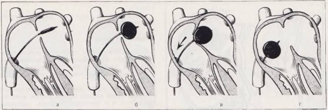
To enlarge the interatrial communication, a Fogarty catheter is advanced through one of the peripheral Veins into the heart. Once the operator has confirmed that the catheter tip is in the left atrium, a balloon located at the tip of the catheter is inflated using a radiopaque contrast agent. The catheter is then pulled back to draw the interatrial septum toward the orifice of the INFERIOR VENA CAVA, and with a sharp tug, the catheter is pulled out of the left atrium. This maneuver tears the interatrial septum, establishing a blood shunt through the newly created defect (Fig. 54).
Fig. 54. Diagram of closed atrioseptostomy. Advancing the catheter into the left atrium and inflating the balloon (a, b). Pulling the interatrial septum down to the inferior vena cava (c), sharply withdrawing the catheter, and creating a defect in the cardiac septum (d).
Typically, the therapeutic effect is immediate, manifesting as a reduction or even complete disappearance of cyanosis and an increase in peripheral arterial oxygen saturation (B. A. Konstantinov, 1969; Yu. S. Petrosyan, V. A. Garibyan, 1972). Other procedures—such as atrioseptectomy, interarterial anastomosis, and pulmonary artery banding—are rarely performed in newborns.
Hypoplasia of the Left Heart
Left heart hypoplasia is one of the most common congenital heart defects, accounting for 7–15% of all congenital cardiac anomalies (V. P. Zhukovsky, 1913; Noon, Nadas, 1958). It is observed exclusively in neonates, as 75% of affected infants die within the first week of life, and the remainder within the first month. The defect is a complex clinical and morphological syndrome consisting of ascending aortic atresia and left ventricular hypoplasia. This collective term also encompasses pre-natal closure of the foramen ovale, as well as atresia or severe stenosis of the mitral valve, which typically occur in association with one another and lead to underdevelopment of the left heart chambers and the aortic valve.
The Pathogenesis of this defect involves a decreased Blood flow through the foramen ovale into the left ventricle and an increased shunting of blood through the ductus arteriosus during placental circulation. The general morphological signs of the hypoplastic left heart syndrome are: 1) severe hypoplasia of the left chambers
of the heart, its valvular apparatus, and the aortic tract; 2) Hypertrophy of the right chambers with an enlarged pulmonary artery and a patent ductus arteriosus; 3) secondary endocardial fibroelastosis.
The ascending aorta is sharply narrowed, its lumen is preserved, and the valve cusps are completely fused to one another. The mitral ring has a small diameter, and its leaflets are underdeveloped or fused. The left ventricle has thick walls and a slit-like cavity. The endocardium is dense, coarse, and whitish-yellow.
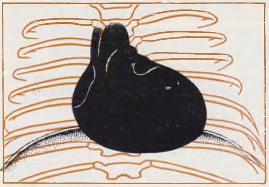
Hemodynamics are disrupted early in placental circulation. The blood flow pathway can be represented as follows: venae cavae —> right atrium —> right ventricle —> pulmonary artery —> ductus arteriosus —> aortic arch (retrograde) —> descending aorta (Fig. 55).
Fig. 55. Hemodynamics in hypoplastic left heart syndrome.

After birth, the direction of circulation remains unchanged. Blood is pumped into the lungs under high pressure, while venous drainage is impeded due to the small capacity of the underdeveloped left heart chambers. Consequently, arterial pulmonary Hypertension with venous congestion develops immediately after birth. Blood from the left atrium can only drain into the right atrium, where arterial and venous streams mix. A mandatory condition for the child's survival, in Addition to a patent ductus arteriosus, is the presence of an atrial septal defect (patent foramen ovale) or anomalous pulmonary venous drainage. The entire pumping function is performed by the right ventricle. The hemodynamics in left heart hypoplasia are characterized by a single functioning (right) ventricle, pulmonary hypertension with venous congestion, and arterial hypoxemia.
Clinical presentation. The defect is more frequently observed in boys. The disease manifests with a sudden onset—shortness of breath during the first day of life. Tachypnea is accompanied by progressive circulatory failure. Cyanosis appears later and, although generalized, never reaches severe degrees (Noon, Nadas, 1958).
Objective examination reveals markedly accelerated (80–100 breaths per minute) and irregular breathing, with retraction of the compliant areas of the chest and nasal flaring. Notable findings include weak peripheral pulses, which are undetectable even in the carotid vessels. In most cases, heart murmurs are absent.
Cardiac Auscultation reveals embryocardia, while bronchial breathing and an Abundance of moist rales of various calibers are audible in the lungs from the first days of life. The Liver enlarges rapidly, whereas edema and ascites usually do not have time to develop.
The Electrocardiogram is dominated by right ventricular strain patterns. Left axis deviation with combined hypertrophy is less common. In such cases, autopsy reveals left ventricular fibroelastosis.
Radiological examination shows cardiomegaly within the first hours after birth. The cardiac silhouette is abnormally rounded and progressively increases in size (Fig. 56). Angiocardiography reveals an enlarged right ventricle and a descending aorta that is a direct continuation of the ductus arteriosus. Following the pulmonary capillary phase, the right heart chambers opacify. Visualization of the left chambers and the ascending aorta is not achieved.
Fig. 56. Chest radiograph of an infant with hypoplastic left heart syndrome and ascending aortic atresia. The cardiac silhouette is abnormally shaped and severely enlarged.

No effective treatment has been found to date.
Coarctation of the Aorta
This defect is a congenital narrowing of the aortic lumen, up to its complete interruption, typically located in the region of the aortic isthmus near THE ORIGIN OF the patent ductus arteriosus.
Coarctation of the aorta is fairly common among infants in their first weeks of life, although it is also observed in adult patients, accounting for about 10 — 15% of all congenital cardiovascular malformations (Mehrizi, 1964). According to Sloan and Cooley (1953), aortic coarctation occurs once in every 500 autopsies. It is twice as common in boys (Campbell, Polani, 1961).
The Anatomy of the defect can vary significantly, which explains the large number of proposed classifications.
For the practicing physician, it is sufficient to distinguish between two MAIN TYPES OF the defect — postductal and preductal (Fig. 57).
Fig. 57. Classification of various types of aortic coarctation: postductal (adult) coarctation (a). Preductal (infantile) coarctation (b, c, d).

In postductal or "adult" coarctation of the aorta, the narrowing is located distal to the opening of the ductus arteriosus (botallian duct) or the ligamentum arteriosum and is usually focal in length. Patients invariably exhibit Left Ventricular Hypertrophy, and approximately 25% have a bicuspid aortic valve. Circulatory impairment is caused by a mechanical obstruction. Collateral vessels develop early to bypass the site of narrowing. Two distinct hemodynamic regimes emerge in the upper and lower halves of the body. The arterial duct may be obliterated or patent.
In preductal or "infantile" coarctation of the aorta, the narrowing is situated proximal to the opening of the ductus arteriosus. The latter typically has a large diameter and appears to be a continuation of the pulmonary artery into the descending aorta. Preductal aortic coarctation may be localized, extensive, or extend into the aortic arch.
In the latter variant, 80% of cases present with additional severe cardiac defects — ventricular septal defect, atrioventricular canal, single ventricle, transposition of the great vessels, etc. (Keith e. a., 1968). With the infantile type of aortic coarctation, systemic hypertension does not develop, and collateral circulation is absent. At the same time, patients with this variant of the defect establish a right-to-left shunt: from the pulmonary artery into the descending aorta. Consequently, the right side of the heart experiences severe overload, as it receives the entire cardiac output of the left heart plus an additional fraction of its own output equal to the shunt volume. The Pulmonary Circulation is characterized by high blood pressure. Thus, the "adult" type of the defect features arterial hypertension of the upper body, whereas the "infantile" type features pulmonary hypertension.
Clinical presentation. Coarctation of the aorta manifests clinically during the 1st year of life in approximately half of the children. About 2/3 of them have the preductal type of coarctation.
Despite fundamental differences in hemodynamics and prognosis between patients with preductal and postductal variants of aortic coarctation, their symptoms are quite similar. In the overwhelming majority of cases, aortic coarctation in infants resembles a prolonged respiratory illness with progressive heart failure.
In approximately 80% of all cases, the initial symptom of aortic coarctation is dyspnea (Marks e. a., 1953), which may appear as early as 48 — 72 hours after birth. In other instances, the defect manifests as feeding difficulties, irritability, coughing, and failure to thrive. Occasionally, parents report episodes of pain in the child (resembling abdominal colic). Objective examination reveals normal coloration of the skin and visible mucous membranes.
In preductal aortic coarctation, cyanosis of the lower half of the body can sometimes be detected. Dyspnea is invariably pronounced, and intercostal retractions are present. With the postductal variant, prominent development of the shoulder girdle can be noted as early as the first weeks of life.
In roughly half of all patients, a murmur cannot be auscultated; in the other half, it is systolic in nature, varying in intensity and localization. The murmur is frequently caused by a ventricular septal defect or another associated malformation.
In most cases, dyspnea is accompanied by other symptoms of heart failure: cardiomegaly, hepatomegaly, and peripheral edema. Congestive rales in the lungs are infrequent and usually observed in the terminal phase of the disease.
Important information is provided by palpating pulses and measuring blood pressure in the upper and lower extremities. Pulses in the arms are typically normal or bounding, whereas femoral pulses are absent. In preductal coarctation, pulses in the legs are preserved. Blood pressure in the upper extremities may reach 180 — 200 mm Hg. In the postductal variant, blood pressure in the legs is effectively zero.
Electrocardiographic examination of infants in the first 2 months of life most commonly reveals right axis deviation in both anatomical variants. The electrocardiogram is of vital diagnostic importance in cases where a left axis deviation or a normal electrical axis is determined as early as the first weeks of life, which provides grounds to suspect postductal aortic coarctation in the child.
X-ray examination reveals cardiomegaly and increased pulmonary vascular markings. Rib notching and other radiographic signs characteristic of coarctation in adults appear after the first year of life. The overall configuration of the cardiac silhouette and its contours can vary significantly.
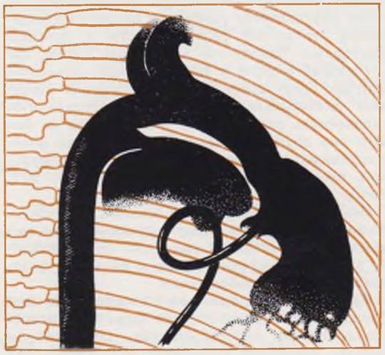
Coarctation of the aorta should be suspected in an infant with heart failure refractory to cardiac glycosides, accompanied by a discrepancy in pulses and blood pressure between the arms and legs. In these cases, cardiac catheterization and aortography can be avoided. In unclear cases, cardiac probing and radiopaque imaging are indicated to clarify the diagnosis and identify associated anomalies (Fig. 58).
Fig. 58. Aortogram of an infant with postductal aortic coarctation, grade II heart failure, respiratory failure.
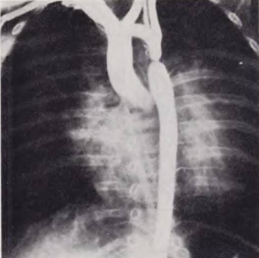
The prognosis for this defect in infants is invariably serious. If left untreated, 80% of children with heart failure die during the first year of life (Gammelguard e. a., 1959). The prognosis is particularly grave in the preductal variant of coarctation due to the high prevalence of associated
malformations and the irreversibility of pulmonary hypertension.
Treatment in all cases begins with the administration of cardiac glycosides. However, they are of limited efficacy in this defect, and only surgery offers the child a chance of survival (V. I. Frantsev, 1966).
In the postductal variant, resection of the coarctation followed by an end-to-end anastomosis provides a lasting cure. Mortality ranges from 2 — 8%.
For the preductal variant, a palliative procedure has been proposed — narrowing the patent ductus arteriosus followed by its eventual ligation and restoration of the aortic lumen. However, even today, high mortality rates are still observed following such surgeries.
Patent Ductus Arteriosus
A patent ductus arteriosus is considered congenital if it continues to function in an infant one week after birth (Gasul, 1966). A ductus with a diameter greater than 2 cm is incapable of spontaneous closure. This anomaly is well known to practicing physicians, as it is quite common and accounts for 10 to 30% of all congenital heart defects (Abbott, 1936). In older children, the hemodynamics and Clinical presentation of a patent ductus arteriosus have been studied in detail (B. V. Petrovsky, A. A. Keshisheva, 1963; F. Kh. Kutushev, L. R. Plotnikova, 1967).
The course of this defect in newborns has distinct features, the chief among them being the presence of circulatory failure (Taussig, 1955). A patent ductus arteriosus produces early, severe clinical manifestations in one out of every eight patients (Prée e. a., 1962).
The defect described is frequently found in combination with others; however, this chapter will focus exclusively on cases of an isolated ductus arteriosus.
Hemodynamics in patent ductus arteriosus during the prenatal period have been thoroughly covered (V. I. Burakovsky, B. A. Konstantinov, 1970) and will not be reviewed here. In pathological situations, a left-to-right shunt is established through it from the aorta into the lesser circulation. The magnitude of this shunt is determined by the diameter of the ductus and The ratio of vascular resistance in the systemic circulation to that in the lungs.
In the first weeks of life, the shunting occurs predominantly during systole, as the diastolic gradient between the circulatory systems is small during this period. As pulmonary vascular resistance decreases, the murmur increases and begins to span the entire cardiac cycle (Rudolph e. a., 1958). The arteriovenous blood shunt overloads the lesser circulation and the left heart chambers, reducing the efficiency of pulmonary blood flow, which leads to circulatory decompensation.
Hemodynamics in patent ductus arteriosus are characterized by pulmonary hypervolemia, overload of the left heart chambers, and the development of left-sided heart failure.
Clinical presentation. The symptoms of the defect in a newborn resemble a subacute respiratory infection accompanied by circulatory failure. The first signs—feeding difficulties, low-grade fever, and cough—may appear on the 3rd or 4th day of life. In the early days, a diagnosis of pneumonia or Bronchitis is usually made. Objective examination reveals progressive circulatory failure, manifested by tachypnea, tachycardia, hepatomegaly, and moist rales in the lungs. When crying, the infant may turn cyanotic (especially the lower half of the body), as a temporary reverse blood shunt is easily induced at this age.
Auscultation reveals either no murmur or a short systolic murmur to the left of the sternum. The heart is invariably enlarged, and the pulmonary vascular pattern is accentuated. Electrocardiography most commonly demonstrates a normal electrical axis and combined ventricular hypertrophy.
The principal symptoms of patent ductus arteriosus are identified by examining the pulse and pulse pressure (Krovetz, 1962). Characteristic findings include a bounding and high pulse (pulsus celer et altus) and a pulse pressure exceeding 40 mmHg.
The prognosis is largely determined by the age at which circulatory failure first appears. In newborns, functional closure of the ductus occurs in the vast majority of cases within the first 24 hours, whereas anatomical obliteration is completed by the 2nd to 3rd month of life (Gessner, 1965).
Treatment. Therapy must be initiated promptly with digitalization. In A number of cases, aortography via axillary artery puncture can subsequently be performed to clarify the diagnosis (G. I. Alekseev, 1968).
Conservative therapy for circulatory failure in newborns with a patent ductus arteriosus is frequently ineffective. Pate and Ainger (1963) reported the death of 44 out of 45 infants treated with digitalis. In such cases, we recommend early surgery. The method of choice is ligation of the ductus with two silk ligatures. The operation leads to recovery.
Pulmonary Valve Atresia with Intact Ventricular Septum
In this defect, the right ventricle has no outflow tract and is separated from the pulmonary bed by Valves fused into a solid membrane or by a muscular band. The anomaly occurs in approximately 0.8% of all congenital heart defects, almost exclusively in infants during their first weeks of life (Gasul, 1966).
Two Variants of the defect are distinguished (Fig. 59). In the first, which accounts for the vast majority of observations, the right ventricle has a minimal internal cavity and markedly thickened walls. In the second variant, the cavity of the right ventricle is significantly enlarged, and the tricuspid orifice is gaping.
Fig. 59. Circulation diagram in pulmonary atresia with intact ventricular septum: with a small right ventricular cavity (a), and with a large right ventricular cavity (b).

Hemodynamics are impaired as early as the Initial Stages of placental circulation. When the right ventricular cavity is small, blood flows from the right atrium through the foramen ovale into the left heart, and the arterial ventricle undergoes marked hypertrophy. When the cavity of the venous ventricle is prominent, tricuspid regurgitation is invariably present, through which this ventricle periodically empties. Subsequently, as in the first variant, blood is shunted into the left heart through the foramen ovale. Pulmonary circulation in the newborn is maintained via the ductus arteriosus and bronchial vessels. Following birth, the right ventricle with its blocked outflow tract generally continues to maintain pressure in the venous atrium higher than in the arterial atrium, preventing the foramen ovale from closing. The subsequent outcome for the newborn depends on the timing of ductal closure.
In the first variant of the defect, hemodynamics resemble tricuspid atresia, whereas in the second, they resemble severe isolated pulmonary stenosis. Hemodynamically, both variants are characterized by chronic arterial hypoxemia, progressive restriction of pulmonary blood flow, and a single functioning (left) ventricle.
Clinical presentation. The primary and earliest symptom of the defect is cyanosis, which appears in the first days of the child's life, is generalized in nature, and progresses rapidly. Cyanosis is quite quickly followed by tachypnea. A week later, signs of right-sided heart failure emerge—hepatomegaly, peripheral edema, and a gallop rhythm at the cardiac apex. Early decompensation is particularly characteristic of the variant featuring a large right ventricular cavity and tricuspid insufficiency. In these cases, pulsation of the jugular veins and liver can frequently be observed.
The described defect is characterized by a moderate increase in cardiac dimensions during the first few days, followed by progressive enlargement. The defect is most often atypical.
On the electrocardiogram, the electrical axis may be deviated to the right, to the left, or remain normal. An increased P2 wave is consistently observed. In chest leads, the first variant more frequently shows left heart hypertrophy and overload, whereas combined hypertrophy is possible in the second.
Radiological examination reveals varying degrees of cardiomegaly, with a highly variable cardiac silhouette, alongside a diminished pulmonary vascular pattern.
The principal role in the topographical diagnosis of the defect belongs to selective angiocardiography from the right atrial cavity.
Differential diagnosis is challenging (see Table 17). The prognosis for pulmonary atresia is grave. Approximately one-third of infants die by the end of the second week of life, and half by the end of the first month (Keith et al., 1968).
Surgical treatment is indicated. Among the various surgical procedures for pulmonary atresia with a well-developed right ventricular cavity, closed transventricular valvulotomy is the most appropriate approach (Robinson, 1965), whereas an interarterial anastomosis is preferred when the venous ventricle is hypoplastic.
In newborns with a small patent foramen ovale, Rashkind balloon atrioseptostomy may prove to be an effective initial palliative procedure.
Tricuspid Atresia
This defect accounts for 1.5% to 3% of all congenital heart diseases. The right venous orifice is completely obliterated, and the venous atrium communicates with the arterial atrium through a patent foramen ovale. The right ventricle is markedly hypoplastic and may communicate with the left ventricle via a ventricular septal defect. In 80% of all cases, pulmonary stenosis, pulmonary trunk atresia, or hypoplasia is present. Transposition of the aorta and pulmonary artery is very frequently associated with tricuspid atresia. Pulmonary blood flow is maintained via the ductus arteriosus.
The hemodynamics are very similar to those seen in pulmonary atresia.
Clinical presentation. In the majority of patients, cyanosis appears within the first week of life and is typically pronounced. Moderate and late-onset cyanosis is characteristic of patients with a large interatrial communication and pulmonary stenosis. Besides cyanosis, noteworthy findings on the electrocardiogram include a left axis deviation (levogram), left ventricular hypertrophy, and left bundle branch block. The heart is not enlarged, and signs of heart failure are absent for a relatively long time. Despite the latter circumstance, neck vein pulsation and liver edge pulsation are readily appreciable. Auscultation findings are non-specific.
Diagnosis in a newborn with marked cyanosis, left ventricular overload on the electrocardiogram, a normal heart size, and a characteristic clear retrosternal space on a radiograph in the second oblique position is practically straightforward (see Table 18). In many cases, angiocardiography is not required to confirm the diagnosis. The prognosis is graver the earlier and more intense the cyanosis.
Treatment. Recently, a palliative procedure—Rashkind balloon atrioseptostomy (Yu. S. Petrosyan, V. A. Garibyan, 1972)—has proven effective in newborns; it increases the diameter of the interatrial communication and significantly reduces the degree of arterial hypoxemia.
Total Anomalous Pulmonary Venous Drainage
Anomalous drainage of the Pulmonary veins into the systemic Venous system occurs in 1.5% to 3% of all newborns with congenital cardiovascular malformations (F. I. Romashov, 1965; Smith et al., 1961).
Anomalous pulmonary venous drainage is frequently observed in complex cardiac anomalies such as a two-chambered heart, single ventricle, transposition of the great vessels, and truncus arteriosus. In such cases, newborns present with asplenia syndrome. These anatomical combinations are incompatible with long-term survival.
Of greater interest to the clinician are the so-called isolated forms of total anomalous pulmonary venous drainage. Their anatomy is highly diverse. The most comprehensive classification of these defects, based on the level at which the anomalous veins empty into the systemic circulation, was proposed by Darling et al. (1957). In order of decreasing frequency, the authors distinguish four anatomical types: 1) supracardiac (accounting for half of all cases of total anomalous pulmonary venous drainage); 2) cardiac; 3) infracardiac; and 4) mixed.
In the supracardiac type, the pulmonary veins drain into the left or right SUPERIOR VENA CAVA. In the cardiac type, they drain into the coronary sinus or the right atrium, and in the infracardiac type, into the inferior vena cava, portal vein, or ductus venosus. Finally, in the mixed type, the pulmonary venous orifices may open simultaneously into the coronary sinus and the left brachiocephalic vein.
Despite the variety of anatomical variants, the hemodynamics in this defect share many common features. They are characterized by marked pulmonary plethora combined with a low effective pulmonary blood flow. Systemic cardiac output is reduced, and its magnitude depends on the volume of right-to-left shunting at the atrial level (most commonly through a patent foramen ovale). The condition is also characterized by moderate arterial hypoxemia, marked right heart overload, and the early onset of respiratory and circulatory failure. Pulmonary hypertension is present from the first days of the child's life.
Clinical presentation. The initial manifestations of the defect—such as dyspnea, cough, and feeding difficulties—appear in the vast majority of infants during the first weeks and months of life. In the infracardiac variant, clinical signs emerge from the very first days of life. Cyanosis and heart failure develop early. However, cyanosis never reaches extreme degrees and is always secondary to dyspnea. In infracardiac forms, a highly characteristic diagnostic sign is observed: despite severe decompensation, heart size remains normal. In other variants of the defect, as mentioned above, the onset is less acute. Many infants never leave the hospital after birth due to an endless succession of "respiratory" infections.
Physical examination reveals marked growth failure (hypotrophy), mild and intermittently intensifying cyanosis, and a prominent precordial bulge. The heart is enlarged; a systolic murmur, an accentuated second sound, and its splitting are auscultated in the second left intercostal space. A soft diastolic murmur, caused by increased blood flow through the superior vena cava, may be audible to the right of the sternum. Moist, fine, congestive rales and areas of bronchial breathing are heard in the lungs. Signs of heart failure escalate following the onset of dyspnea and become severe at the peak of the clinical picture (hepatomegaly, edema, distended neck veins, and a tense fontanelle).
The electrocardiogram consistently demonstrates a right axis deviation (dextrogram), along with right ventricular hypertrophy and overload.
Radiological examination reveals cardiomegaly, aortic hypoplasia, and prominence of the pulmonary artery segment. The pulmonary vascular markings are accentuated. When the pulmonary veins drain into the left superior vena cava, a characteristic "figure-of-eight" cardiac silhouette may be observed. Radiopaque angiocardiography performed from the right ventricle or pulmonary artery with serial imaging is of decisive diagnostic value, as it allows visualization of the pulmonary blood flow and the exact site where the anomalous pulmonary veins empty into the systemic venous system. The prognosis is extremely grave, with approximately 75% to 80% of children dying before reaching 1 year of age.
Treatment begins with digitalization and anti-pneumonic therapy. Once the topographic diagnosis is established, surgery is indicated. Surgical intervention consists of creating an anastomosis between the left atrium and the common pulmonary venous collector. Mortality rates for these operations remain high due to the frequent Development of Respiratory failure and left ventricular weakness. Presumably, better outcomes in newborns can be expected from a palliative procedure—Rashkind atrioseptostomy—which relieves pulmonary congestion in such patients (Serrato et al., 1968). Successful operations for infracardiac forms of the defect thus far remain limited to isolated case reports (Cooley, 1962; Jegeir et al., 1967).
Double Aortic Arch
This developmental anomaly manifests in newborns with signs of respiratory distress due to tracheal compression between the aortic arches and the resulting narrowing of its lumen.
Embryogenesis. In human embryos, six pairs of aortic arches develop, connecting the ventral and dorsal aortic roots. Each of these pairs traverses a specific branchial (pharyngeal) arch and possesses right and left branches. During embryogenesis, the first, second, and fifth aortic arches either degenerate or transform into their respective arterial vessels. The fourth left arch persists as the aorta, while the right forms the proximal portion of the Subclavian Artery. When the right branch persists—usually passing posterior to the esophagus—the aortic arch is split into two trunks that encircle the trachea and esophagus. If the left branch atrophies and disappears while the right branch of the fourth aortic arch persists, a right-sided aortic arch of the anterior (Assmann) type is formed. In this case, the aortic arch passes anterior to the trachea from left to right and is usually associated with dextroposition of the aorta or other congenital heart defects (Tetralogy of Fallot, Eisenmenger complex). Tracheal compression is less frequent with this type of anomaly than with a double aortic arch. The latter occurs as a complete or incomplete duplication, wherein the right branch of the fourth embryonic aortic arch fails to develop into a full right-sided aortic arch, and the left branch does not completely disappear. Instead, a rudimentary diverticulum of varying size remains, from which the left subclavian artery, ductus arteriosus, or ligamentum arteriosus originates. As a result, a vascular ring is formed around the trachea and esophagus by the right-sided aortic arch, the aortic diverticulum, the ductus arteriosus (or its Connective Tissue remnant), and the pulmonary artery. This anomaly is known as the posterior (Akin) type of right-sided aortic arch (V. Ionáš, 1960).
Clinical presentation. Symptoms depend on the degree of tracheal and esophageal compression and consist of dysphagia and respiratory impairment. In most cases, breathing is wheezing or stridor-like. Inspiratory dyspnea may be observed. Cyanosis can be transient or persistent. Some infants require continuous oxygen therapy. Newborns tend to keep their heads thrown back, as this position facilitates airflow through the narrowed trachea (St. Dimitrov, 1960). A persistent dry cough is frequently noted. When the recurrent laryngeal nerve is compressed, the cough acquires a metallic, barking quality, and the infant's voice becomes hoarse. Feeding exacerbates the symptoms. Regurgitation and vomiting occur frequently; vomitus may contain fresh or altered blood, which is a sign of esophagitis. Aspiration pneumonia soon develops and frequently leads to the demise of such patients. Dysphagic symptoms may be milder during the first month of life and become more pronounced upon the Introduction of solid foods. Patients are highly susceptible to respiratory infections. The onset of laryngotracheitis significantly impairs breathing and can provoke severe hypoxia.
Diagnosis is established through radiological and instrumental examinations. A plain lateral radiograph of the trachea may reveal narrowing of its lumen at the level of the third and fourth thoracic vertebrae. Barium swallow examination demonstrates posterior indentation of the esophagus at the same level. Tomography can provide valuable data. During tracheobronchoscopy, the tracheal lumen appears compressed in the anteroposterior direction due to anterior wall impingement, and transmitted aortic pulsations are clearly visible. Contrast-enhanced imaging of the trachea is usually unnecessary. Angiography allows the determination of THE POSITION OF the aortic arch, its branches, and the dimensions of the trunks in cases of a double arch.
Treatment. Surgical intervention is indicated for infants presenting with severe respiratory distress and dysphagia. Without surgery, such patients rarely survive beyond 1 to 2 years of age. Death may occur suddenly or result from recurrent pneumonia. In the most severe cases, surgery is performed during the neonatal period, requiring a careful balance between surgical risks and the severity of symptoms, primarily respiratory failure.
Surgical management involves ligation and division of the narrower arch, typically the anterior one. In cases of incomplete aortic arch duplication, the procedure entails ligating the ductus arteriosus or the ligamentum arteriosus that compresses the trachea. If respiratory compromise is caused by tracheal compression from a right-sided aortic arch displaced to the left by a branch of the subclavian artery, ligation and division of the latter may be performed, as collateral circulation adequately maintains perfusion to the upper extremity (V. Jonaš, 1960).
Last update: 10/08/2026
Editorial and Educational Adaptation: This material has been compiled based on the primary/original source text. The project team performed an editorial review, corrected technical inaccuracies, structured sections, and adapted the content for an educational format.
What was processed:
- elimination of formatting defects (OCR errors, structural breaks, corrupted characters);
- editorial organization of content;
- standardization of terminology in accordance with academic sources;
- verification of factual statements against the original source text.
All mentions of the author, publication year, and origin of the primary text have been preserved in accordance with the source.