NEONATAL SURGERY - 1976
2. SPECIAL SECTIONS
8. Malformations of Individual Organs and Systems
Chylothorax and Chyloperitoneum
The leakage of lymphatic fluid into the pleural (chylothorax) and abdominal (chyloperitoneum) cavities in newborns is a rare but life-threatening condition. Chylothorax in children was first described by Bartolett in 1633. A literature review of neonatal chylothorax (16 cases) was published by Bavitch (1962). Tischer compiled 38 cases of spontaneous chylothorax in infants During the first 3 months of life from worldwide literature spanning 50 years (1917 — 1966).
Pathogenesis. The condition is usually caused by congenital or acquired defects of the major Lymphatic vessels, which develop between the 4th and 5th weeks of embryonic life. Injuries to the Thoracic duct can result from inflammatory processes or birth trauma. Among 9 patients with this pathology observed in our clinic, 2 had a malformation of the lymphatic vessels, and in one infant, the onset of chylothorax was associated with a complicated delivery. Symptoms in these patients appeared during the neonatal period. In the remaining children, lymphorrhea into the pleural cavity was the result of surgical trauma or inflammation.
Clinical presentation. The Diagnosis of chylothorax and chyloperitoneum is based on physical examination findings, clinical data, radiographic evaluation, and diagnostic paracentesis or thoracentesis. The leading symptom of chylothorax is respiratory distress caused by lung compression and mediastinal shift. Physical and radiographic examinations reveal signs of fluid accumulation in the pleural cavity, most commonly on the right side. Chyloperitoneum manifests as abdominal distension, giving the abdomen a flattened appearance ("frog belly"). Sometimes, children suffer from bowel dysfunction. Percussion reveals the presence of fluid in the Abdominal cavity. The diagnosis for both conditions becomes definitive following puncture and aspiration of characteristic opalescent chylous fluid resembling diluted milk. The fluid contains a high percentage of protein (4 — 9%) and neutral fat (6% or more), with a specific gravity of 1012 — 1028. Upon standing, it separates into two layers: a thick upper layer and a liquid lower layer. Microscopic examination shows a predominance of lymphocytes. The loss of significant amounts of protein, fat, and electrolytes profoundly impacts the child's condition. Patients with severe forms of the disease are anaemic and develop hypoproteinaemia. Along with signs of malnutrition, there is usually a normal or even increased body weight due to the accumulation of free fluid. In rare cases, chylous fluid is found in both the pleural and abdominal cavities simultaneously. This condition is caused by diffuse Malformations of the lymphatic vessels or diaphragmatic defects.
Differential diagnosis. Chylothorax must be differentiated from exudative or purulent Pleurisy. Unlike the latter, the presence of chylous fluid in the pleural cavity is rarely accompanied by signs of inflammation, as Lymph possesses bactericidal properties. The diagnosis is definitively established after aspiration. Similar symptoms are caused by a congenital diaphragmatic hernia presenting with "asphyctic strangulation." To rule out the latter, it is recommended to inject 30 ml of air into the ABDOMINAL CAVITY AND take a radiograph with the infant lying on their right side. Opacification of one lung field, which may suggest a pleural effusion, is also observed in total Atelectasis, lung agenesis, and pulmonary aplasia. Unlike chylothorax, however, mediastinal shift in these cases occurs toward the side of the opacification. The same side also shows a reduced thoracic volume with narrowed intercostal spaces and an elevated diaphragmatic dome. To diagnose chyloperitoneum, ascites—which may occur secondary to Liver and Kidney diseases or abdominal tumors—must be ruled out. A reliable diagnosis in such cases is possible only through diagnostic abdominal paracentesis.
Treatment. Periodic aspiration of chylous fluid from the pleural or abdominal cavity is combined with intravenous infusions of protein preparations, Blood, and plasma. Adequate Nutrition is particularly vital, supplementing breast milk with cottage cheese and kefir, or using concentrated formulas with elevated protein and fat content for artificial feeding. The evacuated chylous fluid can be reintroduced into The Stomach or rectum. Bauersfeld (1937), V. I. Frantsev (1963), and others attempted intravenous administration of chylous fluid but noted the risk of fatal anaphylactic Shock. A safer method of continuous infusion of pleural contents into the bloodstream has been described via pleurovenous shunting using a Holter valve (Holter, 1964). In A number of cases, spontaneous lymph leakage into the pleural cavity ceases spontaneously after several aspirations or following pleural drainage (Pallay, 1970). Chylous fluid clots much more poorly than blood; therefore, the efficacy of conservative therapy is primarily related to The formation of pleural adhesions. To stimulate adhesion formation, a 40% glucose solution, talc, or iodolipol is introduced into the pleural cavity.
If conservative therapy yields no effect within 1 — 2 months, Surgical treatment is indicated. Prolonged puncture treatment is dangerous due to the patient's progressive emaciation. Surgical intervention consists of ligating the thoracic duct, which can be performed at any level since lymph drainage is maintained via collateral lymphatic pathways and lymphovenous anastomoses (B. V. Ognev, 1950). Additionally, the Pleura and the bed of the duct are treated with a 5–10% tincture of iodine to promote adhesions and better obliterate the site of the defect. Suturing duct defects in newborns is technically challenging. To facilitate locating the lymphatic duct, the biological contrast method proposed by B. V. Ognev is recommended. In cases where the cause of lymphorrhea cannot be identified, treatment aimed at obliterating the pleural cavity is performed.
Searching for lymphatic vessel defects within the abdominal cavity is rarely successful: only isolated reports have been published in which the leaking lymphatic vessel was identified and ligated (S. Ya. Doletsky et al., 1970; Vasko, Tapper, 1967).
In a case reported by G. A. Ostromoukhova (1969), an infant recovered following laparotomy, subsequent abdominal punctures, and air insufflation into the peritoneal cavity.
Mortality rates for both conservative and Surgical Treatment of chylothorax in children reach 20 — 25% (Randolph, Gross, 1957; Louhimo e. a., 1966).
Developmental Disorders of the diaphragm lead to the formation of defects, followed by the displacement of abdominal Organs into the thoracic cavity and The Development of diaphragmatic hernias. Diaphragmatic hernias occur on average in 1 out of every 1,700 — 2,500 newborns (S. Ya. Doletsky, 1960; G. A. Bairov, 1968).
The Embryogenesis OF THE diaphragm and the MAIN TYPES OF diaphragmatic hernias are schematically presented in Fig. 60.
Class="center">Fig. 60. Embryogenesis of the diaphragm. Classification of diaphragmatic hernias.
The diaphragm develops from three main sources. Its ventral part is formed by the septum transversum, while the paired dorsolateral parts arise from pleuroperitoneal folds. Initially, the septum transversum is a loose mass of mesoderm (a, b); its cardiac surface is covered with a layer of mesothelium, and its caudal surface gradually transitions into the glandular tissue of the liver. As differentiation proceeds, the BOUNDARIES OF THE septum transversum become more distinct.
Pleuroperitoneal folds arise along the margins of the protrusions that cover the common cardinal Veins (b, c). The septum transversum and pleuroperitoneal folds grow toward each other and fuse to form the main part of the diaphragm. The median-dorsal portion of the diaphragm is formed from the remaining segments of the primordial mesentery. Reinforcement of the diaphragm occurs through the marginal ingrowth of Muscle from the body walls (d).
Impaired Development of the various sources that form the diaphragm leads to the formation of diaphragmatic hernias.
Hernias of the anterior diaphragm are associated with underdevelopment of the septum transversum: Ia — anterior hernias (true); Ib — phrenicopericardial hernias (false); Ic — retrograde phrenicopericardial hernias (false). Impaired development of the pleuroperitoneal folds or their failure to fuse with the septum transversum explains the formation of false diaphragmatic hernias (defects): IIa — left-sided slit-like defect (Bochdalek foramen); IIb — large right-sided defect; IIc — Aplasia of the left hemidiaphragm.
Developmental disorders at The final stage of diaphragmatic formation—its reinforcement through muscular ingrowth—lead to true hernias: IIIa — localized eventration on the right; IIIb — significant eventration on the left; IIIc — eventration (relaxation) of the left hemidiaphragm. The sketches illustrate the radiographic manifestations of various types of hernias.

Hernias of the Diaphragm Proper
Depending on the presence or absence of a hernial sac, hernias of the diaphragm proper are divided into true (when a hernial sac is present) and false (when a hernial sac is absent). Left-sided localization of diaphragmatic hernias prevails over right-sided. In true diaphragmatic hernias, the hernial ring is often difficult to identify and poorly defined; therefore, strangulation of the Contents of the hernial sac in true hernias typically does not occur. In false hernias, the hernial ring is in most cases formed by a dense tendinous or muscular edge, and strangulation of the hernial contents is frequent.
Clinical manifestations and radiographic findings. The clinical manifestations of diaphragmatic hernias result from the displacement of abdominal organs into the thoracic cavity. The time of onset and severity of symptoms depend on the size of the hernia sac, the timing of organ displacement, and the infant's compensatory reserves. Frequently, especially in false hernias, symptoms appear within the first hours, days, or weeks of life and tend to progress rapidly. When a significant portion of the intestine and other abdominal organs shifts into the chest, they compress the lung and displace the Mediastinum. In such patients, respiratory distress comes to the fore. Early and most characteristic signs include episodes of cyanosis and asphyxia that worsen during feeding, crying, or agitation (Vos et al., 1971). These are accompanied by coughing, dyspnea, and potential cardiac dysfunction due to the Displacement of the Heart and great vessels. These episodes of respiratory and cardiac impairment last for several minutes and often diminish or resolve when the infant is turned onto the affected side. A characteristic physical finding is a scaphoid abdomen. Percussion over the corresponding hemithorax reveals tympanitic or, conversely, dull percussion notes. Auscultation demonstrates diminished breath sounds, and it is often possible to hear peristaltic murmurs, bowel gurgling, etc. The Heart is typically displaced to the contralateral side. General disorders (failure to thrive, infantilism, developmental delay) are not characteristic of newborns.
Variability of symptoms is a notable feature. For instance, repeated examinations before and after feeding in most cases reveal distinct Changes in the clinical picture, which is explained by the shifting of intestinal contents and changes in bowel filling.
Radiographic data are decisive for establishing the diagnosis. The most comprehensive understanding of the pathology is provided by radiographs in two projections. Characteristic radiological signs of a diaphragmatic hernia include areas of opacification and radiolucency in the lung fields, along with the displacement of The Heart and mediastinum to the opposite side (Fig. 61). In doubtful cases, the administration of a contrast agent into the intestine is indicated. The clinical manifestations and radiographic picture described above are common to true diaphragmatic hernias. Let us examine the Clinical Features of specific types of diaphragmatic hernias in greater detail.
Fig. 61. Radiograph of a newborn with a left-sided false diaphragmatic hernia—Bochdalek's hernia (a). Air-filled bubbles representing intestinal loops and the stomach are visible in the thoracic cavity above the diaphragm. The heart is displaced to the far right. A small amount of gas is present in the abdominal cavity. Asphyxial strangulation. Emergency surgery. Recovery.
Radiograph of an infant with a hiatal hernia (b). Barium suspension fills the Esophagus, stomach (with the cardia located in the mediastinum), and jejunum.


True hernias. In newborns, true hernias often go undiagnosed. In such cases, the herniated area typically does not exceed 1/3 of the surface area of the diaphragmatic dome. True hernias are more common on the right side. The hernia sac restricts the migration of organs into the thoracic cavity. Compression of the Lungs and mediastinum may be minimal or absent, which accounts for the clinical manifestations being absent in the majority of patients during the neonatal period. Such hernias are diagnosed during incidental or routine radiographic examinations based on the presence of stable alterations in diaphragmatic configuration.
With complete eventration of the entire diaphragmatic dome (relaxation) in newborns, severe Hypoxia frequently occurs. Radiographic examination reveals a high-standing diaphragmatic dome with limited excursion. When performing Cytology/practical/136.html">DIFFERENTIAL DIAGNOSIS OF diaphragmatic relaxation in neonates, one must keep in mind the possibility of diaphragmatic muscle paresis due to birth trauma. Dynamic observation of the patient resolves any doubts. In cases of paresis, muscle tone is restored in the majority of patients within the first weeks of life, the diaphragm descends, and normal function resumes. True diaphragmatic hernias in newborns generally do not require surgical intervention.
False hernias frequently cause complications in the first days and weeks of life, necessitating emergency surgery based on vital indications (Kumhaar, 1968; Lister, 1971; White, Suruk, 1972). In most cases, false hernias are associated with a posterior slit-like diaphragmatic defect (Bochdalek's foramen) localized in the costolumbar region. The defect may be parietal, in which case the parietal Peritoneum transitions directly into the parietal pleura. Frequently, the defect is separated from the chest wall by a thin muscle bundle.
The clinical presentation is pronounced as early as the first days of life. Characteristic features include episodes of cyanosis, vomiting, and Cardiac Arrhythmias. Hernial strangulation caused by meteorism (asphyxial strangulation) is common and can be fatal to the infant if emergency surgery is not performed. Some patients gradually adapt to the presence of the anomaly. In these instances, symptoms gradually subside, leaving only intermittent dyspnea, occasional pronounced episodes of cyanosis, and vomiting (especially after feeding). In rare cases, the hernia may be completely or partially reducible. Under these conditions, the infant remains virtually healthy during periods when the intestine is located in the abdominal cavity, while pathological symptoms arise only when the hernial contents migrate into the pleural cavity. Radiographic examination of patients with false hernias reveals a "total" displacement of abdominal organs into the chest. Contrast examination of the intestine using a barium suspension AIDS in precise topographical diagnosis.
A significant diaphragmatic defect (pleuroperitoneal canal) encompasses more than 1/3 of the dome's surface area and is located in the lateral or central division. The defect typically has a triangular-oval shape.
Clinical manifestations are similar to those of slit-like defects, although the symptomatology is more vivid because the kinking of organs at the hernia ring occurs in different directions, resulting in more pronounced trauma. The diagnosis is refined using radiographic imaging Methods.
Complete absence of one diaphragmatic dome (aplasia) is extremely rare. Infants die immediately after birth because breathing is virtually impossible in such cases.
Treatment. The majority of patients with false diaphragmatic hernias require urgent surgical intervention. This applies primarily to newborns exhibiting symptoms of asphyxial strangulation. Delaying surgery in these patients generally leads to a fatal outcome. In cases of diaphragmatic aplasia, immediate intubation and Artificial ventilation are indicated right in the delivery room. Emergency surgery is indicated for hernia strangulation accompanied by a progressive worsening of the patient's condition, escalating symptoms of intestinal obstruction, and respiratory and cardiac impairment.
The duration of preoperative preparation must be minimized as much as possible, since correcting general systemic disorders is only truly feasible after the organs have been returned to their normal anatomical position.
Surgical technique. The method of choice is a transabdominal approach with a transverse incision of the subcutaneous tissue in the left epigastric region, followed by vertical Separation of the muscle fibers. This approach is adequate, ensures minimal trauma to the intestine, and upon opening the abdominal cavity and inspecting the diaphragmatic dome through its defect, a metal catheter is introduced into the pleural cavity through which 40–50 ml of air is injected via a syringe. This maneuver equalizes the pressure in the abdominal and pleural cavities, significantly facilitating the subsequent reduction of intestinal loops.
Following organ reduction and inspection of the lung (which is hypoplastic in the majority of patients), the diaphragmatic defect is closed with a single row of interrupted sutures. Closure of the defect is facilitated if a muscular ridge is well-developed on the chest wall side. In the absence of such a ridge, the edge of the defect is secured to the chest wall using interrupted sutures passed around the rib.
Some authors (Hard, 1969) recommend splitting the diaphragmatic dome during plication in newborns and interposing a portion of the latissimus dorsi muscle between the resulting layers. However, due to its complexity and traumatic nature, this method has not gained widespread acceptance. In cases of extensive defects in certain patients, The Use of alloplastic Materials is indicated. A fundamentally important technical step in these cases is separating the prosthesis from the pleural cavity using a pedicled flap of peritoneum. Otherwise, the child will experience prolonged pleuritis until complete epithelialization of the defect occurs.
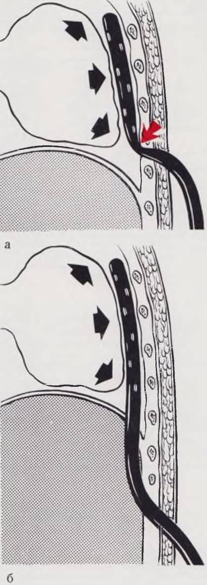
Complete evacuation of air and attempts at instantaneous re-expansion of the lung in such patients are hazardous; overexpansion of the lung leads to alveolar rupture, the development of Pneumonia, and potential tears in the lung tissue. The operation is concluded by placing a drainage tube into the pleural cavity, which is brought out through the diaphragm and the lateral abdominal wall (S. Ya. Doletsky). Such transabdominal drainage prevents kinking of the drain and promotes a more complete re-expansion of the lung (Fig. 62). Active suction is not performed. Within 1–3 days, the lung expands independently and fills the pleural cavity. The abdominal wall wound is then closed tightly in layers.
Fig. 62. Drainage of the pleural cavity. A drain inserted through the intercostal space (a) kinks when the lung expands. Drainage through the abdominal cavity (b) is not complicated by drain kinking.
In individual patients, closing the abdominal wall after reducing the organs into the abdominal cavity may be difficult. In such cases, Meeker (1958) recommends performing a gastrostomy for the purpose of abdominal cavity decompression. We have operated on 30 infants with false diaphragmatic hernias during the neonatal period, with 3 deaths. Subsequently, the operated children develop practically the same as healthy children.
Hiatal hernias
Hiatal hernias come in various types (Fig. 63). Differential diagnosis is performed to rule out failure of gastric descent (Fig. 64).
Clinical picture. In hiatal hernias extending into the thoracic cavity—specifically the mediastinum—only the stomach herniates, which in most patients does not severely compress the mediastinal organs and lungs. The clinical picture of hiatal hernias is dominated by manifestations of esophageal kinking or gastric compression at the hernial orifice. Regurgitation and vomiting are the most frequent symptoms of hiatal hernias in newborns. The vomitus may contain streaks of blood. During feeding, when the esophagus becomes overfilled, episodes of cyanosis may occur during vomiting. The diagnosis is refined through radiological examination. Plain radiographs in two projections reveal an air bubble in the posterior mediastinum (Fig. 61, b). Administration of a barium suspension clarifies The Nature of the pathology.
Fig. 63. Classification of hiatal hernias (according to S. Ya. Doletsky).
Hiatal hernias always feature a hernial sac and are classified as uncomplicated (I) and complicated (II). Uncomplicated hernias include those with herniation of only the stomach into the sac (A): with an elevated esophagus (a), paraesophageal hernia (b), complete displacement of the stomach (c), and with herniation of other organs into the sac (B): Small Intestine (d), Large Intestine (e), liver (f), omentum (g), etc. Complicated hernias (II) are characterized by esophagitis (h), leading to stenosis (i), esophageal shortening (j), formation of an ulcerous diverticulum capable of perforating into the mediastinum (k), and others. Hiatal hernias can be sliding or fixed.

Fig. 64. Variants of incomplete descent of the stomach ("short esophagus").
Mild degrees of incomplete descent are frequently observed in newborns (70% of total cases according to Waterstone). They tend to resolve spontaneously with postural management and represent an example of relative immaturity (a). In moderate degrees (2 — 4 cm) of gastric non-descent (b), surgical treatment is possible by relocating the esophageal hiatus above the gastric mucosa zone combined with fundoplication. For significant degrees of non-descent (c), replacement of the supradiaphragmatic part of the stomach with an intestinal graft is employed. Incomplete descent of the stomach is typically complicated by esophagitis.

Treatment. Once the diagnosis is established, surgery is performed regardless of the child's age. Cases of chalasia and achalasia, which are difficult to diagnose definitively, constitute an exception.
Surgical technique. The operation is performed via a transabdominal approach (Pages, 1968; Cahill, 1969; Ekespare, 1971). The stomach is retracted downward and to the left. The left lobe of the liver is mobilized. At the moment of traction, the portion of the stomach previously located in the mediastinum shifts into the abdominal cavity. The next stage involves isolating the hernial sac. In hiatal hernias, the hernial sac is a protrusion of the parietal peritoneum that extends onto the cardiac region of the stomach. Excision of the hernial sac is optional, but a circular incision of the peritoneum in the area of the esophageal hiatus is mandatory. During this step, care is taken not to damage the vagus nerves, as their trauma causes gastric paresis in the postoperative period. The hernial sac is displaced in the oral direction using a dissector, and the abdominal segment of the esophagus is skeletonized and brought down 2 — 3 cm below the esophageal hiatus. The relocation of the esophagus is a fundamentally crucial step of the operation. Leaving it in its natural position adjacent to the aorta may cause a recurrence. The physiological rationale of the Procedure lies in approximating and suturing the medial crura of the diaphragm posterior to the esophagus so that a layer of Muscle tissue lies between the esophagus and the aorta. In individual cases, creating the angle of His by fixing the gastric fundus to the abdominal esophagus or anchoring the stomach to the diaphragm is indicated. The operation concludes with layered closure of the abdominal wall.
We operated on 15 newborns and infants in their first months of life for hiatal hernias with 2 fatalities. No hernia recurrences were observed within a follow-up period of up to 5 years postoperatively.
Hernias of the Anterior Diaphragm
Anterior and phrenicopericardial hernias are distinguished. In anterior hernias, which always have a hernial sac, abdominal organs migrate into the thoracic cavity through the hiatus of Larry (Morgagni hernia). The clinical picture is non-specific. During the neonatal period, these hernias are generally not diagnosed. Surgery in newborns is not indicated.
Phrenicopericardial hernias, unlike anterior ones, are typically false hernias. A defect exists in the tendinous center along the PROJECTION OF THE Pericardium, connecting the pericardial cavity with the abdominal cavity. Loops of the intestine may migrate into the pericardial cavity or, conversely, the apex of the heart may prolapse into the abdominal cavity.
Symptoms of cyanosis, dyspnea, and vomiting predominate in the Clinical presentation of phrenicopericardial hernias. X-ray Examination reveals shadows of intestinal loops projected over the cardiac silhouette.
Surgical treatment is indicated. The surgeon's task in phrenicopericardial hernias is extremely challenging. Simple closure of the defect in these cases frequently leads to recurrence (cardiac pulsations rapidly tear the sutures). Therefore, plastic repair of the defect is indicated. In this procedure, the serous membrane should lie against the heart—most preferably a pedicled flap of parietal peritoneum or the omentum.
Postoperative period. The Organism of an infant born with a diaphragmatic hernia is largely adapted to the anomaly, and this developmental defect becomes a peculiar "norm" for the child. Returning the organs into the abdominal cavity along with the plastic repair of the diaphragmatic defect sometimes leads to severe pathological shifts. Consequently, complications are relatively frequent in children operated on for diaphragmatic hernias. For instance, immediately after surgery, intra-abdominal pressure rises, the diaphragm is displaced upward, and its excursion becomes restricted (Tsuchida, 1969). Hemodynamic disturbances in the INFERIOR VENA CAVA system may occur. Prevention of these complications involves managing intestinal paresis. In case of respiratory impairment, intubation and prolonged mechanical ventilation are indicated. Monitoring of pulmonary status is essential. Fluid from the pleural cavity is drained via a chest tube or by puncture (if no drainage tube is in place).
In Conclusion, timely diagnosis, proficient surgical execution, and proper postoperative management make it possible to save the life and ensure normal development of the majority of newborns with this anomaly. During the neonatal period, false diaphragmatic hernias and hiatal hernias most commonly serve as the indication for surgical intervention.
Abdominal Wall. Abdominal Organs. Perineum
Embryogenesis. The Formation of the anterior abdominal wall in the human embryo from the so-called primary vertebrae, appearing as protrusions of their anterior and lateral body surfaces, begins as early as the end of the 1st to the beginning of the 2nd week of intrauterine life (B. M. Patten, 1959). By the end of the 5th week, the primordia of the rectus and lateral Muscles appear in the upper half of the abdomen, originating from the medial plate of the mesoderm. These primordia grow inferiorly, where they meet the distal PARTS OF THE gubernacula (gubernaculi Testis), extending downward on each side toward the future internal openings of the inguinal canals. By the 7th to 10th weeks of embryonic life, these muscles close the abdominal cavity anteriorly, leaving only the umbilical ring open. In the 3rd month of intrauterine life, the Differentiation of the lateral muscles into three layers—the external oblique, internal oblique, and transversus abdominis—begins.
The development of the fetal intestine proceeds along two parallel pathways: the intestinal tube forms simultaneously with The process of rotation accompanied by the extraembryonic Location OF THE abdominal organs (Fig. 65).
Fig. 65. Normal intestinal rotation and possible pathological variants during the stages of rotation.
Period I of rotation. At the 5th week of intrauterine life, the intestinal tube lies in the sagittal plane on the primary dorsal mesentery (a),
During the 6th to 7th weeks, the growth of the intestinal tube outpaces the growth of the body cavity, As a result of which a portion of the midgut herniates into the umbilical cord (b). A temporary "physiological umbilical hernia" is formed.
The rapidly developing right lobe of the liver "pulls" the umbilical vein (left) along with it, which subsequently transforms into the round ligament of the liver, displacing the prearterial segment of the intestine to the right and downward (c).
Around the 8th week of intrauterine life, the midgut loop located within the umbilical cord rotates 90° counterclockwise into the horizontal plane (d).
II stage of intestinal rotation. By the 10th week of embryonic development, driven by the growth of the abdominal cavity, the intestine gradually begins to shift from the umbilical cord into the abdominal cavity. The returned small intestine lies to the right of the artery (d). Due to the insufficient volume of this abdominal compartment, subsequent loops of the intestine push the loops that previously migrated behind the artery into the left half of the abdominal cavity (e). The loops displaced to the left push the mesentery of the hindgut forward, positioning the splenic flexure and descending colon in their normal anatomical locations. By the 11th week, the entire intestine is located within the abdominal cavity (ж). Rotation continues, culminating in a 270° intestinal rotation from the sagittal position of the beginning of stage I.
III stage of intestinal rotation and fixation. The cecum descends from the upper right quadrant into the right iliac fossa. The intestinal mesentery becomes fixed to the posterior abdominal wall (з). The zones of fixation are outlined by a dashed line. The mesentery of the transverse colon fuses with the greater omentum, forming the gastrocolic ligament (и).
Depending on the specific stage at which normal intestinal rotation arrests, various pathological conditions may develop in the newborn.
An arrest of development during stage II may lead to: k — compression of the duodenal lumen by peritoneal bands fixing the cecum; л — midgut volvulus; м — midgut volvulus combined with concurrent occlusion of the duodenal lumen by peritoneal bands (Ladd's syndrome).
An arrest of development during stage III results in: н — retroperitoneal and retrocecal positioning of the Appendix; о — high-positioned cecum; п — internal hernias caused by abnormal fixation of the mesentery.

Fig. 65

Fig. 66. Stages of normal regression of umbilical cord structures and potential variants of its disruption.
The umbilical cord is a Structure that connects the body of the fetus to the Placenta during the prenatal period. Its constituent elements undergo substantial changes throughout development.
From the perspective of its initial Functions, the yolk sac is a vestigial structure in mammals. It can be considered a part of the primitive gut that remained outside the early embryo's body. The embryonic primitive gut connects to the yolk sac (а) via the vitelline duct (ductus omphaloentericus). Regression of the yolk sac begins in the 6-week embryo. Soon, it reduces into a shrunken vesicle (б). The vitelline duct atrophies and completely disappears.
Following the formation of the embryonic hindgut, a diverticulum emerges in its wall — the allantois (а), the proximal part of which expands during development (б, в) and forms the Urinary Bladder. The allantoic stalk, located within the umbilical cord, gradually regresses to form the urachus. Primary urine passes through it into the Amniotic Fluid. By the end of prenatal life, the lumen of the urachus typically closes, leaving an epithelial cord surrounded by dense fibrous tissue that extends from the apex of the urinary bladder to the umbilicus (г).
By the time of birth, the umbilical vein and umbilical Arteries continue to function among the structures of the umbilical cord. Obliteration of the initial segments of these vessels occurs during the first months after birth.
Impaired normal regression of umbilical cord elements leads to the development of various pathological conditions, which include: complete vitelline duct fistula (д), incomplete vitelline duct fistula (е), vitelline duct cyst (ж), fibrous cord at the site of the vitelline duct leading to intestinal torsion and obstruction (з), Meckel's diverticulum (и), complete urachus fistula (к), incomplete urachus fistula (л), urachal cysts (м, н), and cyst of the ligamentum teres hepatis (о).
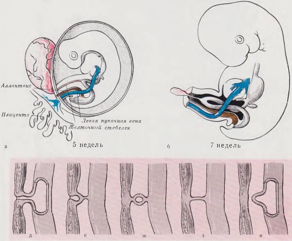
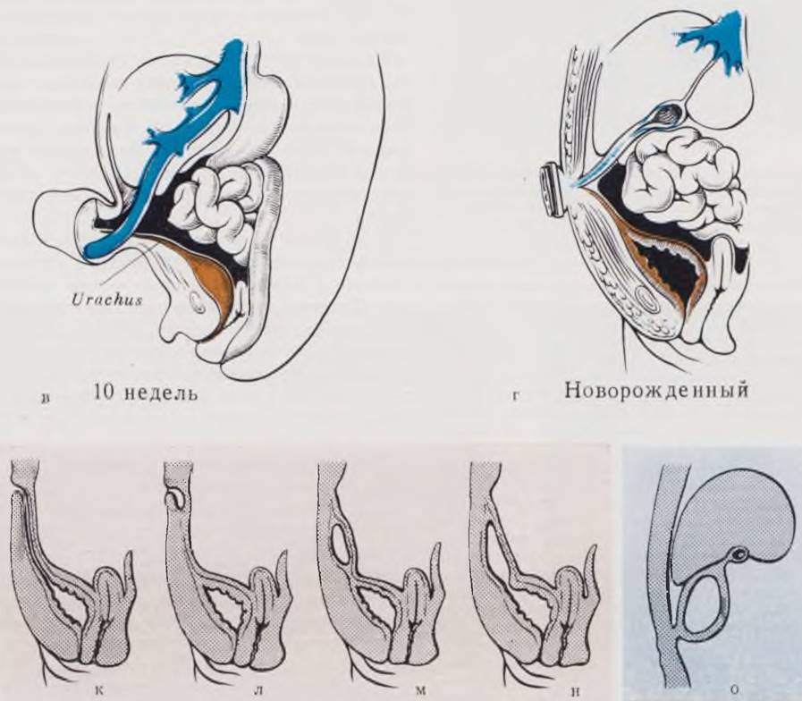
Fig. 67. Newborn with anterior abdominal wall muscle aplasia. Cryptorchidism. Prune Belly Syndrome.

The process of intestinal tube formation is no less complex. As demonstrated by the works of Tandler (1902), N. G. Khlonin (1946), and others, the embryonic intestinal tube up to the 3rd–4th week of life possesses a lumen lined with a single-layered epithelium. Starting from the 4th week, the single-layered epithelium begins to proliferate rapidly, transforms into a Stratified Epithelium, and gradually obliterates the entire lumen of the intestinal tube. This developmental period of the intestine is referred to as the solid or cord stage. It has now been proven that the duodenum and small intestine pass through the solid stage. In the large intestine, only partial epithelial proliferation occurs, which typically does not occlude the lumen. After the 40th–45th day of embryonic development, recanalization of the lumen begins. The epithelial Cells filling the lumen undergo gradual lysis, small vacuoles appear among them and coalesce, and by the 60th day, the intestinal lumen is fully restored. Delayed development of the intestinal tube at the cord stage causes intestinal atresia. Delayed development during the vacuolization stage leads to the formation of membranous atresia or intestinal duplication.
Disruption of the embryonic development of the umbilical cord is of major importance in the genesis of anterior abdominal wall malformations (Fig. 66).
Last update: 10/08/2026
Editorial and Educational Adaptation: This material has been compiled based on the primary/original source text. The project team performed an editorial review, corrected technical inaccuracies, structured sections, and adapted the content for an educational format.
What was processed:
- elimination of formatting defects (OCR errors, structural breaks, corrupted characters);
- editorial organization of content;
- standardization of terminology in accordance with academic sources;
- verification of factual statements against the original source text.
All mentions of the author, publication year, and origin of the primary text have been preserved in accordance with the source.