Post-myocardial infarction angina: pathomechanisms, clinical features, management, and prognosis - Bobrov V. O. 2009
Current concepts of the mechanisms of post-myocardial infarction angina
Mechanisms of formation and development of post-infarction angina, Classification, clinico-angiographic parallels, and Diagnosis
Early post-infarction angina (EPIA) is a clinical syndrome characterized by anginal attacks occurring at rest or upon mild physical exertion. According to various authors, the incidence of EPIA ranges from 15% to 52%. This high Variability in incidence is due to the heterogeneity of patient cohorts, and depends on the timing of pain recurrence within the first 24 hours after acute myocardial infarction (AMI), The Use of thrombolytic therapy, patient age, whether the AMI is primary or recurrent, the presence of small-focal AMI, and pre-infarction angina.
The diagnosis of EPIA can be confirmed by the presence of ST-segment depression of 1 mm or more, T-wave inversion or pseudonormalization during an anginal episode, as well as elevated cardiac enzyme activity. KEY FEATURES OF EPIA include chest pain triggered by physical activity that subsides at rest or upon sublingual nitroglycerin administration, as well as substernal pain occurring during routine strenuous activity after patient discharge. Angina in patients with a prior myocardial infarction (MI) without valvular involvement is highly specific (greater than 95%); consequently, verifying EPIA using electrocardiography solely for diagnostic confirmation is impractical. Stress testing detects EPIA significantly more often (in 58% of patients) than Holter monitoring (27%) or hyperventilation testing (11%) during the late hospital phase of AMI. The low detection rate of residual ischemia via Holter monitoring (HM) may be attributed to the inability of standard chest leads to precisely identify ischemia in the inferior and posterior Regions of the left ventricle (LV).
Diagnostic challenges in EPIA stem from the persistence of ST-segment elevation in some patients during both the acute and subacute stages of myocardial infarction. This elevation can reflect either acute ischemic changes or persistent AMI-related ST-segment shifts, thereby reducing the sensitivity of ECG Diagnostics for EPIA. Therefore, ST-segment changes are not strictly mandatory for establishing the diagnosis, as the prognosis is equally unfavorable in EPIA patients with or without ST-segment alterations.
The absence of well-developed myocardial collaterals in The Setting of long-standing coronary artery disease—often observed in younger patients—promotes the onset of EPIA. Morphologically, this manifests as localized stenosis against a Background of coronary atherosclerosis.
According to ICD-10, EPIA is classified within The Structure of acute forms of coronary artery disease (CAD): acute EPIA is regarded as a form of unstable angina.
Post-infarction angina occurring within the first two weeks of a documented AMI is classified under E. Braunwald’s classification as belonging to the highest risk Class C. During an ECG recording at the moment of an anginal attack, ST-segment depression is often (though not always) observed, driven by more pronounced myocardial ischemia.
According to the proposed working classification, EPIA is categorized under unstable angina (UA). There are 5 patient categories within UA: EPIA, acute coronary insufficiency, stable angina at rest, progressive angina, and new-onset angina.
Some authors divide EPIA into spontaneous angina, exertional angina, and silent ischemia, while others categorize it into pre-infarction new-onset, pre-infarction progressive, and post-infarction angina, further distinguishing patient subgroups with angina at rest, exertional angina, and mixed exertional and rest angina.
A classification of EPIA based on The Nature and localization of ECG changes has been proposed:
1. With ST-segment elevation in the AMI zone.
2. With ST-segment depression in the AMI zone.
3. With ST-segment depression in other zones.
There are several physiological mechanisms underlying The Development of EPIA. The infarct-related coronary artery may remain patent yet significantly stenosed, causing ischemia in viable myocardium. This scenario is typically observed in nearly all patients following thrombolytic therapy. In other instances, the infarct-related coronary artery may be totally occluded, and EPIA arises from inadequate Blood supply to the area adjacent to the affected segment via collateral blood flow originating either from the occluded vessel or from distal branches. Finally, another possible scenario involves the occlusion of the infarct-related artery alongside stenosis of non-infarct-related vessels, manifesting as distal ischemia due to reduced collateral flow from the infarct-related vessel.
The primary factors identified via coronary angiography include residual hemodynamically significant stenosis within the infarct-related coronary artery, diminished collateral BLOOD FLOW IN the region adjacent to the infarcted segment, coronary thrombosis, and coronary spasm.
In recent years, angioscopic studies have provided new insights into the state of the coronary Arteries; however, some authors note that this method only reveals the consequences of vascular disorders, while determining their underlying causes—particularly in EPIA—is severely limited. The advancement of coronary angioscopy has enabled direct color visualization of atherosclerotic plaques and associated coronary thrombi.
The concept that a ruptured plaque serves as the sole basis for thrombosis and subsequent AMI is not always valid. Ruptured plaques have been identified in the coronary arteries of individuals who died from non-cardiac pathologies, yet they showed no clinical signs of AMI.
An atherosclerotic plaque consists of a fibrous cap and a lipid core containing macrophage-derived foam Cells (blood monocyte derivatives) that produce tissue factors. Tissue factors are potent procoagulants that potentially stimulate thrombosis upon contacting blood within the coronary lumen. Mechanical stress concentrates on the fibrous cap, counteracting its significant stretching and direct plaque expansion. The fibrous cap contains smooth Muscle cells that synthesize macromolecules (Collagen, Elastin) forming its matrix. When damaged, the plaque surface—namely its fibrous cap—becomes a source of numerous BIOLOGICALLY ACTIVE SUBSTANCES capable of interfering with the METABOLISM and function of blood elements and the vascular wall itself.
Thus, the Extracellular matrix of the fibrous cap holds the key to understanding unstable coronary syndromes.
Recently, other mechanisms of atherosclerotic plaque destabilization have also been explored. Impaired responses to endothelium-dependent vasodilators have been established in damaged coronary arteries. These findings indicate the presence of vasospasm, which can disrupt blood flow in such vessels, particularly at stenotic sites. Furthermore, it is well known that in the majority of MI cases, thrombotic vascular obstruction develops at the site of plaque rupture.
The results of coronary angiography and ventriculography indicate morphological similarities in AMI between patients with and without EPIA. Patients with EPIA exhibit lesions in the proximal segments of the left coronary artery and its circumflex branch, as well as multi-vessel disease and multiple stenotic segments. Concurrently, a higher incidence of total coronary artery occlusion and coronary thrombosis, yet a lower incidence of severe stenosis, has been observed in EPIA patients. Thrombosis is a hallmark feature of EPIA.
Regarding diagnostic criteria, it must be emphasized that EPIA is a clinical symptom complex; therefore, clinical observation, while subjective, remains the most vital diagnostic modality for this pathology. The cornerstone of clinical diagnostics is 24-48 hour Holter ECG monitoring. Furthermore, Holter ECG monitoring is the most convenient tool for diagnosing Cardiac Arrhythmias—a formidable complication of the subacute myocardial infarction period.
In cardiac intensive care units, stress tests such as bicycle ergometry and transesophageal pacing are recommended to assess coronary reserve.
To evaluate intraventricular hemodynamics and myocardial contractility, ultrasound or radioisotope cardiac scanning is essential, enabling the assessment of segmental myocardial wall motion. Stress echocardiography is also highly indicated.
Recently, dobutamine stress echocardiography has come into clinical practice. Exceptionally high sensitivity (up to 96%) is achieved when evaluating EPIA via thallium or technetium myocardial perfusion scintigraphy. Among all non-invasive techniques, this method provides the clearest diagnosis of such a critical risk factor for sudden cardiac death as post-infarction left ventricular aneurysm.
Data have been obtained indicating the feasibility of coronary angiography in patients with recurrent myocardial ischemia and signs of residual ischemia caused by distal blood flow impairments in the infarct-related coronary artery or multi-vessel coronary disease. This makes it possible to assess coronary artery patency, identify individuals at high risk for fatal outcomes, and determine further management tactics, including physical training, Selection of drug therapy, Surgical Treatment Methods, and others. In addition, using coronary angiography in single infarct-related coronary artery disease, it was found that the prevalence of stenosis is higher in patients with recurrent ischemia than in those without it.
Given the need to prescribe anticoagulants and antiplatelet agents, great importance is attached to studying the cellular and plasma links of Homeostasis, monitoring electrolyte levels (especially in cases of arrhythmias and Heart Failure), plasma lipid profiles, and the state of the vascular wall.
Since the presence of a thrombus in the coronary artery is of primary etiological significance, the recanalization of coronary arteries is being studied as a pathogenetically justified type of treatment. In 1989, E. Braunwald proposed the "open artery" hypothesis as the basis for successful myocardial infarction treatment. In 1978, E.I. Chazov first demonstrated the clinical efficacy of thrombolytic therapy via the intracoronary administration of the thrombolytic agent fibrinolysin. In 1981, K. Rentrop et al. demonstrated the positive effect of intracoronary streptokinase administration in patients with acute myocardial infarction.
The early multicenter thrombolysis trials GISSI and ISIS-2, the results of which were published in 1986, noted the high efficacy of streptokinase in the treatment of acute myocardial infarction. The ISIS-2 trials demonstrated the high efficacy of aspirin, which, when combined with streptokinase, reduced mortality by 42%. The thrombolytic era of myocardial infarction treatment had begun. Consequently, new myocardial states following thrombus lysis were identified.
Myocardial dysfunction in post-infarction ischemia: mechanisms of development
The term "hibernating" myocardium was first proposed by S. Rahimtoola in 1984. This state of myocardial dysfunction (typically of the left ventricle) is caused by its prolonged hypoperfusion with partially or completely absent blood flow.
In acute myocardial ischemia resulting from coronary artery occlusion lasting no more than 15–20 minutes, the impaired left ventricular function rapidly recovers spontaneously. However, if pronounced myocardial hypoperfusion continues for more than 20 minutes, necrotic changes typically occur, followed by the development of cardiosclerosis. After a large-focal myocardial infarction, left ventricular dysfunction is irreversible.
In hibernation, left ventricular dysfunction is more prolonged than in acute myocardial infarction; however, unlike in patients with Post-infarction cardiosclerosis, it is potentially reversible and normalizes or significantly improves after surgical myocardial revascularization—that is, after coronary artery bypass grafting (CABG) or percutaneous transluminal coronary angioplasty (PTCA). In some cases, the function of the hibernated left ventricular myocardium improves after the administration of nitroglycerin.
Myocardial hibernation is an adaptive response characterized by a reduction in heart Muscle Function to a level that achieves a balance between myocardial oxygen demand and blood supply, resulting in the absence of symptoms and signs of myocardial ischemia and the Prevention of myocardial infarction.
Research findings cast doubt on whether prolonged hibernation is a stable and reversible condition. Electron Cell/15.html">Microscopy of myocardial biopsies obtained during CABG surgery has revealed structural changes in areas of hibernating myocardium (hypertrophy or atrophy of individual cardiomyocytes, degenerative cellular changes, extracellular matrix accumulation, and an increase in the number of fibroblasts and macrophages). Therefore, it cannot be ruled out that frequent, prolonged episodes of ischemia may cause degenerative changes in cardiomyocytes and, consequently, cell death via apoptosis (programmed cell death) with subsequent scar formation. Recent studies have shown that hibernating myocardium, as viewed by electron microscopy, is characterized by typical pathological changes: a decrease in the number of sarcomeres, an increase in the number of so-called "small" Mitochondria, progressive Glycogen accumulation, and loss of the sarcoplasmic reticulum, although signs of degenerative damage are absent. Hibernating myocardium also exhibits the expression of Proteins involved in glucose Transport Across the cell membrane, with enhanced expression of fetal GLUT-1 (Insulin-independent) compared to normal GLUT-4 (insulin-dependent). At the same time, despite the presence of significant regional and/or global hypokinesia, oxidative metabolism remains higher than, for example, in scar tissue.
Under conditions of O2 delivery deficit, such as in cardiac ischemia, the balance between ATP production and consumption is disrupted, shifting metabolism from aerobic to anaerobic Glycolysis with the depletion of cellular ATP reserves, impaired membrane permeability, increased lactate production, development of intracellular acidosis, and ion channel dysfunction. This leads to elevated intracellular Na+ and Ca2+ levels, neutrophil activation, which on the one hand promotes the generation of free radicals and on the other causes cell membrane damage. The entire complex of Metabolic Disorders developing in cardiomyocytes ultimately leads to myocardial injury.
The diagnosis of hibernating myocardium is based on the detection of left ventricular wall hypo- and akinesia in an area of reduced perfusion. In patients with recurrent ischemia, hibernation can be detected both in the supply territory of the infarct-related coronary artery and in more remote areas.
Myocardial viability must be demonstrated using a dobutamine stress test performed under echocardiographic guidance. At low doses (5–10 mcg/min), dobutamine restores the contractile function of the hibernating myocardium, whereas in areas of irreversible myocardial damage, function remains as depressed as under resting conditions (E. Picano et al., 1994; M. Ragosta, G. Beller, 1993).
Stunned myocardium is a state of post-ischemic left ventricular dysfunction that persists after reperfusion, despite the restoration of coronary blood flow and the absence of irreversible myocardial injury.
Thus, an important difference between stunned and hibernating myocardium is the presence of normal or near-normal coronary blood flow at rest, while the absence of irreversible myocardial damage distinguishes stunned myocardium from myocardial infarction.
Left ventricular dysfunction caused by myocardial stunning is most commonly encountered following the early administration of thrombolytic agents in the acute phase of myocardial infarction.
The exact mechanisms of myocardial stunning remain unknown. Two main hypotheses have been proposed to explain the Etiology of post-ischemic myocardial dysfunction: the generation of free radicals and calcium overload in cardiomyocytes. Some authors suggest that myocardial stunning is caused by free radicals generated at the onset of reperfusion. Accumulating evidence indicates that cardiomyocyte calcium overload at the onset of reperfusion plays an important role in the Pathogenesis of myocardial stunning after myocardial ischemia in vitro and, potentially, in vivo. A number of studies have shown that various calcium antagonists effectively prevent calcium ion influx into ischemic cardiomyocytes at the onset of reperfusion and attenuate post-ischemic left ventricular dysfunction.
During ischemia-reperfusion periods, oxygen-free radicals are generated from various sources, including mitochondria, cardiomyocyte membranes, endothelial cells, and leukocytes. Free oxygen radicals induce peroxidation of sarcolemmal lipid components, altering membrane permeability and enhancing Ca2+ influx into The Cell. This leads to calcium overload and the activation of Proteolytic Enzymes acting on cardiomyocytes.
Studies of Gene Expression IN stunned myocardium have revealed overexpression of certain proteins (SERCA-2, phospholamban, calsequestrin, calmodulin). This led to the Conclusion that the slow recovery of myocardial contractility following stunning is a prerequisite for the gradual Repair of Damaged structures.
It is most difficult to differentiate myocardial stunning, necrosis, and hibernation, especially in the Cytology/cytology/16.html">Early stages of acute myocardial infarction. In patients with multi-vessel coronary artery disease, thrombotic occlusion of one artery develops, leading to Necrosis of a portion of The Heart muscle and hibernation of the adjacent myocardium. In the event of spontaneous or pharmacologic restoration of Blood flow through the infarct-related coronary artery, a certain portion of the myocardium will remain in a stunned state for several hours or days. Upon restenosis of the infarct-related coronary artery, hibernation may spread to other myocardial areas.
Recently, literature reports have emerged regarding endogenous cardioprotection, convincingly demonstrated in animal experiments and designated as the ischemic preconditioning phenomenon. For example, four episodes of 5-minute coronary artery occlusion followed by reperfusion and subsequent prolonged 40-minute ischemia protected the heart better (infarct size was 1/4 smaller) than in cases without prior short-term ischemic episodes. A notable antiarrhythmic effect was also observed. Based on this, it was concluded that ischemia itself induces an adaptive response to potential recurrent ischemia.
The occurrence of short-term ischemic episodes has been well modeled in other experimental studies in mice, rabbits, dogs, and pigs, and has been observed in human clinical settings. It has also been established that its protective efficacy persists for up to 2 hours. Some researchers attribute the primary mechanism of cardioprotection in ischemic preconditioning to The stimulation of A1-adenosine receptors by adenosine formed via ATP Hydrolysis. Simultaneously, adenosine acts on G-proteins and phospholipase C, resulting in the release of endogenous protective substances. The induction of NO synthase leads to The production of nitric oxide. In addition, protein kinase C translocates from the Cytosol to the sarcolemma, opening ATP-dependent K+ channels, significantly shortening the Action Potential and reducing cellular Ca2+ influx. All of this leads to decreased cardiomyocyte contractility, reduced cellular energy consumption, and lower ATP expenditure.
These same mechanisms explain the "warm-up" phenomenon, in which the first anginal episode is the most severe, while subsequent ones (during the day) present with shorter duration, milder clinical pain syndromes, and a characteristic increase in exercise tolerance. The question regarding The impact of the preconditioning phenomenon on the onset, development, and course of recurrent ischemia, treatment features, and prognosis remains unclear.
Post-infarction left ventricular remodeling
The loss of a portion of functioning myocardium due to infarction, ischemia, or an inflammatory process, as well as chronic cardiac overload, is accompanied by a complex of structural changes involving both damaged and undamaged myocardial areas. These alterations in the structure and geometry of heart chambers, termed "cardiac remodeling" and often preceding clinical manifestations of heart failure, can independently increase systolic and diastolic ventricular dysfunction and negatively impact patients' quality of life and survival. In precise interpretation, "remodeling" refers to The process of structural reorganization in which new material is incorporated or the structure is entirely altered. In a broader sense, cardiac remodeling signifies the process of complex disruption of cardiac Structure and function in response to damaging overload or the loss of viable myocardium. The cardiac remodeling process primarily involves progressive increases in myocardial mass, chamber dilation, and alterations in ventricular geometric characteristics.
The Significance of the relationship between normal and impaired ventricular function and geometry in various cardiovascular diseases involves pressure or volume overload, as well as myocardial ischemic injury.
The term "remodeling" began to be used in the 1980s to denote structural and geometric changes of the left ventricle (LV) developing after acute myocardial infarction (AMI). At that time, cardiac remodeling research was limited to noting existing changes in LV structure and geometry. The mechanisms of remodeling, its determinants, and hemodynamic effects remained unstudied.
Extensive Study of the clinical aspects of cardiac remodeling began in the 1990s following the publication of classical studies by M. Pfeffer and E. Braunwald on LV remodeling after acute myocardial infarction, especially after the SAVE trial, which demonstrated that the use of the ACE inhibitor captopril in the comprehensive treatment of myocardial infarction helps inhibit post-infarction LV remodeling, accompanied by a significant improvement in the clinical course of the disease and prognosis.
Impaired ventricular geometry is inherent to all patients with LV dysfunction and often precedes a decrease in ejection fraction, systemic hemodynamic disturbances, and clinical manifestations. Furthermore, changes in LV geometry represent an early process that initiates and contributes to the development of heart failure.
The process of early LV remodeling after AMI involves topography disruption in both affected and unaffected areas. In the affected area, the spread of the infarct zone with regional expansion and thinning of the infarcted region occurs within the first 24 hours from the onset of the disease. The expansion of the infarct significantly alters LV volume and geometry, lays the groundwork for The formation of acute aneurysm and myocardial rupture, and serves as an important substrate for LV chamber enlargement. LV dilation in the acute period of myocardial infarction can be attributed to a compensatory mechanism that maintains stroke volume and occurs when more than 20% of the myocardium is damaged.
Parietal thinning of the infarcted LV region is a consequence of decreased resistance of the affected myocardium during systolic stress with each heartbeat and is driven by several mechanisms, including myocyte stretching with a decrease in their diameter, and myocyte slippage.
In addition, an increasing number of researchers describe remodeling processes occurring in the unaffected myocardium remote from the necrotic zone. The myocardium distant from the infarct area is subjected to an acute increase in diastolic myocardial stress. As noted above, this is accompanied by myocyte slippage and hypertrophy. The myocardial hypertrophy observed here exhibits features of combined pressure and volume overload.
High myocardial stress in the unaffected myocardial region serves as a stimulus for myocardial hypertrophy and chamber remodeling in the chronic post-infarction phase. Hypertrophy of the unaffected heart muscle is an early physiological response to myocardial injury that accompanies Changes in the affected myocardial zone. The positive effects of the early phase of ventricular hypertrophy include the normalization of systolic function in the damaged wall and the maintenance of stroke volume. At the same time, a growing number of researchers indicate that prolonged exposure to high myocardial stress facilitates the transition from heart muscle hypertrophy to its failure.
Early changes in LV volumes and geometry have important prognostic significance for patients who have suffered a myocardial infarction. It has been proven that a relatively small increase in end-diastolic and end-systolic LV volumes post-infarction increases the risk of death by 4–5 times.
In late remodeling, the predominantly unaffected myocardium is recruited into the process. In small-focal or non-transmural infarctions, ventricular function and geometry may return to normal during the recovery phase, whereas in large infarctions, progressive remodeling occurs with additional volume enlargement and further alterations in LV geometry. J. Erlebacher et al. showed that late LV volume enlargement is common in patients with extensive MI.
The modern literature continues to debate the issue of progressive LV remodeling in the late period after myocardial infarction. Although these progressive changes are well described in experimental studies, until recently there was a lack of convincing clinical studies confirming that LV remodeling after myocardial infarction is a progressive process and that, once initiated, it continues for months without additional ischemic episodes. In recent years, a number of long-term prospective multicenter trials (SAVE, AIRE, SMILE, SOLVD) have been completed, the results of which convincingly demonstrate that the process of LV remodeling after myocardial infarction can continue slowly but progressively over a long period, and early use of ACE inhibitors can favorably influence cardiac remodeling processes, patients' quality of life, and prognosis.
LV geometry plays a central role in its normal functioning and in the process of cardiac remodeling in various cardiovascular diseases. The loss of the normal ellipsoidal shape of the ventricle is an early sign of cardiac injury that precedes and may trigger the development of chronic heart failure. Impairment of regional LV geometry is particularly critical after myocardial infarction; it can contribute to late progressive ventricular dysfunction, which tends to persist for a long time in the absence of additional ischemia and myocardial necrosis.
Cardiac remodeling precedes and accompanies the clinical manifestations of heart failure, and it can independently increase both systolic and diastolic ventricular dysfunction. At a certain stage of the disease, "rebarreled heart syndrome" ("structural cardiomyopathy") may relegate the etiological damaging mechanism to the background and determine the patients' quality of life and prognosis.
Cardiac remodeling should not be viewed as a general stereotypical process. Studying and understanding the physiological and pathogenetic role of cardiac remodeling in each specific case will help avoid unjustified therapeutic interventions and thereby optimize the approach to treating cardiovascular diseases.
Thus, taking into account the predominance of the systolic or diastolic component of heart failure developing after an AMI can allow for a differentiated approach to these heterogeneous groups of patients.
Modern views on myocardial diastolic dysfunction and methods for its assessment
LV systolic dysfunction is currently a well-proven predictor of adverse prognosis in post-AMI patients, but in recent years, LV diastolic dysfunction (DD) has also become associated with the development of HF, progressive LV dilation, and post-AMI mortality. Interestingly, while there are numerous papers on the presence and diagnostic value of myocardial diastolic dysfunction (MDD) in coronary artery disease (CAD), there are no studies on its impact in the early post-infarction period compared to the predictive value of other diagnostic methods. An exception is the work by S.H. Poulsen et al., who made the first attempt to evaluate the predictive value of diastolic function abnormalities compared to heart rate variability (HRV) in patients after their first AMI.
Diastolic dysfunction is an important pathogenetic factor in many cardiac diseases. Moreover, MDD can precede systolic dysfunction in a number of myocardial disorders. A detailed phase-by-phase evaluation of diastolic abnormalities requires the determination of LV pressure, its volume, stiffness constants, and LV relaxation indices.
However, the interpretation of intraventricular hemodynamics parameters obtained during cardiac catheterization has certain limitations. Furthermore, invasive studies are quite hazardous and expensive, and therefore cannot be widely used. Non-invasive investigation of cardiac diastolic function remained impossible until the 1970s, when Methods for Assessing LV diastolic filling emerged, such as echocardiography (EchoCG), Doppler echocardiography, and radionuclide angiography. These methods made it possible to evaluate The rate of LV relaxation, its passive properties, and filling. In this aspect, the 2005 recommendations of the European Society of Cardiology on chronic heart failure are also important, requiring mandatory visual evidence of LV dysfunction and objective signs of LV DD with preserved LV systolic function when dealing with isolated diastolic HF.
Diastole physiology. Normal myocardial diastolic function refers to LV filling that provides Cardiac Output according to the body's needs at a mean pulmonary capillary wedge pressure of less than 12 mmHg. Consequently, the onset of MDD is primarily determined by an increase in pulmonary venous pressure.
Diastole is the period from the end of blood ejection (closure of the semilunar Valves) to the closure of the atrioventricular valves. For the LV, the duration of diastole is the period from aortic valve (AV) closure to mitral valve (MV) closure. Ventricular filling depends mainly on three primary factors: ventricular stiffness, relaxation, pericardial constraint, and ventricular interaction. In addition, heart rate (HR), the state of the left atrium, and the MV are of significance.
LV stiffness (passive-elastic properties) is directly proportional to myocardial mass and its stiffness, and inversely proportional to cavity volume. Myocardial stiffness can increase due to an increased Connective Tissue content with enhanced collagen cross-linking and due to the disruption of muscle fiber arrangement.
There are 3 main factors influencing diastolic relaxation: relaxation load, inactivation (termination of Actin-Myosin cross-bridges), and spatial-temporal asynchrony of tension and inactivation. Among the loads affecting relaxation, end-systolic strain at all levels of cardiac structure can be distinguished, with the accumulation of potential energy of elastically compressed myocardium, which facilitates ventricular filling at the beginning of diastole. In addition, coronary filling load (determined by the degree of coronary bed filling during isometric relaxation) and ventricular filling load (decrease in wall thickness and increase in cavity dimensions during diastole) are of importance.
Inactivation is the termination of actin-myosin cross-bridges in the myocardium. Its impairment may be caused by cardiomyocyte calcium overload and energy depletion.
Another important factor in relaxation is the spatial and temporal asynchrony of tension and inactivation. A certain physiological asynchrony of these processes is also normal. However, in the presence of sclerosis areas and fiber disarray, the degree of regional contraction and relaxation asynchrony increases. Diastole is subdivided into the following phases:
- isovolumetric relaxation time (from aortic valve closure to mitral valve opening);
- LV rapid filling phase;
- LV slow filling phase;
- atrial contraction.
In early diastole, when LV pressure falls below left atrial (LA) pressure, the mitral valve opens and LV diastolic filling begins. The pressure gradient between the left atrium and the left ventricle drives a rapid blood flow from the LA through the mitral valve during early diastole. The magnitude of this gradient is determined mainly by the LA pressure level and the rate of LV pressure decline. The duration of this phase and the rate of LV filling are significantly influenced by the "LA-LV" pressure gradient, the Elastic properties of the heart muscle, passive compliance, and its relaxation rate. LV volume increases rapidly. Approximately 60-80% of the stroke volume enters the LV During the first third of diastole. As pressures in the LA and LV equalize, LV filling slows down; as a rule, LV relaxation ends at this moment. The middle part of diastole is referred to as diastasis. The rate of LV filling during diastasis is determined by the pulmonary venous inflow rate. The volume of LV filling in late diastole (atrial systole) depends on LV compliance and LA contractility, as well as on the LV pressure at the onset of atrial contraction.
Diastolic hemodynamics largely depends on heart rate. Normally, the LA empties significantly during early diastole. In this situation, LA contraction contributes very little to LV filling. Conversely, if early diastolic filling is reduced, more blood remains in the left atrium by the onset of its systole, which leads to an increased contribution of the LA to LV filling.
Interrelationship between systolic and diastolic Functions. Systolic and diastolic myocardial functions are closely interrelated. For instance, more active myofibril contraction facilitates the energy supply required for active relaxation. Moreover, since the right and left heart chambers are contained within a closed system (the pericardial sac), changes in right ventricular volume and pressure exert a corresponding effect on LV diastolic properties. Thus, in acute volume overload, the rise in LV diastolic pressure is determined not only by changes in its elasticity, but also by the Influence of the distended right ventricle and Pericardium. On the other hand, facilitation of muscle fiber relaxation leads to their stretching, which, in turn, improves systolic function via the Frank-Starling mechanism. Consequently, impairments in systolic or diastolic phases are interconnected.
Difference between systolic and diastolic myocardial dysfunction. Myocardial systolic dysfunction is an impairment of myofibril contractility and, consequently, The ability to eject blood from the LV into the aorta. Diastolic dysfunction is the inability to achieve adequate LV filling without a compensatory rise in left atrial pressure. In MDD, LV relaxation and filling do not proceed rapidly, occur with delay, or remain incomplete. Atrial pressure increases compensatorily, and signs of pulmonary or systemic venous congestion may appear. The most frequent cause of MDD is impaired myocardial contractility. However, primary diastolic dysfunction in the absence of any systolic involvement has been diagnosed much more frequently of late. Conditions predominantly associated with MDD are classified as "diastolic diseases." These include: hypertrophic and restrictive cardiomyopathies, myocardial involvement in Diabetes Mellitus, arterial Hypertension, and aortic stenosis; in elderly patients, during myocardial ischemia and fibrosis, as well as structural disorders such as constrictive pericarditis, atrial myxoma, mitral stenosis, and cardiac tamponade. In patients with impaired diastolic filling due to hypertrophy, restrictive cardiomyopathy, ischemia, or pericardial disease, congestion symptoms appear despite normal myocardial contractility. Therefore, the onset of congestive heart failure should not be attributed solely to myocardial systolic dysfunction. The overall prognostic role of MDD and its role in patients with post-infarction ischemia are currently being clarified.
Pathophysiological mechanisms of MDD. Various factors, individually or in combination, can lead to an elevation of LV end-diastolic pressure, LA and pulmonary venous pressures, and clinical manifestations of HF.
The term "stiff myocardium" was first used by A. Dodek (1972) to describe a group of patients with CHD and recurrent episodes of acute left ventricular failure without clinical and radiological signs of cardiomegaly. Increased passive stiffness or decreased compliance of the LV is the result of chronic processes that alter The properties of the ventricular wall. Frequently, the cause of a stiff myocardium is a large scar or LV aneurysm following Transmural myocardial infarction, as well as its diffuse fibrosis in dilated cardiomyopathy. It should be noted that the reduction in LV compliance in a number of pathological processes has a certain compensatory significance, reducing the degree of stretching and overload of the affected myocardium.
Another cause of increased LV passive stiffness is myocardial hypertrophy. This includes both concentric symmetric hypertrophy due to prolonged pressure overload (arterial hypertension or aortic stenosis) or volume overload (valvular regurgitation), and asymmetric hypertrophy, often seen in hypertrophic cardiomyopathy. In patients who have suffered an AMI, the development of local hypertrophy in preserved areas of the myocardium is one of the Components of the long-term LV remodeling process. A significant increase in myocardial mass may be accompanied by impairments in its contractility and distensibility. Thus, hypertrophy directly affects the stiffness of the heart wall, which is explained by quantitative and qualitative changes in collagen fibers, muscle fiber disarray, and other factors. Furthermore, the presence of hypertrophy is accompanied by an increased myocardial sensitivity to ischemia, the development of relative coronary insufficiency, and impaired myocardial relaxation.
Relaxation disorders represent another pathogenetic factor in elevated diastolic pressure. The rate of myocardial relaxation is determined not only by its inotropic function, but also by the ventricular load during the preceding contraction, and depends on heart rate and the synchrony of contraction and relaxation processes. Impairments in myocardial relaxation are closely associated with ischemic injury. It is believed that active diastolic relaxation accounts for about 15% of myocardial Energy Expenditure. On the other hand, the strength and duration of cardiac contractions are primarily determined by intracellular calcium concentration; relaxation occurs when calcium concentration decreases. Impaired calcium ion efflux from the Cytoplasm and their uptake by the sarcoplasmic reticulum due to Hypoxia and insufficient cyclic adenosine monophosphate production can lead to relaxation disorders and HF. An important role is also played by increased aortic pressure and disruption of the synchronous course of contraction and relaxation. Heterogeneity underlies various relaxation abnormalities, for example, in chronic CHD or after AMI; its correction is accompanied by facilitated LV relaxation and filling.
Doppler indices of LV diastolic function. Doppler echocardiography is a method for direct and non-invasive measurement of blood flow velocities, allowing for prospective and dynamic evaluation of cardiac diastolic function. Analysis of diastolic filling is most often performed from an apical four-chamber view, although a two-chamber view can also be used. Doppler examination is performed in two modes: pulsed-wave and continuous-wave. Blood flow across the MV is usually assessed in pulsed-wave mode, which makes it possible to obtain a velocity curve at the level of the mitral leaflets (the MV fibrous ring) and perform a detailed analysis of diastolic phases. The results of the Doppler examination depend largely on the angle between the ultrasound beam and the blood flow directions. The most reliable data are obtained when they are parallel.
Combining pulsed-wave Doppler echocardiography and a phonocardiogram makes it possible to establish the moments of aortic valve closure and MV opening—the beginning and end of The first phase of diastole. There is also an alternative way to measure the duration of this diastolic phase. For this purpose, a continuous-wave Doppler curve is obtained by directing the ultrasound beam so that it intersects the LV outflow tract and the anterior MV leaflet. The end of flow in the LV outflow tract corresponds to the moment of aortic valve closure; the artifact of MV opening is also recorded. On the Doppler echocardiogram, the first phase of diastole corresponds to the LV isovolumetric relaxation time, which depends on the myocardial relaxation rate and the rate of LV pressure decline (compliance).
Blood flow velocity across the MV reflects the pressure gradient between the heart chambers. The Doppler method assesses changes in pressure relationships between the LA and LV. The transmitral flow curve has a characteristic biphasic structure. The onset of the first peak corresponds to the moment of mitral valve opening, its amplitude (E) to the peak early diastolic filling velocity, and its duration (dE) to the length of the second diastolic phase. Acceleration time is the period from the onset of transmitral flow to the moment it reaches its highest velocity (Fig. 1).
An indicator of the deceleration rate of early diastolic filling is the deceleration time—the period from the peak filling velocity to the moment of its maximal decline. This parameter is influenced by myocardial relaxation, passive LV filling characteristics, and the pressure ratio between the LV and LA. In mid-diastole, transmitral blood flow velocity is low (diastasis); as it increases, additional oscillations between peaks E and A correspond to the third phase of diastole.
The velocity of late diastolic LV filling (the fourth phase of diastole) is reflected on the Doppler echocardiogram by the height of peak A (A), and its duration by the length of this peak (dA). Velocity time integrals EI and AI correspond to early and late diastolic flow volumes. Informative indicators of diastolic function include the ratios between the amplitudes and areas of waves E and A (E/A or EI/AI). The ratios of the integrals EI and AI to the total diastolic flow integral TI correspond to the contributions of early filling (EI/TI) and atrial systole (AI/TI) to total LV diastolic filling. Another indicator is the half-filling fraction, which is defined as the percentage of the time integral during the first half of diastole.
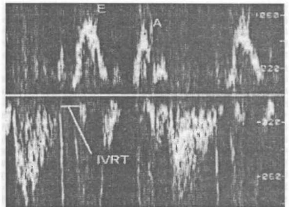
Fig. 1. Normal transmitral blood flow patterns
In addition to parameters characterizing individual phases of diastole, integral indices providing a General Overview of cardiac diastolic properties have become widely used. These include the total duration of transmitral flow, the duration of the period from MV opening to the peak of atrial filling, and the time required for the LA-LV pressure gradient to decrease by half during early diastole. Given that the duration of diastole and its individual phases varies depending on heart rate, these parameters are normalized to the RR interval.
Thus, the researcher has a significant number of indices at their disposal to evaluate cardiac diastolic function. It is considered that the most informative parameters for interpreting diastolic function characteristics and the relative roles of early and late LV filling are: E, A, E/A, EI, AI, EI/AI, EI/TI, AI/TI, acceleration time and deceleration time, as well as the period from MV opening to the peak atrial filling velocity.
All of the listed parameters are obtained by examining subjects in sinus rhythm. The only indicator of diastolic function in patients with atrial fibrillation is the isovolumetric relaxation time.
The volumetric filling rate of the LV depends on the mitral orifice area. It can be assumed that the mitral orifice area (S) equals the area of the mitral ring and therefore remains constant throughout diastole. Taking this parameter into account, normalized indices of LV diastolic filling are obtained: early filling (E × S) and atrial filling (A × S). These parameters, as well as The ratio of early filling to LV end-diastolic volume, correlate closely with corresponding parameters obtained by contrast ventriculography.
Assessment of DD mechanisms by the Doppler method. The Doppler method plays a crucial role in determining the pathophysiological mechanism of LV dysfunction in congestive circulatory failure and in identifying patients with MDD and normal systolic function. Doppler assessment of transmitral flow is an extremely sensitive method for the early diagnosis of myocardial ischemia. Impairments in LV diastolic filling in hypertensive patients precede the appearance of clinical signs, ECG changes, or echocardiographic criteria for myocardial hypertrophy. At the same time, it should be noted that decreased compliance of the hypertrophied LV is, to some extent, a manifestation of its Structural and functional adaptation to increased load.
Normally, transmitral flow reflects predominantly early diastolic LV filling and the presence of a "diastolic reserve" in patients with normal heart rate and pulmonary venous pressure values. Blood flow velocity across the mitral valve depends on many factors that must be considered for the correct interpretation of diastolic function features. Myocardial relaxation, passive compliance, left atrial pressure, blood pressure, PQ interval duration, and heart rate all affect the pattern of transmitral flow. In particular, tachycardia reduces The Role of early diastole, increases preload, and enhances the atrial contribution to LV filling. Under these conditions, the transmitral flow pattern may change, but overall diastolic function and cardiac output are not compromised. However, when the atrium can no longer compensate for reduced early diastolic filling, cardiac output is maintained by increasing pulmonary venous pressure. A further rise in LA pressure makes it possible to increase the role of early diastole. In this case, the transmitral flow pattern resembles a normal one ("pseudonormalization"). A significant increase in LV end-diastolic pressure increases LA afterload and ultimately leads to LA systolic failure. Therefore, in severe MDD and a marked rise in pulmonary venous pressure, "normal" Doppler peak ratios may also be observed.
Thus, an increase in left ventricular diastolic pressure leads to a decrease in the rate of late filling and the A-wave; another cause of the second peak's reduction may also be left atrial systolic weakness. Data show that in severe coronary artery disease (CAD), an E/A ratio greater than 2 corresponds to a left ventricular end-diastolic pressure exceeding 20 mm Hg.
The reduction in early diastolic left ventricular filling and, accordingly, the increased relative role of atrial filling are primarily the result of prolonged myocardial diastolic relaxation and the loss of the "diastolic reserve." Impaired relaxation of the heart muscle is a significant pathophysiological mechanism of left ventricular diastolic dysfunction (LVDD) in acute coronary insufficiency, hypertension, aortic stenosis, hypertrophic cardiomyopathy, and dilated cardiomyopathy. On the doppler echocardiogram, this manifests as a decrease in the E-peak, altered amplitude and interval ratios of transmitral flow (E/A and EI/AI), and an increase in acceleration time and deceleration time (see Fig. 2). A major limitation in adequately assessing myocardial relaxation is that the exact moment of its cessation is unknown. For practical purposes, relaxation is conventionally measured starting from the moment of aortic valve closure. The most sensitive and early Doppler sign of LVDD resulting from impaired relaxation is the prolongation of IVRT.
With a significant decrease in compliance (or an increase in stiffness) of the myocardium, left ventricular pressure rapidly increases already in the early diastole phase, resulting in a high afterload on the left atrium. THE CONTRIBUTION OF atrial systole and the amplitude of the A-wave decrease, while the E/A ratio increases. A sharp reduction in the pressure gradient between the left atrium and left ventricle appears on the transmitral flow graph as a shortened deceleration time, which corresponds to a "restrictive" transmitral blood flow pattern (Fig. 3).
Patient age significantly affects Doppler diastolic parameters, making it necessary to account for age when analyzing individual patient data and comparing them with normative values. Thus, normally, the peak velocity E, the integral EI, as well as the E/A and EI/AI ratios decrease with age, reflecting a reduced contribution of early filling and an increased role of active atrial left ventricular filling.
The E/A ratio varies from 2.08 ± 0.55 in healthy individuals under 30 years of age to 0.84 ± 0.29 in subjects over 70. For individuals under 40, an E/A value below 1.0 is pathological, whereas in those over 70, it is regarded as normal. An EI/AI ratio below 1.5 is considered pathological under age 40 and is observed in half of subjects over 70 without pronounced signs of heart pathology. Consequently, due to increased myocardial mass in the elderly, signs of elevated left ventricular stiffness frequently appear. At the same time, fiber relaxation heterogeneity is accompanied by prolonged IVRT and a decreased E/A ratio. The combination of relaxation abnormalities and reduced compliance, particularly in marked myocardial hypertrophy, can lead to the "normalization" of several Doppler indices of diastolic filling (E/A and EI/TI).
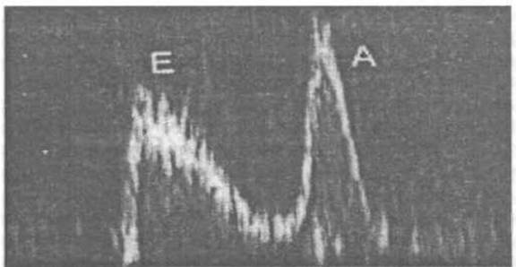
Fig. 2. Transmitral blood flow pattern in impaired left ventricular relaxation
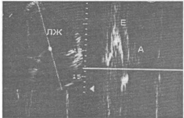
Fig. 3. Restrictive type of intraventricular hemodynamics
Diagnosing LVDD is also difficult in patients with marked left ventricular dilation, significant mitral regurgitation, and aortic regurgitation. For instance, in patients with dilated cardiomyopathy and LVDD, the transmitral flow peak ratio often remains normal. In pronounced mitral regurgitation, the pressure gradient between the left atrium and left ventricle increases during the early diastole phase. Consequently, the amplitude of the first peak increases, making it impossible to detect impaired left ventricular diastolic filling.
Doppler echocardiography makes it possible to determine the hemodynamic state in the pulmonary artery, which is important for assessing diastolic filling. To roughly estimate mean pulmonary artery pressure, the following formula has been proposed: P = (-0.5 × AT) + 80, where AT is the pulmonary artery flow acceleration time in ms.
Since 90% of patients with pulmonary hypertension have tricuspid regurgitation, Doppler echocardiography data on tricuspid blood flow can often be used to determine pulmonary artery pressure using the formula: P2 = 4V2 + P1, where P2 is the right ventricular systolic pressure, V is the maximum tricuspid regurgitation velocity, and P1 is the right atrial pressure (central venous pressure).
Other diagnostic methods for LVDD. LVDD is present in a significant proportion of patients. Detailed phase evaluation of diastolic disorders requires determining left ventricular pressure, volume, and stiffness constants. At the same time, interpreting intraventricular hemodynamics is impossible without simultaneously recording left atrial pressure, which greatly complicates the researchers' task. Radionuclide ventriculography also provides information on left ventricular filling that is comparable in diagnostic value to cardiac catheterization data.
Overall, clinical practice lacks a single "gold standard" for diagnosing LVDD. Alongside echocardiography with Doppler evaluation, pulmonary artery wedge pressure can be measured directly. For research purposes, isovolumic relaxation parameters (dP/dt) and ventricular distensibility—the pressure-volume curve (dV/dp)—can also be compared with echocardiographic and Doppler parameters of diastolic filling. To clarify the causes of LVDD, the ratio of left ventricular end-diastolic pressure to its mass or cavity wall radius is determined, which is particularly reliable for persistent alterations in the pressure-volume relationship. Left ventricular wall thickening indicates hypertrophy or an infiltrative process; otherwise, a "stiff" myocardium should be suspected.
Current management strategies for patients with post-infarction myocardial ischemia
Because the clinical manifestations and pathophysiological mechanisms of post-infarction angina (PIA) and acute myocardial infarction (AMI) are similar, and in accordance with the American Heart Association guidelines for reducing the risk of death in patients with CAD and atherosclerosis, as well as the European Society of Cardiology guidelines in conjunction with the European Societies for the Prevention of Atherosclerosis and Hypertension, patients with PIA are indicated for comprehensive medical treatment including antithrombotic therapy, beta-blockers, and ACE inhibitors. Nevertheless, the administration of long-acting nitrates and calcium antagonists in individually tailored doses remains relevant.
Long-acting or intravenous formulations of nitroglycerin are used as symptomatic therapy and to optimize preload (to a pulmonary artery wedge pressure below 14 mm Hg) and afterload (to reduce systolic pressure by 10–15 mm Hg, optimally below 110 mm Hg, but not lower than 100 mm Hg).
The ability of beta-blockers to improve the prognosis of patients after AMI is due to a reduction in the incidence of sudden cardiac death (SCD) resulting from anti-fibrillatory and anti-ischemic effects, and correlates with the degree of heart rate reduction. Prescribing beta-blockers to patients at high risk of myocardial ischemia is of particular importance. Among these patients, a 25% reduction in mortality is observed. For example, the use of atenolol in the international ISIS-1 trial showed a 15% reduction in mortality during 7 days of treatment following AMI, with the positive effect persisting for one year. Furthermore, the use of propranolol for 25 months post-AMI in the BHAT trial showed a 26% reduction in mortality. Beta-blockers are also effective in patients who have undergone thrombolytic therapy. In the TIMI-II trial, early administration of metoprolol against the background of tissue plasminogen activator (t-PA) therapy demonstrated a significant reduction in recurrent ischemic episodes during the first 6 days compared to later administration (15.4% and 21.2%, respectively). In patients with severe AMI complications (arrhythmias, hypotension, congestive heart failure), beta-blocker therapy reduced post-infarction mortality despite their negative inotropic effect. According to the European Society of Cardiology guidelines, in the absence of contraindications, beta-blockers are recommended for all AMI patients long-term.
The use of calcium antagonists in AMI patients has been heavily criticized, but the risk is primarily associated with short-acting nifedipine formulations. Specifically, the results of a meta-analysis by R. Fletcher (1991) showed that its use in AMI significantly increases the risk of SCD, which is believed to be related to the stimulation of the sympathoadrenal system. Verapamil administered 2 weeks post-AMI for 12–18 months reduced overall mortality and the incidence of recurrent infarctions. The best outcome in the acute phase of the disease was observed in patients without signs of heart failure or atrioventricular block. Patients prescribed verapamil also showed a 20% reduction in the combined endpoint rate (deaths and recurrent infarctions). The randomized placebo-controlled CRIS trial revealed verapamil's efficacy in reducing mortality and recurrent myocardial infarctions, aligning with findings from other studies.
When diltiazem was used post-myocardial infarction, it exhibited a negative effect in patients with heart failure and low left ventricular ejection fraction. In patients without these complications, a 23% reduction in recurrent infarction rates was observed.
The question regarding the impact of long-acting dihydropyridines (amlodipine, isradipine, felodipine) on the course of PIA remains open.
A new class of anti-ischemic drugs—potassium channel openers, represented by nicorandil—has recently been proposed. The drug's efficacy in unstable angina highlights the promise of its further study.
According to the European Society of Cardiology, when ACE inhibitors are administered within the first hours of symptom onset, myocardial infarction mortality is 4–5 per 1,000 patients. However, these data are not definitive and primarily apply to patients with heart failure or recurrent myocardial infarction. For instance, the CONSENSUS II trial, in which enalapril was administered intravenously within 24 hours of AMI onset, was prematurely terminated due to a trend toward increased mortality compared to placebo. Yet, later studies have convincingly proven the safety of early oral ACE inhibitor administration starting with low doses. For example, the results of enalapril use in the SOLVID trial demonstrated a 23% reduction in recurrent myocardial infarction and a 20% reduction in unstable angina, indicating the anti-ischemic effect of ACE inhibitors. The multicenter SAVE trial proved the preventive value of ACE inhibitors during myocardial infarction, showing a 19% relative reduction in mortality, with cardiac mortality decreasing by 21%, hospitalizations for heart failure by 22%, and recurrent infarction by 25%. It is advisable to prescribe ACE inhibitors to patients with an ejection fraction below 45%. The results of the SMILE trial indicate reduced post-AMI mortality under The Influence of ACE inhibitors.
In patients with a left ventricular ejection fraction below 35%, a 22% reduction in mortality was found following trandolapril administration compared to placebo. Ramipril increased survival in AMI patients with heart failure by 27%, while reducing the incidence of recurrent infarction, stroke, and severe heart failure by 18%. The efficacy of short-term ACE inhibitor use in the post-infarction period has been proven: captopril administered within the first 24 hours of AMI onset contributed to a significant reduction in mortality. Similar data were obtained with lisinopril administration, showing an 11% reduction in mortality and composite endpoints (deaths, pronounced heart failure, ejection fraction drop below 35%, and the number of akinetic or dyskinetic echocardiographic segments). These results are generally confirmed by other researchers.
The reduction in mortality among PIA patients treated with ACE inhibitors can be explained by their cardioprotective action (reduction of early and late left ventricular remodeling by decreasing preload and afterload, and neutralizing the Adverse effects of angiotensin II activation and the sympathoadrenal system). The vasoprotective effect may be attributed to the inhibition of cardiomyocyte apoptosis in the peri-infarct zone of AMI and the prevention of interstitial fibrosis and myocardial hypertrophy at later stages.
The vasoprotective effect of ACE inhibitors is mediated by preventing coronary vasospasm through increased bradykinin-stimulated endothelial NO production, coupled with reduced vascular remodeling via the inhibition of smooth muscle cell proliferation. The anti-ischemic action of these drugs is driven by an antithrombotic effect—achieved by lowering plasminogen activator inhibitor levels—and an antiatherogenic effect, which stems from suppressing free radical formation in the vessel wall, decreasing its permeability to LDL, and inhibiting the transformation of macrophages into foam cells.
The rationale for administering heparin and other antithrombotic agents during the early stages following a myocardial infarction lies in their ability to prevent rethrombosis and reocclusion of the infarct-related artery. According to a meta-analysis of 20 trials conducted in the pre-thrombolytic era (McMahon et al.), post-AMI mortality decreased by 17±7 %, and the rate of recurrent infarctions dropped by 22±10 %.
In patients who did not undergo thrombolytic therapy, the mortality rate was 8.7 % among those receiving heparin, compared to 12.9 % in the untreated control group. Other studies reported mortality rates of 10.9 % following intravenous heparin administration, 11.2 % with subcutaneous administration, and 10.1 % without heparin treatment.
When high doses of heparin are administered intravenously within 6–12 hours of symptom onset, studies show that even without thrombolytic therapy, heparin combined with acetylsalicylic acid can fully restore patency in the infarct-related artery if given within 2 hours of AMI onset. Intravenous heparin is necessary following alteplase (t-PA) therapy, but is unnecessary when using streptokinase. Clinical trials have demonstrated the superiority of low-molecular-weight heparin over unfractionated heparin in AMI patients, as well as the ability of hirudin, when combined with thrombolysis, to achieve faster reperfusion of an occluded infarct-related artery than heparin.
Currently, the following antiplatelet agents are utilized:
- cyclooxygenase inhibitors, specifically acetylsalicylic acid (ASA);
- ADP receptor antagonists, including ticlopidine, autoprost, and clopidogrel;
- platelet glycoprotein (GP) IIb/IIIa receptor antagonists, such as c7E3 Antibodies (abciximab), synthetic Peptides (integrilin), synthetic non-peptides (lamifiban, tirofiban), and orally active agents (xemilofiban, fredafiban);
- thromboxane receptor antagonists—ridogrel, pirmagrel, and daltroban (solutroban);
- other agents, including von Willebrand factor antagonists, Thrombin receptor antagonists, and NO Donors.
Administration of ASA has been associated with a 23 % reduction in mortality, which reaches 42 % when combined with streptokinase, and up to 53 % when administered within 6 hours of AMI onset. Furthermore, ASA therapy reduced the rate of non-fatal reinfarctions by 49 %.
The therapeutic benefits of ticlopidine and tirofiban (a GPIIb/IIIa platelet receptor blocker) have been well established. The risk of myocardial infarction decreased from 5.7 % to 4.3 %, The Need for emergency percutaneous transluminal coronary angioplasty (PTCA) dropped from 5.4 % to 4.2 %, and coronary artery bypass grafting (CABG) rates fell from 20.2 % to 1.8 % (although minor hemorrhagic complications were observed). The greatest efficacy was noted during the first 24 hours of the disease.
A series of trials (CARPORT, CAPTURE, EPILOG, RAPT) evaluating other antiplatelet drugs have yielded conflicting results that warrant further investigation.
Regarding the use of indirect anticoagulants in the management of post-infarction myocardial ischemia, clinical studies have failed to demonstrate any significant advantages of warfarin.
Because thrombosis is a mandatory prerequisite for the development of post-infarction angina, thrombolytic therapy is essential. At the same time, administering thrombolytic agents during the first hours of anginal pain in AMI patients helps improve myocardial contractility, despite a 15–30 % risk of subsequent reocclusion.
In unstable angina (UA), thrombolytic agents are rarely prescribed, and even less frequently in post-infarction angina. Due to the small patient cohorts involved, these findings remain contradictory regarding both clinical progression and angiographic data. Thrombolytic therapy for post-infarction angina is primarily reserved for patients experiencing recurrent myocardial infarction to prevent and treat ischemic events. Large-scale, double-blind, randomized trials have revealed no significant differences in the incidence of post-infarction angina between the treatment and control groups.
Coronary angiography should be strongly recommended (especially for younger patients) for those with persistent angina during the early stages of myocardial infarction despite pharmacotherapy, those presenting with rest ischemia, patients whose angina and myocardial ischemia are triggered by low physical exertion, or those in whom Holter monitoring detects ischemia without an accompanying increase in heart rate.
According to several authors, surgical management is associated with a more favorable prognosis in patients with post-infarction angina. However, it must be kept in mind that both isolated medical therapy and surgical interventions remain palliative and symptomatic approaches.
Given the complex and diverse pathophysiological mechanisms underlying atherosclerosis—the primary driver of CORONARY HEART DISEASE—successful treatment of post-infarction angina can only be achieved through a combined, multimodal approach.
When comparing PTCA outcomes in patients with progressive angina, post-infarction angina, and stable angina, the least favorable results were observed in the post-infarction angina group. These included sudden cardiac death, complications requiring emergency CABG, and long-term consequences such as hospital readmission, recurrent myocardial infarction, and subsequent CABG surgery.
According to the guidelines of the European Society of Cardiology, CABG is indicated when medical therapy fails or when coronary angiography reveals critical lesions, such as left main coronary artery stenosis or three-vessel disease complicated by left ventricular dysfunction. In such patients, surgical intervention improves long-term prognosis. Nevertheless, some authors report that early postoperative mortality following CABG in patients with post-infarction ischemia remains relatively high.
Concluding the review of therapeutic measures for post-infarction angina, it should be emphasized that depending on the patient's specific clinical and functional status, the treatment regimen must be supplemented with other appropriate medications.
Beyond the acute in-hospital management of post-infarction angina, long-term follow-up care is of paramount importance. Optimizing the timeline and extent of work capacity recovery in these patients presents significant challenges. At the same time, this issue holds substantial practical and economic value, particularly with the ongoing Implementation of health insurance systems.
Approaches to the management of patients with diastolic dysfunction. The primary goal of heart failure treatment driven predominantly by diastolic dysfunction is the elimination of its underlying etiologic factors (such as cardiac tamponade, mitral or aortic stenosis, constrictive pericarditis, metabolic disorders, or toxic exposures) and the correction of primary disease-related complications (myocardial ischemia, arterial hypertension, and rhythm or conduction disturbances). Based on the aforementioned mechanisms of diastolic heart failure development, Pathogenetic Therapy can be directed toward the following goals:
- reducing preload and ventricular chamber dimensions during chamber dilation (using peripheral vasodilators and Diuretics);
- heart rate reduction to improve LV diastolic filling (beta-blockers);
- interventions aimed at halting the progression and promoting potential regression of myocardial hypertrophy (adequate antihypertensive therapy, ACE inhibitor administration);
- enhancement of myocardial relaxation (primarily calcium channel antagonists).
The management of patients with isolated LVDD is individualized, dictated by the underlying condition, and differs from the treatment of patients with impaired LV systolic function. The presence of a normal ejection fraction renders the use of cardiac Glycosides inappropriate and even detrimental, as they contribute to calcium overload in myocardial cells. Other positive inotropic agents must be employed with extreme caution.
Reducing preload and cardiac chamber dimensions is most effectively achieved through dietary sodium restriction, nitrates, diuretics, and ACE inhibitors (ACEIs). The presence of myocardial ischemia serves as an additional indication for nitrate therapy, whereas dilated cardiomyopathy calls for ACEIs. It is crucial to avoid overly prolonged and excessive preload reduction. In patients with LVDD, a moderate increase in preload acts as a compensatory mechanism to maintain an adequate stroke volume. Undeniably, the presence of edema and pulmonary congestion necessitates the administration of diuretics and peripheral vasodilators. Nevertheless, treatment with these agents requires caution, and its duration must be determined on an individual basis.
In the presence of atrial fibrillation, restoring sinus rhythm should be attempted whenever feasible. This ensures late diastolic LV filling via atrial contraction. To reduce heart rate and improve LV diastolic filling, beta-blockers and calcium channel antagonists (such as verapamil or diltiazem) are particularly useful, as they concurrently decrease myocardial oxygen demand.
The administration of medications that facilitate LV filling and relaxation while mitigating myocardial hypertrophy helps improve therapeutic outcomes in cardiac patients with diastolic myocardial dysfunction.
Thus, myocardial diastolic dysfunction is a highly prevalent pathology that plays a pivotal role in the pathogenesis of heart failure. The core mechanisms of LVDD involve increased myocardial stiffness and impaired relaxation. Doppler echocardiography is a sufficiently straightforward, non-invasive method that allows for the assessment of LV diastolic function and serial monitoring over time. The parameters of the transmitral flow velocity curve reflect the relative contribution of diastolic phases to LV filling. Alterations in these indices serve as an early and sensitive marker of myocardial diastolic dysfunction. The evaluation of diastolic filling indices must account for patient age, heart rate, LV contractility, and preload. Patient management should take into consideration the underlying disease and the severity of diastolic filling abnormalities.
Risk markers for sudden cardiac death in patients with post-infarction angina. Approximately 10–20% of patients who suffer an acute myocardial infarction (AMI) die within the first year, predominantly during the first month. According to the classification by L. Hinkle and N. Thaler, post-AMI mortality can be categorized as arrhythmic (with or without the presence or progression of heart failure due to circulatory failure) and unclassified (non-cardiac).
Arrhythmic death is defined as mortality occurring against the background of sudden loss of consciousness and disappearance of the pulse in the absence of prior circulatory collapse. Death due to circulatory failure is typically characterized by a sudden or progressive decline in the heart's pumping function immediately preceding circulatory arrest. From a practical standpoint, it is essential to differentiate arrhythmic death from ischemic death resulting from coronary artery occlusion and the onset of fatal ventricular fibrillation (VF).
In the former scenario, one is dealing with a sustained, long-standing proarrhythmic substrate that can be identified using various diagnostic modalities. Circulatory arrest in these patients stems from the spontaneous onset of ventricular tachycardia (VT), which transforms into VF in 70% of cases. Less commonly, death ensues directly from VF without preceding VT.
In the latter scenario, fatal arrhythmias arise exclusively in connection with ischemic episodes (the proarrhythmic substrate), posing significant challenges in identifying patients at high prognostic risk while they are clinically stable. The most frequent cause of circulatory arrest in such patients is VF without preceding VT. Currently, three main groups of sudden cardiac death (SCD) predictors are distinguished in patients with AMI:
- rhythm and conduction disturbances (VT, conduction and repolarization abnormalities);
- LV dysfunction (decreased ejection fraction, clinical signs of heart failure);
- persistent myocardial ischemia (ST-segment depression during stress testing, ischemia in regions remote from the myocardial infarction zone).
The combination and summation of various risk factors play a major role in the development of SCD. Specifically, it has been established that the highest risk of SCD (up to 80% mortality within 2 years) occurs in patients exhibiting multiple adverse prognostic signs. The most unfavorable prognosis is observed in patients with residual myocardial ischemia accompanied by LV dysfunction and VT. A number of studies have noted a correlation between VT and arrhythmic death in post-infarction patients, notably in the Multicenter Post-Infarction Research Group study. The Beta-Blocker Heart Attack Trial (BHAT) likewise established a significant association between complex ventricular arrhythmias and SCD.
Non-invasive dynamic ECG monitoring is an essential and cost-effective risk stratification method in post-infarction patients. The Emergence of VT more than 48 hours after the onset of AMI indicates the presence and persistence of a pathophysiological substrate for life-threatening arrhythmias. Frequent extrasystoles (exceeding 10 per hour) are observed in 75% of patients and carry no prognostic significance. The prevalence of VT ranges from 20% to 70%. Such wide variations in prevalence reflect differences in the studied populations, the duration of Holter monitoring, and the timing of recording in the post-infarction period. In patients who underwent Holter monitoring prior to death, signs of ischemia were detected in approximately 50% of cases (in the presence of coronary atherosclerosis).
According to N. Hinkle and Thaler, myocardial ischemia is the cause of death in 60% of patients with SCD, while in only 20–30% of SCD patients is death attributed to mechanisms other than VT and VF.
Both the Multicenter Post-Infarction Research Group study and the Beta-Blocker Heart Attack Trial (BHAT) established a clear link between VT and SCD. Numerous investigations have demonstrated a correlation between ventricular late potentials, inducible arrhythmias triggered by programmed electrical stimulation, and arrhythmic death.
Prior to hospital discharge following an AMI, ventricular late potentials are detected in roughly one-third of patients. Most researchers view ventricular late potentials as a criterion for the inductibility of sustained monomorphic VT (though not VF). One-year follow-up of post-AMI patients revealed that VT and VF occurred in 19% of patients who had ventricular late potentials recorded prior to discharge, compared to only 4% of those without them. The spontaneous disappearance of ventricular late potentials during follow-up in patients who tested positive at discharge does not improve the prognosis.
Induction of VT during electrophysiological testing post-AMI has low predictive accuracy. This is because predicting spontaneous VT and VF in patients without prior arrhythmic episodes necessitates relying exclusively on sustained monomorphic VT, the incidence of which ranges from 6% to 12%. The prognostic value of polymorphic VT for predicting VT and VF is low and is relevant only in survivors of cardiac arrest.
It remains unclear whether ventricular late potentials and electrophysiological stimulation represent dependent or independent risk factors for arrhythmias in post-infarction patients. The presence of VT increases the risk of both arrhythmic and non-arrhythmic cardiac death in this patient cohort. The lack of a predominant association between arrhythmic death and arrhythmic events compared to non-arrhythmic death may simply indicate that VT serves as a marker of myocardial ischemia rather than the direct cause of mortality.
Antiarrhythmic therapy in post-AMI patients with VT does not reduce the frequency of arrhythmic events compared to placebo. Cordarone (amiodarone), utilized as a coronary vasodilator and antianginal agent, also exhibits antiarrhythmic efficacy comparable to beta-blockers (demonstrating marked anti-ischemic activity, a reduction in arrhythmic manifestations, SCD, and recurrent infarctions).
Myocardial ischemia as a trigger for ventricular arrhythmias is well documented. Both transient ischemic episodes detected during ambulatory monitoring and complex ventricular arrhythmias have been established as independent additive risk factors for SCD. Although extensive data point to a close relationship between myocardial ischemia and the development of ventricular arrhythmias, such a correlation is not universally supported by other studies.
C. Jespersen et al., utilizing Holter monitoring during the late hospital phase of myocardial infarction and one month post-discharge, found no correlation between exercise-induced ST-segment depression and VT. Other authors have similarly reported a lack of this correlation; however, these studies were conducted in the delayed post-infarction period in patients who had undergone coronary artery bypass grafting (CABG) or percutaneous transluminal coronary angioplasty (PTCA).
At the same time, Tsuji et al. established a reliable correlation between exercise thallium scintigraphy data and VT, aligning with Gottlieb and colleagues, who noted a high incidence of arrhythmias during myocardial ischemia. Animal experiments demonstrated that the presence of ischemia makes the heart more arrhythmogenic. Furthermore, patients with RPI are more prone to developing VF during electrophysiological stimulation compared to patients without a prior myocardial infarction.
Coronary revascularization in patients recovering from AMI eliminated the potential for arrhythmia-induced VF (though not VT). This can primarily be attributed to the fact that VF typically occurs in patients with "conduction chaos" linked to myocardial ischemia, which can be resolved via CABG surgery. Conversely, VT is primarily associated with "re-entry" circuits resulting from scar tissue in the LV wall, a state independent of revascularization Procedures.
It should be noted that a rather frequent risk factor for SCD in post-AMI patients is transient supraventricular tachycardias (SVTs), which serve as an indicator of the severity of impaired myocardial contractility rather than a direct sign of fatal outcomes, and are typically identified in patients with more profound myocardial damage. Behar et al. demonstrated in their studies that the paroxysmal form of SVT is a risk factor exclusively in patients with heart failure. However, S.M. Jaspersen and co-workers found no correlation between the manifestations of HF and SVT, although patients with severe heart failure were excluded from their study.
As noted above, while VT and VF are most frequently observed in the early post-infarction period, several other authors report an increased incidence of SVT in the later post-infarction period. Given that SVTs can trigger malignant VTs, some authors suggest this phenomenon may be a direct cause of SCD. Nevertheless, Holter monitoring (HM) rarely captures the transformation of SVT into VT. It remains unclear whether SVT acts as a trigger for VT, whether they share a common pathogenesis, and ultimately, whether SVT can independently lead to circulatory collapse, myocardial ischemia, and death.
According to Jaspersen et al., Holter monitoring revealed no correlation between exercise-induced ST-segment depression and the frequency of SVT episodes in post-AMI patients. However, under normal daily activity, a significant increase was observed in the number of individuals exhibiting ST-segment depression during exertion.
Traditionally, left ventricular function is regarded as the single most important predictor of cardiac death following AMI.
Comprehensive analyses of the relationship between heart failure and cardiac arrhythmias indicate that VT, ventricular late potentials, arrhythmias, and HF are independent risk factors for SCD.
Thus, HF is a manifestation of a large scar tissue mass capable of slowing conduction and establishing re-entrant circuits. The exact nature of arrhythmia triggers remains understudied, though re-entry circuits provoked by ischemia are likely candidates.
Heart failure during the acute phase of a myocardial infarction may not necessarily stem from compromised cardiac pump function due to a reduced mass of viable myocardium, but could instead result from transient myocardial dysfunction caused by reversible myocardial ischemia.
Recently, several authors have demonstrated the prognostic significance of HRV following an AMI. Typically, HRV decreases during the first 2 weeks post-AMI, increases over 6–12 months, yet remains below normal levels. The risk of mortality in patients with reduced HRV is 5.2 times higher than in those with preserved HRV.
HRV assessment holds greater prognostic value for predicting SCD than detecting a reduced LVEF. Because HRV reflects the autonomic modulation of sinus node activity, analyzing this non-invasive parameter allows clinicians to evaluate the Autonomic Nervous system's impact on the heart. This evaluation can be performed using spectral analysis, which separates sympathetic influences from parasympathetic ones.
According to the Task Force of the European Society of Cardiology and the North American Society of Pacing and Electrophysiology, a reduction in HRV following myocardial infarction may indicate diminished vagal tone, leading to a predominance of sympathetic mechanisms and cardiac electrical instability. Literature data regarding HRV status in patients with post-infarction myocardial ischemia remain contradictory. For instance, C. Rai et al. found no statistically significant changes in 24-hour HRV parameters in patients with RPI. Conversely, A. Pozzati et al. noted a decrease in HRV parameters preceding ST-segment depression, which predicted SCD. Spectral analysis of HRV in post-AMI patients revealed an overall reduction in the power of spectral components, accompanied by an increase in low frequencies and a decrease in high frequencies during 24-hour monitoring. These findings may point to a sympathetic-vagal imbalance leaning toward sympathetic hyperactivity and reduced vagal modulation.
A method for the dynamic Assessment of the QT interval has been developed. As is well known, the QT interval corresponds to the duration of the myocardial cell action potential. It is hypothesized that varying QT interval lengths reflect the heterogeneity of myocardial fiber repolarization. It has been established that a corrected QT interval dispersion greater than 80 ms increases the risk of monomorphic VT and worsens the prognosis for AMI patients. Because the prolongation of this interval is associated with increased post-infarction mortality, and calculating the standard corrected QT interval can obscure repolarization abnormalities, evaluating the R-R/QT ratio has been proposed. Alterations in this ratio have been detected in patients with primary VT, as well as in those with malignant VT following AMI. The question of how the corrected QT interval changes in relation to RPI, along with its predictive value for SCD, requires further investigation.
Patients with AMI exhibited a reduction in baroreflex sensitivity (BRS), which is most pronounced in individuals at high risk for arrhythmic complications. Baroreflex sensitivity is expressed as the stimulus-response curve relating changes in heart rate and systolic BP induced by phenylephrine. Patients with sustained monomorphic VT induced during programmed electrical stimulation showed a significant reduction in BRS. A sensitivity of less than 3 ms·mmHg-1 is considered depressed. No statistically significant correlation was found between HRV and BRS, as they reflect different aspects of autonomic activity. The post-infarction scar forms a potential substrate for complex ventricular tachyarrhythmias, characterized by electrical heterogeneity, altered refractoriness, and abnormal conduction properties that can ultimately lead to SCD.
The exact pathophysiological mechanisms by which ventricular and supraventricular arrhythmias transform into malignant forms remain understudied to this day. Accumulated data suggest that one of the triggers for malignant arrhythmias in myocardial infarction patients is transient painful and painless ischemic episodes, which can elevate the risk of SCD. Given its profound social significance and clinical relevance, this issue warrants further research.
The impact of post-infarction angina on the autonomic nervous system. Heart rate variability (HRV) analysis has recently been recognized as the most informative non-invasive method for the quantitative assessment of autonomic cardiac regulation. Specifically, it has been proven that HRV metrics reflect impairments in the autonomic control of heart function, and their reduction serves as an adverse prognostic sign in patients recovering from a myocardial infarction.
Nevertheless, it is already a well-established fact that both the presence of post-infarction ischemia and decreased HRV post-AMI are independent risk factors for mortality and life-threatening arrhythmias.
The pathophysiological essence of reduced HRV lies in its integrated reflection of functional regulatory imbalance between the sympathetic and parasympathetic Divisions of the ANS. Furthermore, decreased HRV indicates heightened sympathetic activity. HRV reflects The Cardiovascular system's complex response to various stimuli, including plasma catecholamine levels, baroreflex activation, and sympathetic and vagal tone. Reduced HRV indices should be viewed not merely as a marker of impaired vagal influence on Cardiac Activity, but also as an indicator of generalized autonomic nervous system dysfunction inextricably linked to the extent of myocardial damage. Autonomic imbalance and excessive sympathetic activation lead to increased myocardial oxygen demand and a lowered ischemic threshold in surviving viable myocardial regions. RPI, in turn, exacerbates LV dysfunction and myocardial electrical instability. The role of HRV analysis in risk stratification for post-myocardial infarction patients is intimately tied to the genesis of ventricular arrhythmias. Numerous clinical studies emphasize that the onset of life-threatening arrhythmias is a multifactorial phenomenon that cannot be explained by a single risk factor alone. The arrhythmogenic substrate—represented by the electrical heterogeneity of adjacent myocardial areas—can be detected via ventricular late potential analysis. Autonomic modulation of this substrate is reflected in changes to HRV parameters. A probable triggering factor for arrhythmia within the arrhythmogenic substrate is transient myocardial ischemia, the occurrence of which is likewise associated with diminished vagal activity and sympathetic nervous system activation.
In this context, findings from the multicenter ATRAMI (Autonomic Tone and Reflex After Myocardial Infarction) study are particularly intriguing. It was established that increased sympathetic ANS activity, as determined by 24-hour Holter ECG monitoring in myocardial infarction patients, serves as an informative independent predictor for both overall mortality and the risk of SCD. In recent years, the METHOD OF DETERMINING HRV—based on the mathematical analysis of normal RR intervals from ECGs recorded during Holter monitoring—has gained widespread acceptance for assessing ANS regulatory function. As demonstrated in the classic study by Kleiger et al., reduced HRV in myocardial infarction patients is a highly informative marker of an unfavorable prognosis and an elevated risk of fatal (including SCD) and non-fatal complications.
Dynamic monitoring of patients with post-infarction angina: prognostic factors. The long-term prognosis for patients who have suffered a myocardial infarction remains unfavorable. According to various authors, first-year mortality alone ranges from 10% to 20%. The primary factors determining prognosis are the severity of LV dysfunction, the extent of coronary artery disease, the presence of ischemia, and ventricular rhythm disturbances.
The prognostic significance of AMI complicated by RPI requires further investigation. For instance, after analyzing the examination results of 453 AMI patients, R. Silva and co-workers hesitated to draw definitive Conclusions regarding the prognostic value of early RPI. However, they noted that atherosclerotic plaque instability following thrombolytic therapy, frequently accompanied by signs of thrombosis, serves as the physiological basis for relapse or recurrent AMI.
In young patients presenting with a primary AMI and no RPI, short-term and long-term prognoses are generally favorable. Regarding pre-infarction angina, data from V. Bertolle et al. over a 12-month period revealed that mortality among patients with RPI was higher than in those with pre-infarction angina (7.7% vs. 1.1%, respectively), whereas no significant difference was found in the incidence of subsequent AMI or angina requiring revascularization. At the same time, a series of new reports have emerged concerning the impact of pre-infarction angina on the clinical course and prognosis of AMI. Some authors indicate that the in-hospital and subsequent prognosis for these patients is worse compared to those without angina. Conversely, findings from other researchers demonstrate that pre-infarction angina improves in-hospital and one-year prognosis due to the development of ischemic preconditioning.
Significant prognostic risk factors identified through pre-discharge exercise testing include physical exertion and ST-segment depression. Nonetheless, some authors do not consider this factor prognostically significant for predicting cardiac events when compared to patients without RPI.
In cases of RPI, the in-hospital recurrence rate of AMI is 22%, compared to 4.8% in the control group. Furthermore, the 5-year survival rate was 68%, with recurrent AMI occurring in 26% of patients, versus 86% and 12% in the control group, respectively. When analyzing treatment efficacy for RPI, positive outcomes were achieved in 66.4% of patients following medical therapy, 81% following CABG, and 91.7% following PTCA. The culmination of myocardial ischemia, scar formation, and associated LV dysfunction is ventricular tachycardia (VT)—the leading cause of death in RPI patients. According to R. Cambel, if a patient exhibits episodes of VT against the backdrop of myocardial scarring, the risk of cardiac death within the next 2 years reaches 40–50%.
Data from the V-HeFT II trials demonstrated that patients with an LVEF greater than 30% and a ventricular premature complex (VPC) frequency exceeding 30 per hour had a lower 3-year survival rate (48%) than patients with an LVEF under 30% and a VPC frequency below 30 per hour (3-year survival rate of 55%). The poorest survival was observed in patients exhibiting a severe combination of LV dysfunction and myocardial electrical instability (43%).
Impaired coronary blood flow and coronary cardiosclerosis result in myocardial dysfunction. S. Vlay et al. report impaired segmental myocardial contractility as a predictor of SCD.
The presence of an LV aneurysm is also of great significance for predicting RMI. For instance, the CAST study (1994) demonstrated that during a 16-month follow-up of 2,404 patients with various VTs, those with an LV aneurysm had a significantly lower survival rate (82%) compared to those without it (91%), whereas no statistically significant difference in survival was found between patients with an LV aneurysm and those with left ventricular dysfunction according to echocardiography.
Multivariate analysis has shown that among patients with RMI, the high-risk group for SCD comprises individuals with lesions in the proximal segment of the anterior interventricular branch and multivessel coronary artery disease combined with a low LV ejection fraction. According to Schwartz K.M. et al., the degree of residual stenosis in patients with RMI averaged 87%, compared to 71% in those without RMI. With stenosis exceeding 75%, the rate of re-occlusions and recurrent AMI within a month of disease onset reached 31%, whereas with stenosis under 60%, it was zero. Other authors report that three-vessel disease involving the infarct-related anterior interventricular branch led to mortality in 70% of patients during the first year post-AMI. Meanwhile, a number of researchers observing patients in a 3-year prospective study noted that fatalities post-AMI were more frequently associated with left main coronary artery disease, non-fatal large-focal AMI with right coronary artery disease, and the absence of RMI with left anterior descending coronary artery disease.
Nademanee K. et al. demonstrated that transient myocardial ischemia lasting more than 60 minutes per day on Holter monitoring increases the risk of subsequent coronary events up to 70%.
According to Kleiger et al., reduced HRV is a highly informative prognostic marker for SCD and fatal AMI complications, indicating heightened sympathetic activity of the autonomic nervous system. Data from a population-based study by the South American Multicenter Post-Infarction Research Group established that low HRV parameters during Holter monitoring strongly correlate with the risk of SCD. This correlation is more pronounced than that of other risk factors, such as LV ejection fraction, frequency of ventricular extrasystoles, and exercise tolerance.
Thus, the key clinical and functional determinants of an adverse prognosis in patients with AMI include: inadequate clinical stabilization (persistent intense anginal attacks and a high total duration of ischemia exceeding 60 minutes per day according to Holter monitoring), transient myocardial ischemia during stress testing, multi-vessel coronary disease or left main coronary artery disease, LV dysfunction and aneurysm, and high-grade ventricular arrhythmias. One of the most critical indicators of an unfavorable clinical course and prognosis in patients with AMI is the development of RMI: RMI and SMI. Timely diagnosis of risk factors for an adverse course of AMI is essential for effective therapeutic interventions and should be the focus of further research.
Last update: 08/08/2026
Editorial and Educational Adaptation: This material has been compiled based on the primary/original source text. The project team performed an editorial review, corrected technical inaccuracies, structured sections, and adapted the content for an educational format.
What was processed:
- elimination of formatting defects (OCR errors, structural breaks, corrupted characters);
- editorial organization of content;
- standardization of terminology in accordance with academic sources;
- verification of factual statements against the original source text.
All mentions of the author, publication year, and origin of the primary text have been preserved in accordance with the source.