BIOCHEMISTRY AND MOLECULAR BIOLOGY - W. ELLIOTT - 2002
CHAPTER 17. PROTECTIVE ENZYMATIC DEFENSE MECHANISMS OF THE ORGANISM
The Immune System is the primary mechanism protecting us against pathogenic agents (see Chapter 25). However, the Organism must defend itself against many other threats as well. This chapter focuses on defense mechanisms mediated by specialized enzymatic reactions. While they share little in common at first glance and may appear as a random collection of processes, all of them are vital reactions fulfilling a common purpose: protecting the organism.
Blood clotting
Blood clotting (or coagulation) aims to form clots that prevent Hemorrhage by sealing wounds in damaged Blood Vessels. For obvious reasons, this physiological response must be rapid and massive, even though the initial chemical signal is very weak. Hence, an efficient mechanism for signal Amplification is required.
We have already seen how biochemical signal amplification is achieved through a reaction cascade (see p. 170). The Essence of such amplification is that an enzyme produced in the preceding enzymatic reaction activates another enzyme, which in turn activates the next, and so on. As a result, the number of Enzymes and products involved in the process increases in an avalanche-like manner. Blood clotting can be conveniently viewed as comprising two processes: the cascade-like activation of the clot-forming enzyme and The formation of the clot itself. Cascade activation is based on limited proteolysis: inactive proteinase precursors are activated when specific peptide bonds within their molecules are cleaved (cf. the formation of Trypsin from trypsinogen; see p. 75). All necessary proteinase precursor Proteins are initially present in the blood, awaiting a signal of vascular wall damage. Until that moment, they remain inactive.
What triggers clot formation?
Blood clotting is carried out through two mechanisms.
1. Under normal conditions, blood vessels are lined internally by a layer of endothelial Cells. If this layer is disrupted, underlying structures, including Collagen fibers, become exposed. When The properties of the vessel's inner surface change, the immediate response is the "application of a temporary patch" to the surface area via the aggregation of nearby Blood Platelets (thrombocytes). The trigger for clot formation is the adsorption of several blood proteins onto the damaged surface, leading to the mutual autocatalytic activation of two proteinases. One of them is called factor XII (the nomenclature here can be confusing: a factor may be either an enzyme or an enzyme cofactor, and the numbering does not correspond to the order of Participation in the process). Factor XII initiates a three-stage reaction cascade, resulting in the activation of yet another proteinase, factor X. This factor, in turn, activates prothrombin, converting it into the main blood-clotting enzyme: the proteinase Thrombin. Thrombin is directly involved in forming blood clots, or thrombi. For those who already know that enzyme names typically end in -ase, we should note that traditional names have been retained for long-known proteinases: Pepsin, trypsin, Chymotrypsin, and thrombin.
2. All Components of the intrinsic mechanism of blood clotting described above reside within the blood itself. Therefore, blood can clot even outside the body—for instance, when poured into a Glass test tube, The surface of which mimics a damaged vessel wall for the blood.
However, there is also another, extrinsic mechanism of blood clotting, in which the signal originates from damaged Tissues. This signal is a protein complex known as tissue factor. Its mode of action involves activating a proteinase, which subsequently activates factor X, after which the process follows the pathway described above. Both ways of initiating clotting are illustrated in Fig. 17.1.
Class="center">Fig. 17.1. Intrinsic and extrinsic mechanisms of blood clotting. For simplicity, the names of numerous proteinases and other factors involved in this process (thirteen in total) are omitted. All these proteinases are strictly specific. A deficiency in factor VIII leads to the disease hemophilia A
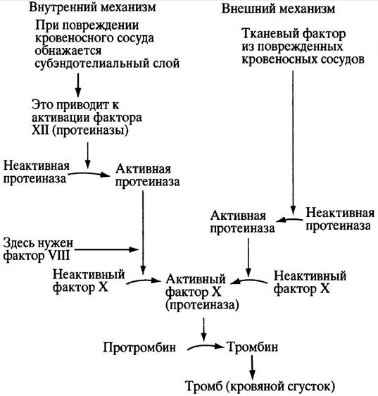
In laboratory conditions (in vitro), blood clots faster via the second of these mechanisms. However, a well-known disorder is hemophilia A, in which the intrinsic blood-clotting mechanism is impaired due to the absence of factor VIII, which is required for the proteolytic activation of factor X. One might think this defect could be compensated for by the extrinsic mechanism, in which factor VIII is not involved. Yet, under physiological conditions, both mechanisms operate harmoniously and interdependently as a single unit.
How does thrombin cause blood clots to form?
Blood contains a protein called fibrinogen. Its molecule consists of two rod-shaped subunits, each formed by three polypeptide chains. Near the N-terminal Regions of the chains, the subunits are linked by three Disulfide Bonds. Close to these regions, two of the three chains contain negatively charged peptide fragments—fibrinopeptides—which prevent the association of fibrinogen monomers (Fig. 17.2). Thrombin converts fibrinogen into fibrin by cleaving off the fibrinopeptides. This exposes sites that allow spontaneous aggregation of fibrin through intermolecular non-covalent interactions. The ends of a fibrin monomer bind to complementary sites in the middle of a neighboring monomer, so that polymerization generates an unstable Structure known as a soft clot (Fig. 17.3). Once the side chains of adjacent monomeric fibrin molecules are linked by covalent cross-links, the soft clot is transformed into a dense, insoluble hard clot.
Fig. 17.2. Conversion of fibrinogen monomers into fibrin monomers. Each half of the monomeric fibrinogen consists of three polypeptide chains, two of which contain negatively charged fibrinopeptides
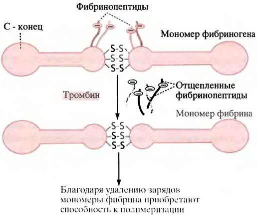
Fig. 17.3. Formation of fibrin strands. The strands form through the spontaneous polymerization of fibrin monomers and their subsequent enzymatic cross-linking (the locations and number of cross-links are largely random). Fibrinogen monomers cannot polymerize because they carry a significant negative charge, and potential association sites are shielded by fibrinopeptides

The cross-links are amide bonds formed during a transamidation reaction between a glutamine residue in one polypeptide chain and a Lysine residue in another:
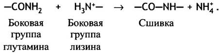
The resulting clot integrates fibrin strands and Blood Cells trapped within them into a single whole.
Regulation of Blood clotting
If blood clotting is not confined to the site of vascular injury, it can cause irreparable damage to the organism because it is autocatalytic in nature and could spread throughout the entire Circulatory system. However, A number of protective mechanisms prevent this. First, in the blood
There are proteinase inhibitors (including antithrombin) that inhibit the clotting process. Second, vessel walls contain the sulfopolysaccharide heparin, which enhances the action of these inhibitors. Third, There is a specialized proteinase called plasmin, which dissolves blood clots. Plasmin itself is formed from inactive plasminogen under the action of tissue plasminogen activator, or TPA (Tissue Plasminogen Activator), which is released from damaged tissues. Recently, it has begun to be used for therapeutic purposes. Although TPA is present in tissues in minute amounts, it has become fully accessible because it can now be produced using Introduction/32.html">Genetic Engineering techniques (see Chapter 24). All these defense mechanisms prevent the spread of blood clotting, restricting it to the area immediately adjacent to the vessel injury site that triggered the clot formation. The regulation of blood clotting is extremely complex. It is hardly surprising that disorders in the blood clotting system are the cause of death for many people.
Rat Poison, Blood Clotting, and Vitamin K
The widely used rat poison warfarin prevents blood clotting, causing rodents to die from internal bleeding triggered by minor and normally inevitable blood vessel injuries. In medicine, warfarin is used to prevent blood clots after a stroke. Structurally, it resembles vitamin K (derived from the German word Koagulation) and acts as its competitive inhibitor (Fig. 17.4).
Fig. 17.4. Structures of vitamin K (a) and warfarin (b). Vitamin K is essential for blood clotting, whereas warfarin acts as its antagonist.
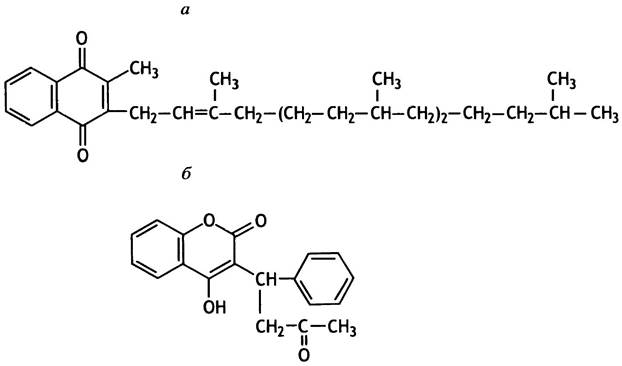
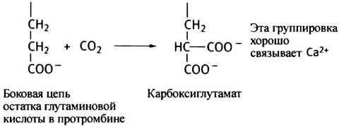
Vitamin K is required for The conversion of prothrombin into thrombin. It serves as a cofactor in an unusual enzymatic carboxylation reaction of a glutamic acid residue in prothrombin.
The Biological Significance of this modification is that the carboxyglutamate residue effectively binds Calcium Ions, which are necessary for thrombin activation. Other factors involved in the cascade are modified in a similar manner.
Defense Against Foreign Substances Entering the Body
Large foreign molecules are handled by the immune system (see Chapter 25). Here, we will focus on low-molecular-weight compounds that are not products of pathogenic organisms.
The modern human body is exposed to a vast array of foreign chemical compounds known as xenobiotics (from the Greek xenos, meaning strange or foreign). These include drugs, pesticides, herbicides, and other chemical industry products, as well as plant-derived Terpenes, Alkaloids, and Tannins. Many of these substances are poorly soluble in Water but highly soluble in fats. They accumulate in the hydrocarbon layer of membranes and in the vacuoles of fat cells, and they are not excreted in the urine. To prevent the harmful effects of such accumulation, these substances must be rendered water-soluble. There are several ways to modify xenobiotics to facilitate their elimination from the body, but the primary role in this process is played by the Liver Enzymes of the cytochrome P450 family. The most typical reaction catalyzed by these enzymes is the hydroxylation of both aliphatic and aromatic groups within xenobiotic molecules. Subsequently, Other Enzymes replace the hydrogen in the hydroxyl groups with highly polar residues (usually glucuronic acid), making the substance sufficiently soluble to be eliminated in the urine.
Let us now examine these processes in greater detail, beginning with the cytochrome P450 system.
Cytochrome P450
Like the Cytochromes of the Respiratory Chain (see p. 123), cytochrome P450 (referred to simply as P450 for brevity) is a hemeprotein. Its name indicates that it is pigmented (from the English Pigment) and that the absorption maximum of the P450 complex with carbon monoxide lies at 450 nm. Carbon monoxide has no direct role in the physiological function of P450; it is added merely to facilitate the determination of P450 content based on the intensity of the absorption spectrum. P450 is located on the inner surface of The Endoplasmic reticulum membrane. The reactions it catalyzes can generally be described by the following equation:
АН + О2 + NADPH + Н+ —> А-ОН + Н2О + NADP+.
Such reactions are called monooxygenase reactions because only one oxygen atom from the oxygen molecule (O2) is incorporated into the modified substance, while the second atom is reduced to water using NADPH. P450 is also referred to as a mixed-function oxygenase because it not only hydroxylates the substrate but also reduces O2 to H2O. The transfer of electrons from NADPH to Fe3+ in the heme of the cytochrome is carried out by P450 reductase, which is also bound to the endoplasmic reticulum membrane.
We have previously encountered a similar type of enzyme during the hydroxylation of phenylalanine (see p. 197). Note the difference between oxidation and oxygenation. Oxidation implies the removal of electrons, whereas oxygenation involves the incorporation of an oxygen atom from an O2 molecule into the substrate molecule (not to be confused with the Oxygenation of Hemoglobin, where an O2 molecule reversibly binds to the iron atom in the heme).
It is remarkable that the P450 system is capable of oxidizing a vast array of compounds that living organisms only began encountering with the advent of the modern chemical industry. One can only speculate about the evolutionary utility of this system, yet it allows us to survive under these newly created conditions. It is quite possible that its initial function was directed against plant terpenes, alkaloids, and Other Compounds that protect plants from being consumed by animals. The versatility of the P450 system is determined by two factors: first, each enzyme within this system possesses a fairly broad substrate Specificity within a certain class of compounds; and second, the substrate specificity profiles of different enzymes, although distinct, partially overlap.
Secondary Modification: Attachment of Polar Residues to the Products of P450-Catalyzed Reactions
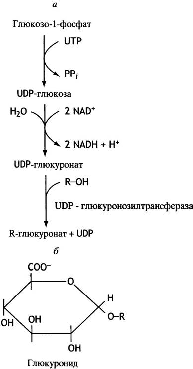
Figure 17.5 illustrates how the hydroxylation products of hydrophobic xenobiotics acquire water solubility through the attachment of a glucuronic acid residue. This residue is transferred from glucuronyl-UDP, which is formed via The oxidation of UDP-glucose in the endoplasmic reticulum membranes (see p. 87). The enzymes responsible for the secondary modification of xenobiotics are located in the same compartment.
Fig. 17.5. The glucuronidation system (a) and The structure of a glucuronide (b).
The Liver's Response to Xenobiotics
When a xenobiotic (such as phenobarbital) enters liver cells, a rapid expansion of the endoplasmic reticulum surface begins. Simultaneously, the glucuronidation and P450 systems are induced: relevant genes are switched on, and the synthesis of these system components is amplified many times over. Once the xenobiotic has been completely eliminated, everything returns to normal. This rapid response helps prevent the potential hazards associated with The entry of foreign substances into the body. However, it creates difficulties for Chemotherapy. The efficacy of many drugs is limited by The rate of their metabolic degradation, especially since the capacity of detoxifying enzyme systems increases the longer these drugs remain in the body. Unfortunately, such a powerful detoxification system has a downside. Most drugs are chemically inert and bind to their target molecules via non-covalent interactions. Yet, upon oxidation by cytochromes P450, they are frequently converted into reactive compounds that cause numerous adverse effects. And this applies not just to Pharmaceuticals! The completely harmless benzopyrene present in tobacco smoke enters the smoker's liver and Lungs, where it is oxidized into chemically active carcinogenic compounds that react with DNA.
Xenobiotics are not the only targets for cytochromes P450; they also participate in the degradation of heme and Steroid Hormones. In the Cell/35.html">Mitochondria of the adrenal cortex, cytochromes P450 catalyze individual steps in the synthesis of steroid hormones from Cholesterol.
Multiple Defense Mechanisms
The organism can be protected against xenobiotics by preventing their accumulation within cells. The Plasma Membrane of numerous cells, including various human tissues, contains a glycoprotein responsible for pumping A wide variety of substances out of The Cell driven by ATP Hydrolysis energy. The range of such compounds is quite broad and includes, in particular, many drugs and cytotoxic agents. The biological significance of this transport system is underscored by the fact that it mediates The transport of steroid hormones out of adrenocortical cells. Although the substances transported in this manner vary in structure, they all share amphipathic properties (possessing both polar and hydrophobic groups) and are lipid-soluble.
Protection Against Endogenous Proteinases
Chapter 4 discussed how the Digestive System avoids the destructive action of its own Proteolytic Enzymes. However, proteinases are ubiquitous throughout the body, making this problem relevant well beyond the gastrointestinal tract.
Here, we will focus on just one aspect of this issue involving the enzyme Elastase from neutrophils. Neutrophils are phagocytic white blood cells that concentrate around infected and inflamed tissue areas. There, neutrophils begin to secrete elastase, which degrades adjacent Connective Tissue. The action of elastase targets one of its primary components—the protein Elastin.
In the lungs, air enters bubble-like structures called alveoli, which increase the surface area for gas exchange between air and blood. Neutrophils are present in the alveoli and release elastase; however, its destructive effect on lung structures is prevented by the protein inhibitor α1-antitrypsin (α1-antiproteinase), which is secreted into the bloodstream by the liver. This inhibitor suppresses The activity of numerous proteinases, including trypsin, but is particularly effective against elastase. Antitrypsin binds tightly to the enzyme, blocking its catalytic center.
α1-Antitrypsin levels in the blood are quite sufficient to protect the pulmonary alveoli from elastase. However, a genetic defect is known in which blood concentrations of antitrypsin are significantly reduced. As a result, elastase can partially degrade lung tissue, manifesting as an enlargement of the alveoli and a reduction in the gas exchange surface area. This condition leads to The Development of pulmonary emphysema.
Tobacco smokers are also prone to emphysema for two reasons. First, the irritation caused by tobacco smoke attracts neutrophils to the lungs, thereby increasing elastase levels there. Second, smoke components inactivate α1-antitrypsin by oxidizing its functionally crucial Methionine residue to a sulfoxide (S —> S = 0). This is sufficient to prevent α1-antitrypsin from inactivating elastase and to promote the proteolysis of lung tissue, leading to emphysema. Although other antiproteinases exist, α1-antitrypsin is arguably the most important.
Defense Against Reactive Oxygen Species
Molecular oxygen serves as the terminal electron acceptor in the mitochondrial respiratory chain (see Chapter 7). Its reduction is described by the equation:
O2 + 4e- + 4H+ —> 2H2O.
At first glance, there is no reason to worry about any protective measures since both oxygen and water are entirely harmless. However, oxygen reduction can lead to the formation not only of water but also of other products that are highly hazardous to the organism. The transition from Anaerobic METABOLISM to the utilization of oxygen as an electron acceptor was a major evolutionary milestone. Danger arises during the univalent (one-electron) reduction of oxygen, which yields a reactive anion radical known as superoxide (radicals are molecules possessing an unpaired electron):
O2 + e- —> O2-.
The unpaired electron finds a partner by attacking a covalent bond in another molecule.
Superoxide is generated in the body for several reasons. Under normal conditions, the reduction of molecular oxygen in the respiratory chain is catalyzed exclusively by cytochrome c oxidase, which transfers all four electrons required for water formation. Nevertheless, the inevitable leakage of electrons from intermediate carriers to oxygen leads to The production of some O2-. Furthermore, Mitochondrial DNA Mutations can cause blockages in electron transport pathways, causing electrons to be diverted into superoxide formation (for example, via the direct oxidation of ubiquinone by oxygen). Such mutations are particularly dangerous because mitochondria lack a DNA Repair system (see p. 259).
Another source of superoxide is the spontaneous oxidation of hemoglobin. Normally, hemoglobin (Hb) reversibly binds oxygen: Hb + O2 —> HbO2. However, oxyhemoglobin (HbO2) has a certain probability of converting into methemoglobin (containing Fe3+ instead of Fe2+) and superoxide O2-.
It is also well established that ionizing radiation induces superoxide formation.
In addition to superoxide, the body produces another hazardous substance, hydrogen peroxide (H2O2). This occurs during the oxidation of certain metabolites by oxidases (respiratory chain cytochrome c oxidase is not among them).
When phagocytes engulf bacterial cells, they consume large amounts of oxygen. It is reduced via NADPH to generate superoxide. Once inside the vacuole, superoxide is converted into hydrogen peroxide, which assists in destroying the bacterial cell. Neutrophils, which accumulate in excess in inflamed joints, are also capable of releasing superoxide, thereby contributing significantly to the development of Arthritis.
The amount of superoxide produced in the body is negligibly small. Yet, even this is enough to drag a vast number of other molecules into undesirable reactions. The reason is that superoxide is a radical, meaning that one of the products of its reactions with normal molecules must necessarily be a radical as well. And since virtually all radicals are extremely reactive, this product will in turn enter into some other reaction, generating yet another radical. Thus, the process is a chain reaction and has the potential to continue indefinitely. It can be halted either through the recombination of radicals to form normal molecules (which is unlikely due to the low concentration of radicals) or via the anomalously low reactivity of daughter radicals.
The exact physiological defects caused by such chain reactions are not yet fully understood, but it is believed that superoxide is implicated in Aging, cataract formation, myocardial infarction, and other pathologies. There are three primary defense mechanisms against superoxides—one chemical and two enzymatic.
Vitamins C and E as Scavengers of Oxygen Radicals
A purely chemical approach to neutralizing superoxide and halting chain reactions involves The Use of antioxidants. These are substances that react with free radicals to yield radicals of very low reactivity, thereby terminating the chain process. The primary natural chain-breaking quenchers are ascorbic acid (Vitamin C) and α-tocopherol (vitamin E). The former is water-soluble and the latter is lipid-soluble, allowing them to act in tandem to protect both cytosolic components and Membrane Lipids. These two substances are not the only antioxidants; similar activity is exhibited by, for example, β-carotene and uric acid. Another effective antioxidant is bilirubin, the breakdown product of heme produced by heme oxygenase (see p. 370). Interestingly, heme oxygenase is activated by products of partial oxygen reduction (an accumulation known as oxidative stress, which can be triggered, for instance, by ionizing radiation). However, it remains unclear whether bilirubin production can be regarded as a protective response of the organism.
Enzymatic Elimination of Superoxide by Superoxide Dismutase
Apparently, all animal tissues contain the enzyme superoxide dismutase. Its highest concentrations are found in mitochondria, and it is also present in Lysosomes and Peroxisomes (see Chapter 16). Superoxide dismutase is found not only within cells but also in Blood Plasma, Lymph, and synovial fluid. This enzyme catalyzes the reaction:
2О2- + 2Н+ —> Н2О2 + О2.
Hydrogen peroxide, in turn, is broken down by another enzyme, catalase:
2Н2О2 —> 2Н2О + О2.
Hydrogen peroxide is produced not only as a byproduct of superoxide dismutation, but also through the action of oxidases. FAD-containing oxidases catalyze reactions that can be generally represented as follows:
АН2 + O2 —> А + Н2O2.
These include, notably, xanthine oxidase, which is involved in purine metabolism (see Chapter 18).
Hydrogen peroxide is dangerous because, in the presence of heavy Metal Ions (such as Fе2+), it decomposes to form highly reactive hydroxyl radicals (not to be confused with harmless hydroxyl anions) that attack DNA and other Biomolecules:
H2O2 + Fе2+ —> Fе3+ + ОН- + ОН-.
Both enzymes—superoxide dismutase and catalase—protect body tissues from radical attacks. However, there is another enzyme that breaks down hydrogen peroxide, namely Glutathione peroxidase. Its protective function is particularly crucial for the Brain, which contains low levels of catalase.
Defense Strategy via Glutathione
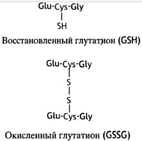
Glutathione is a thiol-containing tripeptide, y-glutamyl-cysteinyl-Glycine (GSН; Fig. 17.6). It is present in most cells and Functions primarily as a reducing agent, for instance, in maintaining protein Cysteine residues in a reduced state. Glutathione performs this protective function without the involvement of enzymes. Another protective role of glutathione is related to the reduction of hydrogen peroxide (as well as organic hydroperoxides R-O-OH).
Fig. 17.6 STRUCTURE OF THE reduced (GSH) and oxidized (GSSG) forms of glutathione. Glu, Cys, and Gly are the three-letter Abbreviations for glutamate, cysteine, and glycine, respectively. The single-letter system is used to denote extended Amino acid sequences.
This reaction is catalyzed by glutathione peroxidase:
Н2O2 + 2GSH —> GSSG + 2Н2O.
Oxidized glutathione (GSSG) is subsequently reduced by NADPH in a reaction mediated by glutathione reductase:
GSSG + NADPH + Н+ —> 2GSH + NADP+.
The integrity of erythrocytes relies on glutathione, which reduces ferrihemoglobin (methemoglobin) back to ferrohemoglobin and destroys peroxides. This explains the physiological importance of the Pentose Phosphate Pathway in red blood cells, which supplies the NADPH (see Fig. 13.1) required for glutathione reduction.
Individuals with a deficiency in glucose-6-phosphate dehydrogenase—the initial enzyme of The pentose phosphate pathway—typically maintain sufficient enzyme activity under normal conditions. However, when peroxides accumulate within their cells (for example, following the administration of the antimalarial drug primaquine), a shortage of NADPH arises, preventing the reduction of GSSG. This ultimately leads to damage to the erythrocyte plasma membrane and subsequent hemolysis.
In this chapter, we have focused primarily on animal defense mechanisms. However, plants also possess them, often in greater variety—likely because they lack an immune system and can neither flee nor chase away predators. As a random example, certain plants produce the enzyme chitinase, which degrades the chitinous exoskeleton of insects.
Chapter 17 Questions
1. Blood Coagulation is a cascade process. What is its biological significance?
2. Explain how thrombin participates in blood clot formation.
3. Spontaneous polymerization of fibrin monomers leads to the formation of a soft clot. How is it converted into a more stable structure?
4. What is The Role of vitamin K in blood coagulation?
5. What is the function of cytochrome P450?
6. Why is NАDРН required in oxygenation reactions?
7. What is the role of glucuronyl-UDР in the elimination of water-insoluble compounds from the body?
8. What is multiple defense?
9. Why does smoking cause pulmonary emphysema?
10. What is superoxide?
11. What mechanisms protect the body from the harmful effects of superoxide?
Last update: 06/08/2026
Editorial and Educational Adaptation: This material has been compiled based on the primary/original source text. The project team performed an editorial review, corrected technical inaccuracies, structured sections, and adapted the content for an educational format.
What was processed:
- elimination of formatting defects (OCR errors, structural breaks, corrupted characters);
- editorial organization of content;
- standardization of terminology in accordance with academic sources;
- verification of factual statements against the original source text.
All mentions of the author, publication year, and origin of the primary text have been preserved in accordance with the source.