ECOLOGICAL BIOCHEMISTRY - Textbook - V. M. Isaienko 2005
Chapter 10. BIOTRANSFORMATION OF XENOBIOTICS
10.1. Oxidation, reduction, degradation, and conjugation of xenobiotics
Reactions involved in the BIOTRANSFORMATION OF XENOBIOTICS, including environmental pollutants, can be broadly divided into four classes: 1) oxidation; 2) reduction; 3) degradation; 4) conjugation.
Oxidation reactions in microsomes. Among A wide variety of xenobiotic oxidation reactions, the principal ones are: 1) oxidation of alcohols and aldehydes; 2) oxidation of amines, hydrazines, and aziridines; 3) oxidation of aromatic alkyl-substituted compounds; 4) hydroxylation of ring structures; 5) aromatization of alicyclic compounds; 6) epoxidation; 7) oxidation or oxidative substitution of organic sulfur; 8) oxidative N-dealkylation.
It should be noted that the oxidative biotransformation of xenobiotics may lead to The formation of toxic, mutagenic, or carcinogenic compounds.
Among the cellular systems involved in The oxidation of xenobiotics, microsomes occupy a special place. Microsomal Enzymes that utilize molecular oxygen to oxidize xenobiotics are classified into Monooxygenases, Dioxygenases, and oxidases.
Monooxygenases incorporate one oxygen atom from a hydrogen donor (DH2) into the substrate (S) and reduce the second atom to Water (Fig. 10.1).
Class="center">
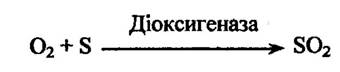
Dioxygenases incorporate two oxygen atoms into the substrate:
Oxidases reduce an oxygen molecule to hydrogen peroxide or two water molecules without incorporating oxygen into the substrate:

Monooxygenases most frequently catalyze the hydroxylation of xenobiotics. They are divided into two groups: those lacking metals and those containing one or more metal atoms (metalloenzymes). The latter typically contain copper atoms (tyrosinase, dopamine-β-monooxygenase, etc.) or iron atoms (phenylalanine hydroxylase, etc.). This group also includes monooxygenases that contain cytochrome P450 as a prosthetic group. In turn, they can be divided into three subgroups depending on their subcellular localization (Fig. 10.1).
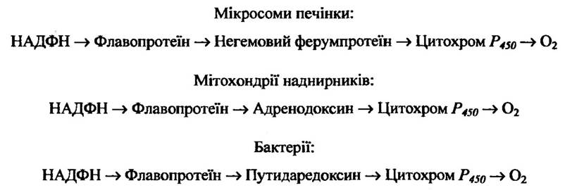
Fig. 10.1. Localization and mechanisms of O2 reduction by cytochrome P450-containing monooxygenases
In mammals, The Central Role in the biotransformation of xenobiotics belongs to hepatocyte microsomes, which account for up to 25% of The Cell dry mass.
Mammalian Liver microsomal monooxygenases contain at least NADPH-cytochrome P450 oxidoreductase, NADH-cytochrome b5 oxidoreductase, and cytochrome P450.
The catalytic activity of NADPH-cytochrome P450 oxidoreductase depends on the method of extraction from microsomes. For instance, if this enzyme system is solubilized with detergents, it becomes directly capable of reducing cytochrome c, and in the presence of Lipids, cytochrome P450. When solubilized with Trypsin, this oxidoreductase reduces cytochrome c but not cytochrome P450, even upon The addition of phospholipases. Because the enzyme has a high affinity for cytochrome c, it is often referred to as NADPH-cytochrome c oxidoreductase. However, it is known that cytochrome c is localized primarily in Mitochondria and is present in very small amounts in microsomes and Cytosol. Therefore, cytochrome c serves as a standard artificial electron acceptor for this oxidoreductase.
The primary function of NADPH-cytochrome P450 oxidoreductase in the microsomal hydroxylating complex is to transfer electrons to cytochrome P450 and, consequently, to drive the oxidation of xenobiotics. This enzyme may also participate in nitroreduction reactions, Fatty acid oxidation, N-oxidation of amines, etc.
Microsomes also contain an NADH-dependent electron transport system, the initial component of which is NADH-cytochrome b5 oxidoreductase (along with FAD), located in the membrane adjacent to cytochrome P450.
It has been shown that under certain conditions, cytochrome b5 is capable of reducing not only NADH but also NADPH. The transfer of electrons from the NADH-dependent chain to the NADPH oxidation chain can occur via three pathways:
1) directly from the NADH-specific enzyme to cytochrome P450;
2) from cytochrome b5 of the NADH oxidation chain to cytochrome b5 of the NADPH oxidation chain;
3) from cytochrome b5 of the NADH oxidation chain to cytochrome P450.
Cytochrome P450 is a hemoprotein in which hemin (the Fe3+–P450(Fe3+) chelate complex) is converted into protoheme (the Fe2+–P450(Fe2+) chelate complex) As a result of electron acceptance from a donor. In addition to the liver, it is also localized in the microtomes of the Lungs, Kidneys, Spleen, intestinal mucosa, Placenta, and certain other Organs. This cytochrome has been found in mammals, some bird species, amphibians, invertebrates, Bacteria, and higher plants.
Cytochrome P450 in the hydroxylating complex interacts with a substrate and molecular oxygen, accepting electrons from the corresponding Donors.
At The First stage, cytochrome P450(Fe3+) interacts with a substrate (S)—a xenobiotic—yielding the cytochrome complex
P450(Fe3+) — substrate. At the Second Stage, the complex is reduced in the NADPH-specific Electron Transport Chain to form the P450(Fe2+) — substrate complex. At the next stage, atmospheric oxygen interacts with the P450(Fe3+) — substrate complex, forming the P450(Fe3+) — substrate — O2 complex. Subsequently, molecular oxygen within the oxygenated complex is activated via its reduction. The oxygenated complex is reduced in a single-electron transfer reaction (P450(Fe2+) → P450(Fe3+)). The second electron comes from the NADH oxidation chain, and its transfer involves cytochrome b5 (NADH — cytochrome b5 — oxidoreductase). At the Fourth Stage, the resulting complex breaks down into oxidized cytochrome P450 and the oxidized substrate; one oxygen atom is transferred to the substrate, while the other is reduced to water. At the final, fifth stage, the unstable P450(Fe3+) — oxidized substrate complex dissociates, releasing cytochrome P450(Fe3+), which re-enters the reaction cycle.
One of the possible pathways for the hydroxylation of xenobiotics involving cytochrome P450 is shown in Fig. 10.2.
Fig. 10.2. Scheme of the xenobiotic hydroxylation reaction involving cytochrome P450:
cyt. P450 — cytochrome P450; cyt. b5 — cytochrome b5
Xenobiotics capable of inducing the hydroxylating complex with cytochrome P430 can be divided into three groups: 1) phenobarbital, diphenylhydantoin, pesticides, and certain others that increase The activity of NADPH — cytochrome P450 — oxidoreductase and cytochrome P450 itself; 2) polycyclic Hydrocarbons and some other substances that do not affect the activity of NADPH — cytochrome P450 — oxidoreductase or the reduction rate of cytochrome P45; 3) Steroids, their synthetic analogues, and related substances that enhance the activity of NADPH — cytochrome P45 — oxidoreductase and increase the reduction rate of cytochrome P450 without altering its amount.
Some xenobiotics are able to inhibit the METABOLISM of other foreign compounds and, consequently, prolong their action. Such compounds include β-diethylaminoethyldiphenylpropylacetate hydrochloride (SKF525A), 2,4-dichloro-6-phenylphenoxyethylamine (DPEFA), N-methyl-3-piperidyl-(N',N) diphenyl carbamate (MPDC), iproniazid, chlorcyclizine, glutethimide, and others. Inhibitors generally act directly on xenobiotic-oxidizing enzymes, unlike Inducers, which affect their synthesis.
Non-microsomal oxidation. In addition to microsomal oxygenases, enzyme systems capable of catalyzing xenobiotic oxidation reactions have been found in mitochondria, soluble cell fractions, and Blood Plasma. The main reactions include Oxidative Deamination, alcohol and aldehyde oxidation, and the aromatization of alicyclic compounds. Some of these enzymes can also catalyze reverse transformations, i.e., the reduction of xenobiotic oxidation products.
During oxidative deamination, aliphatic amines are oxygenated by amine oxidases into the corresponding aldehydes with the elimination of ammonia (Fig. 10.3).
Fig. 10.3. Oxidation reaction of aliphatic amines during oxidative deamination
Some of these enzymes differ in their substrate Specificity, inhibitor action, and cellular localization. They are localized in the liver, kidneys, intestinal mucosa, and blood plasma.
Monoamine oxidase, localized in mitochondria, catalyzes the oxidative deamination of primary, secondary, and tertiary aliphatic amines. Primary amines are metabolized to the corresponding acid or alcohol via an aldehyde. Secondary and tertiary amines are more resistant to deamination and predominantly undergo dealkylation to form primary amines, or are excreted unchanged. In addition to exogenous amines, monoamine oxidase also deaminates natural amines such as 5-hydroxytryptamine, catecholamines and their methyl derivatives, as well as certain Other Compounds.
Diaminooxidase has been found in many Cells, particularly in the liver, kidneys, and intestinal mucosa, but it is localized primarily in mitochondria. Diaminooxidase oxidatively deaminates diamines (Histidine, cadaverine, putrescine, etc.) by removing one molecule of ammonia.
This enzyme system does not deaminate diamines with nine or more carbon atoms. Instead, they are deaminated by monoamine oxidase, which, in turn, does not deaminate lower diamines.
Several amine oxidases have been found in blood plasma. For instance, spermine oxidase oxidatively deaminates spermine and other Polyamines, whereas benzylamine oxidase acts on benzylamine and mescaline.
Alcohol dehydrogenase, localized in the soluble fraction of the liver, kidneys, and lungs, oxygenates primary alcohols such as ethanol, n-butanol, fluoroethanol, benzyl alcohol, and cyclohexanol. For instance, in the case of ethanol, the following reaction takes place (Fig. 10.4).
Fig. 10.4. Dehydrogenation reaction of ethanol by alcohol dehydrogenase
It is worth noting that ethanol oxidation can also be carried out by a specific microsomal ethanol-oxidizing system (utilizing NADPH) as well as (to some extent) by catalase (utilizing hydroperoxide).
The reverse reaction can also occur, in which aldehydes and ketones (acetaldehyde, acetone, cyclohexanone, etc.) are reduced to alcohols.
Mammalian alcohol dehydrogenase has a low affinity for methanol, which is metabolized mainly by peroxidases, specifically xanthine oxidase and catalase.
Secondary alcohols in the animal body are oxidized to ketones by alcohol dehydrogenase, though their oxidation rate is significantly lower than that of primary alcohols. Higher secondary alcohols and tertiary alcohols are oxidized extremely slowly.
Aliphatic and aromatic aldehydes are oxidized to the corresponding carboxylic acids, which can subsequently undergo further transformation via β-oxidation. Enzymes catalyzing such reactions in mammals include aldehyde oxidase, xanthine oxidase, and NAD-dependent aldehyde dehydrogenase.
Aldehyde oxidase and xanthine oxidase are localized in the soluble fraction of liver cells. These enzymes catalyze the oxidation of aldehydes formed during the deamination of endogenous amines (adrenaline, noradrenaline, 5-hydroxytryptamine) and xenobiotic amines (benzaldehyde, acetaldehyde, and salicylaldehyde). Fig. 10.5 illustrates the benzaldehyde oxidation reaction.

Fig. 10.5. Oxidation of benzaldehyde by aldehyde oxidase
The NAD+-dependent aldehyde dehydrogenase catalyzes the oxidation of chloral hydrate to trichloroacetic acid (Fig. 10.6).

Fig. 10.6. Dehydrogenation reaction of chloral hydrate by aldehyde dehydrogenase
Mitochondria contain an enzyme system that catalyzes The conversion of cyclohexanecarboxylic acids into aromatic acids. This process can yield benzoic acid (Fig. 10.7).
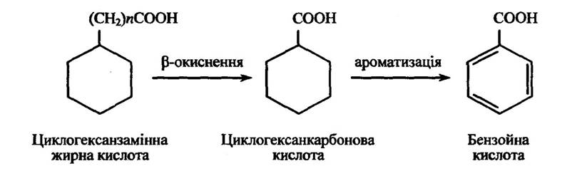
Fig. 10.7. Oxidation reaction of cyclohexane-substituted Fatty acids with an even value = 2,4,6 ...)
If cyclohexane-substituted fatty acids (general formula C6H11(CH2)nCOOH) contain an odd number n, they are converted into cyclohexaneacetic acid, which is subsequently metabolized via oxidation of the cyclohexane ring.
The enzyme system catalyzing the aromatization of acyclic compounds has been detected in liver and Kidney mitochondria; it is most active in rabbits and guinea pigs, and completely inactive in mice, dogs, and humans.
Reduction reactions in microsomes. These reactions are less common in xenobiotic biotransformation than oxidation processes. Nevertheless, their contribution to overall xenobiotic biotransformation is substantial.
Microsomal nitrate reductases catalyze the reduction of aromatic nitro compounds; their enzymatic activity depends on numerous factors, primarily the availability of NADH and NADPH in the medium, exhibits low substrate specificity, and reduces nearly all aromatic amines. Flavoprotein and cytochrome P450 participate in the reduction of nitro compounds.
The initial stage of substrate reduction is likely associated with The transport of reduced equivalents from the flavoprotein, resulting in the formation of intermediate nitro derivatives and hydroxylamine (Fig. 10.8).

Fig. 10.8. Reduction reaction of aromatic nitro compounds by nitrate reductase
At the same time, cytochrome P450 apparently transfers electrons to intermediate compounds formed following the generation of nitrobenzene anion radicals. The reduction of amines by nitroreductase occurs in two stages: reduction of substrates to hydrazo compounds and their reductive Cleavage (Fig. 10.9).

Fig. 10.9. Reduction reaction of nitroso compounds by azoreductase
Nitro compounds in microsomes can be reduced via NADPH-cytochrome P450 oxidoreductase; NADPH-cytochrome b5 oxidoreductase, cytochrome P450, and flavoprotein may also participate. One hypothesis is that NADPH-cytochrome P450 oxidoreductase transfers electrons to the substrate via flavins, which fully or partially reduce cytochrome c.
Hepatic microsomes contain localized N-oxidoreductases that reduce N-oxides to their corresponding amines. They exhibit activity even in the absence of NADPH-cytochrome oxidoreductase and cytochrome P450.
The reduction of nitroso compounds by nitroreductases in microsomes has also been investigated. The activity of these enzymes is highest in The Liver and significantly lower in the kidneys, lungs, and Small Intestine mucosa.
Epoxide reductase catalyzes the reduction of aromatic epoxides. Its activity requires NADPH, and this process is mediated by cytochrome P450-dependent enzymes.
Liver microsomes also contain an enzyme that, in the presence of NADPH and oxygen, removes a halogen from its aliphatic moiety while simultaneously reducing the substrate. For instance, chloroform is reduced as shown in Fig. 10.10.
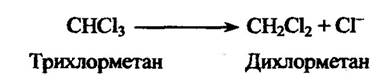
Fig. 10.10. Chloroform reduction reaction
Fluorine removal is also possible. A typical example of this process is the dehalogenation of halaton, which results in the elimination of a fluorine atom (Fig. 10.11).
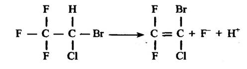
Fig. 10.11. Halaton reduction reaction
The reduction of ketones to alcohols is catalyzed by ketone reductases. This reaction is generally reversible.
Non-microsomal reduction. A number of enzymes involved in xenobiotic reduction reactions are also located outside the microsomes. For example, nitroreductases and ketone reductases are found in the Cytoplasm, while N-oxidoreductases are present in mitochondria and blood plasma, among others. Many enzymes that catalyze reduction reactions are found in microorganisms.
The reduction of double bonds is observed in the metabolism of various unsaturated aliphatic or alicyclic compounds capable of becoming saturated. These compounds include monocyclic Terpenes. An example of the double bond reduction in a cyclohexadiene ring is the conversion of α-phellandrene (menthadiene) into phellandronic acid (p-menthane).
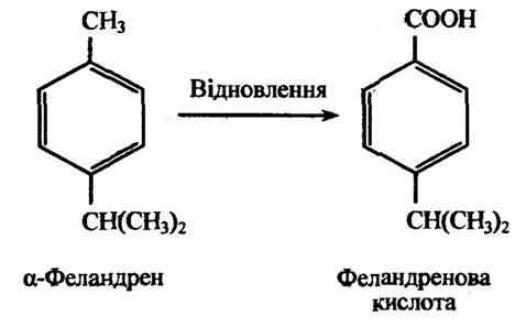
Fig. 10.12. β-Phellandrene reduction reaction
The reduction of a double bond in the side chain occurs during the conversion of pulegone to menthol (Fig. 10.13).
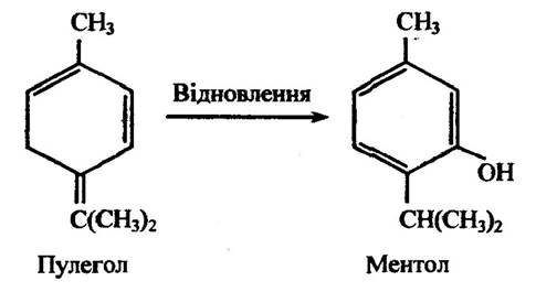
Fig. 10.13. Pulegone reduction reaction
One type of non-microsomal reduction is the reduction of disulfides to thiols. In this process, diethyl sulfides are reduced to ethyl mercaptan (Fig. 10.14).
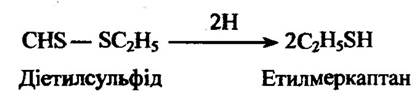
Fig. 10.14. Diethyl sulfide reduction reaction
Hydroxamic acids can be reduced to the corresponding amides. For example, salicylhydroxamic acid is converted into salicylamide (Fig. 10.15).
In turn, salicylamide forms conjugates with 5-bromosalicylamide, which is produced during the reduction of the 5-bromo derivative of salicylhydroxamic acid.
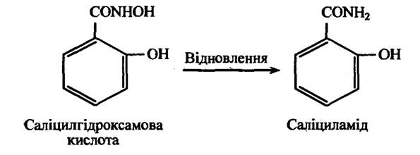
Fig. 10.15. Salicylhydroxamic acid reduction reaction
Sulfoxides and N-oxides are also capable of being reduced. For instance, dimethyl sulfoxide is reduced to dimethyl sulfide, and trimethylamine N-oxide to trimethylamine (Fig. 10.16).
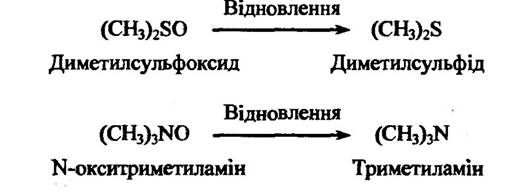
Fig. 10.16. Reduction reaction of dimethyl sulfoxide and trimethylamine N-oxide
Another type of non-microsomal reduction is the reductive dehydroxylation of hydroxamic acids, catechols, and certain aliphatic hydroxyl derivatives.
During aromatic dehydroxylation, one of the pathways involves the removal of the 4-hydroxyl group from the phenolic ring. For instance, the aromatic dehydroxylation of homoprotocatechuic acid (3,4-dihydroxyphenylacetic acid) proceeds with the formation of meta- or parahydroxyphenylacetic acids (Fig. 10.17).
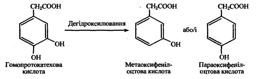
Fig. 10.17. Dehydroxylation reaction of homoprotocatechuic acid
Similarly, 3,4-dihydroxycinnamic acid (caffeic acid), 3,4-dihydroxyphenylalanine (L-DOPA), rutin and quercetin Flavonoids, and other pyrocatechol derivatives can undergo dehydroxylation.
It has been established that, in addition to the dehydroxylation of 4-substituted pyrocatechol derivatives, the loss of a phenolic hydroxyl group can also occur in xanthurenic acid, which is capable of converting into 8-hydroxyquinaldic acid (Fig. 10.18).
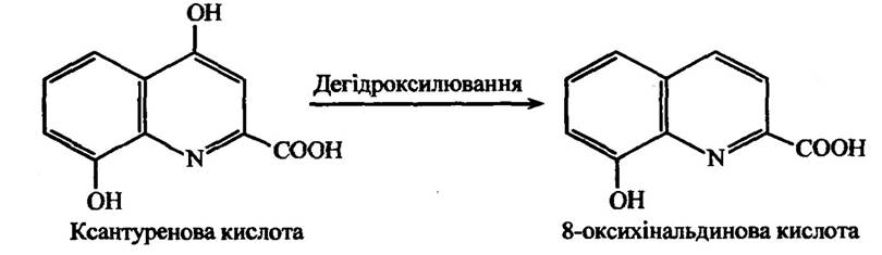
Fig. 10.18. Dehydroxylation reaction of xanthurenic acid
Aliphatic dehydroxylation of noradrenaline, much like aromatic dehydroxylation, leads to the formation of methoxyphenylacetic and homovanillic acids alongside other metabolites (Fig. 10.19).
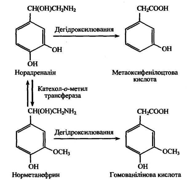
Fig. 10.19. Dehydroxylation reaction of noradrenaline
Another type of dehydroxylation is N-dehydroxylation. A typical example of this process is the conversion of N-hydroxyurethane to urethane (Fig. 10.20).

Fig. 10.20. Dehydroxylation reaction of N-hydroxyurethane
A similar process can also occur during the conversion of 4-hydroxyaminobiphenyl (transformed into 4-aminobiphenyl), N-hydroxy-4-acetylaminostilbene (transformed into 4-acetylaminostilbene), N-hydroxy-2-N-acetylaminofluorene (transformed into 2-acetylaminofluorene), and other compounds.
Reduction processes involving atoms with variable valence also contribute to reduction reactions during xenobiotic biotransformation.
Dehydration (cleavage) reactions. Examples of xenobiotic dehydration reactions include the Hydrolysis of esters. These reactions are catalyzed by widespread enzymes such as cholinesterases, aliesterases, arylesterases, and others. Certain Alkaloids—such as atropine, cocaine, and others—are subject to hydrolysis. Specifically, cocaine is hydrolyzed by plasma enzymes of rabbits rather than humans (Fig. 10.21).
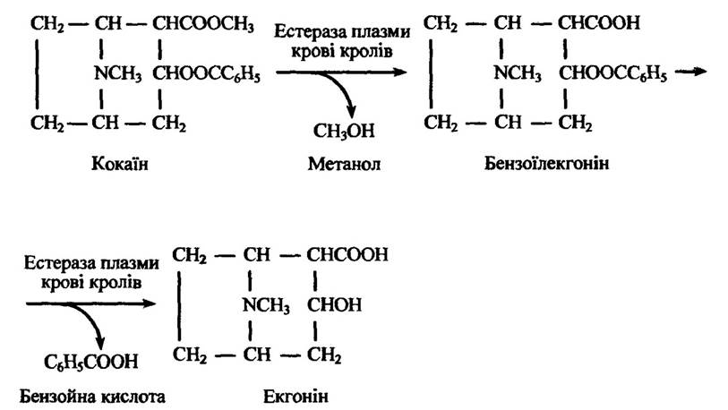
Fig. 10.21. Hydrolysis reaction of cocaine
Liver microsomal esterases are capable of hydrolyzing a number of esters, notably meperidine (Fig. 10.22).
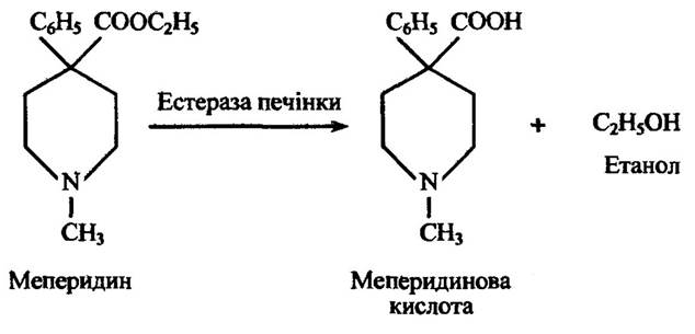
Fig. 10.22. Hydrolysis reaction of meperidine
Amides are more stable in the animal Organism than their corresponding esters. Therefore, their hydrolysis proceeds more slowly.
The rate of hydrolysis of aliphatic amides depends on the chain length of the alkyl group. For instance, phenylacetamide is hydrolyzed relatively quickly, whereas acetanilide undergoes only minimal hydrolysis and is largely excreted unchanged from the body.
The hydrolysis of aromatic amides also depends on their chemical nature and THE POSITION OF substituents on the aromatic ring. For example, benzamide (C6H5CONH2) is completely hydrolyzed, whereas p-aminobenzamide is only 20% hydrolyzed, and p-hydroxybenzamide by just 4%.
Hydroxylated acids can be metabolized not only via reduction, as discussed earlier, but also by undergoing hydrolysis to yield an aromatic carboxylic acid. For instance, benzohydroxamic acid is capable of hydrolyzing to benzoic acid (Fig. 10.23).
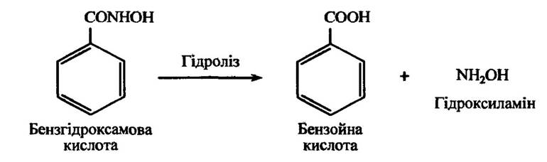
Fig. 10.23. Hydrolysis reaction of benzohydroxamic acid
Aromatic acid hydrazides undergo similar hydrolysis. An example of such a process is the hydrolysis of benzohydrazide to benzoic acid (Fig. 10.24).
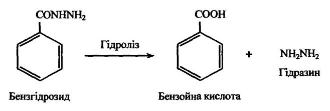
Fig. 10.24. Hydrolysis reaction of benzohydrazide
Carbamates are also capable of hydrolyzing to yield carbamic acid and an alcohol (Fig. 10.25).
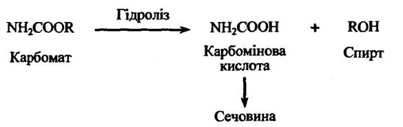
Fig. 10.25. Hydrolysis reaction of carbamate
Ethyl carbamates, notably the hypnotic drug hedonal (methylpropynylcarbinyl carbamate), undergo hydrolysis in a similar manner.
Substituted urethanes can also undergo hydrolysis; specifically, ethyl chloramate hydrolyzes to trichloroacetic acid.
Nitriles or aromatic cyanides are metabolized primarily via aromatic hydroxylation. However, they can also undergo hydrolysis to yield the corresponding carboxylic acid. For instance, the hydrolysis of benzonitrile leads to the formation of benzoic acid (Fig. 10.26).

Fig. 10.26. Hydrolysis reaction of benzonitrile
A number of heterocyclic compounds undergo hydrolysis through the Cleavage of the heterocyclic ring. Hydantoins are evidently the simplest such compounds. The hydrolysis reaction of hydantoin is illustrated in Fig. 10.27.
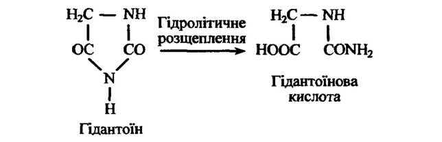
Fig. 10.27. Hydrolytic cleavage reaction of hydantoin
Benzoxazoles are capable of hydrolyzing via the cleavage of the oxazole ring to form ortho-formamidophenol derivatives, with subsequent formation of ortho-aminophenol derivatives (Fig. 10.28).
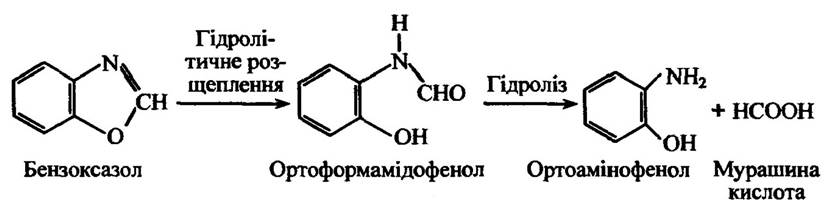
Fig. 10.28. Hydrolytic cleavage of benzoxazole
Indole is first hydroxylated to yield indoxyl and then isatin, in which hydrolytic cleavage of the pyrrole ring occurs, ultimately resulting in the formation of anthranilic and formic acids (Fig. 10.29).
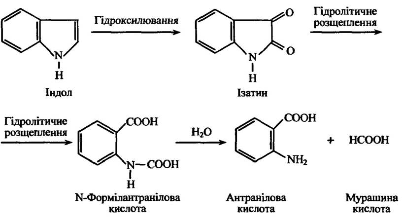
Fig. 10.29. Metabolism reactions of indole
A similar opening of the heterocyclic ring occurs during the metabolism of coumarin. Coumarin undergoes hydroxylation to yield 3-hydroxycoumarin, followed by its hydrolytic cleavage to ortho-hydroxyphenylacetic and/or ortho-hydroxylactic acid (Fig. 10.30).
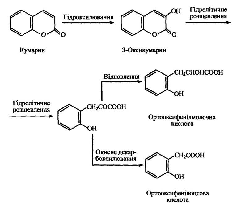
Fig. 10.30. Metabolism reactions of coumarin
Flavonoids can also undergo heterocyclic ring opening, the site of which is determined by the animal species and the substituents in the pyran ring. The hydrolytic cleavage reactions of diosmetin and quercetin are shown in Fig. 10.31.
Aromatic Compounds can also undergo ring opening via pathways different from those already discussed. An example is the metabolism of benzene, which leads to the formation of muconic acid (Fig. 10.32).
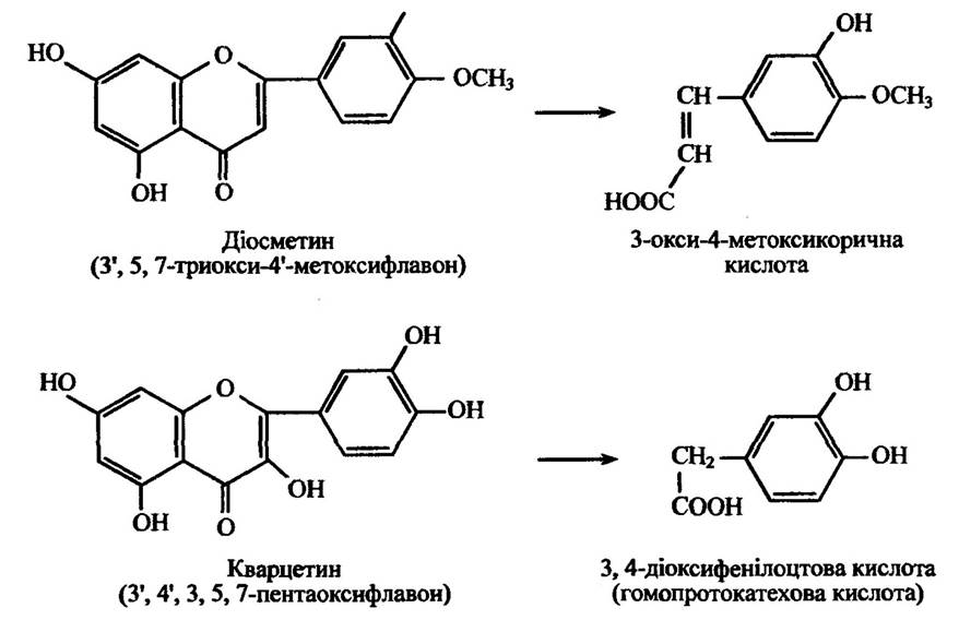
Fig. 10.31. Metabolism reactions of diosmetin and quercetin
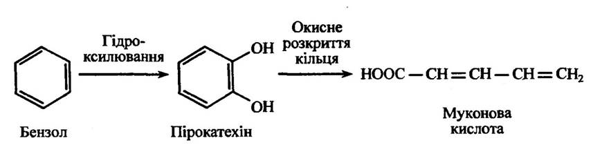
Fig. 10.32. Metabolism of benzene with the formation of muconic acid
There are a variety of ways in which cyclic structures can be cleaved. Of substantial importance is the biodegradation of Lignin, a natural polymer whose content in wood accounts for 20–30% of its dry mass.
At the first stage of lignin degradation by microorganisms, demethylation of the methoxyl groups of the guaiacyl and syringyl units of the lignin polymer occurs. This results in a decreased methoxyl group content and an increased number of ortho- and diphenolic residues. At the next stage, the fungal dioxygenases catalyze the ring-opening of these aromatic lignin residues, yielding aliphatic carboxylic groups.
Arsenobenzene derivatives are capable of being metabolized through molecular cleavage in an oxidation reaction, initially forming arsenoxides and subsequently arsonic acids. An example of such reactions is the cleavage of the antisyphilitic drug arsphenamine (Fig. 10.33).
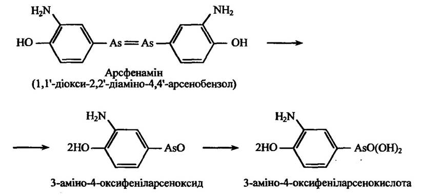
Fig. 10.33. Oxidative cleavage reaction of arsphenamine
Unlike arsenobenzene derivatives, azobenzene derivatives are cleaved not by oxidation, but rather through a reduction process.
One of the ways compounds undergo degradation is cyclization. Examples of such processes include the transformation of ortho-aminophenylethanol and various ortho-nitrophenyl derivatives (such as ortho-nitrophenylacetylene, ortho-nitrophenylpropionic acid, etc.) (Fig. 10.34).
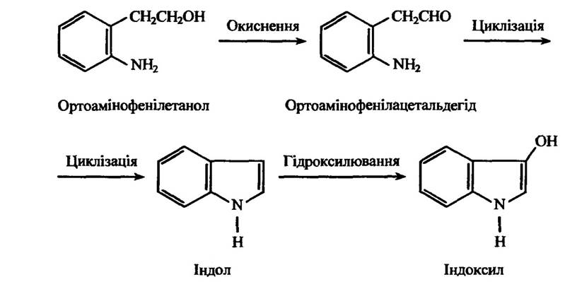
Fig. 10.34. Cyclization reaction of ortho-aminophenylethanol
ortho-Coumaric (ortho-hydroxy-trans-cinnamic) and ortho-hydrocoumaric (ortho-hydroxyphenylpropionic) acids undergo cyclization in an analogous manner.
Certain microorganisms are capable of dehalogenating chlorine-containing xenobiotics, which include a wide range of substances such as pesticides and a number of natural metabolites of lower plants.
The most significant pathways are oxidative, reductive, and hydrolytic dehalogenation. Oxidative dehalogenation proceeds via mechanisms of dehydrohalogenation, oxidative dehalogenation with the formation of double bonds, and dehalogenation-hydroxylation involving oxygen molecules mediated by monooxygenases and dioxygenases.
Monooxygenases can participate in the Microbial Degradation of chlorinated phenoxyalkanoic acids and are capable of cleaving fluorine from aromatic rings during hydroxylation. Dioxygenases can cleave aromatic rings, thereby facilitating the dehalogenation of aromatic compounds.
An example of chlorine removal is the dehydrochlorination of the insecticide 2,2-bis(para-chlorophenyl)-1,1,1-trichloroethane (DDT), which proceeds very slowly to yield 2,2-bis(para-chlorophenyl)-1,1-dichloroethylene (DDE) (Fig. 10.35).
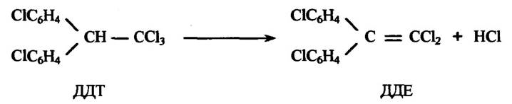
Fig. 10.35. Dehydrochlorination reaction of DDT
Alkyl halides (such as bromochloromethane, methylene dichloride, methylene dibromide, etc.) undergo hydrolytic dehalogenation by liver and kidney enzymes, yielding free halide ions (Fig. 10.36).

Fig. 10.36. Hydrolytic dehalogenation reaction of bromochloromethane
Some aromatic compounds can form phenols through halogen substitution (Fig. 10.37).
Aliphatic and aromatic halogenated hydrocarbons can be converted into glutamic derivatives and mercapturic acids via halogen atom substitution when interacting with glutamate in the presence of liver enzymes.
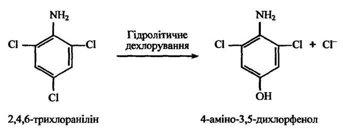
Fig. 10.37. The hydrolytic dechlorination reaction of 2,4,6-trichloroaniline
Microsomal liver enzymes mediate the reductive dehalogenation of carbon tetrachloride to chloroform in the presence of NADPH and O2 (Fig. 10.38).

Fig. 10.38. The reductive dehalogenation reaction of carbon tetrachloride
A similar dehalogenation process occurs with anesthetics such as halothane, methoxyflurane, and several others.
Polychlorinated biphenyls are relatively stable toxic compounds. It is known that microbial degradation of biphenyl proceeds via catabolic pathway systems similar to those of other aromatic hydrocarbons.
Benzypyrene is a persistent polycyclic aromatic compound whose degradation yields carcinogenic hydroxy and epoxy derivatives. It does not undergo mineralization in activated sludge systems, although several microbial cultures capable of partially breaking down this compound via hydroxylase systems have been described.
Petroleum industry wastewater is treated biologically after most of the oil is removed using coagulants. The extent of microbiological degradation of oil spills depends on various factors, the primary ones being the COMPOSITION OF THE oil and the availability of nutrients for microorganisms, particularly nitrogen and phosphorus.
Synthetic Surfactants used in industry and everyday life are also biodegradable. Their breakdown typically begins with the oxidation of terminal methyl groups, followed by the cleavage of linear side chains via β-oxidation. Ring-containing molecular structures generally decompose only after the complete degradation of the side chain.
Conjugation reactions. In addition to the aforementioned oxidation, reduction, and degradation reactions of xenobiotics, their biotransformation also involves conjugation reactions, as noted earlier. These are BIOSYNTHETIC PROCESSES IN which xenobiotics or their metabolites combine with endogenous compounds (such as glucuronic acid, sulfate, acetyl-CoA, Glycine, and various aromatic and aliphatic amines) to form conjugates. In this process, endogenous compounds attach to the Functional groups of xenobiotics (hydroxyl, amino, carboxyl, epoxy groups, halogen atoms, etc.). The resulting conjugates generally become more polar and less lipid-soluble, which facilitates their excretion from organisms.
In many conjugation reactions, endogenous compounds are transferred with the participation of Coenzymes. Prominent among these are uridine diphosphate coenzymes, specifically uridine diphosphate-α-D-glucose (UDPG) in the formation of β-glucosides, and uridine diphosphate-α-D-glucuronic acid (UDPGA) in the formation of β-glucuronides.
Glucoside conjugates are formed in insects, Mollusks, plants, and bacteria by transferring glucose from UDPG to a xenobiotic.
For phenols, conjugate formation proceeds as shown in Fig. 10.39.
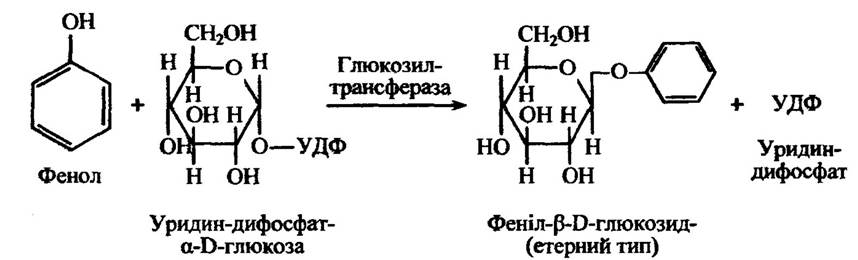
Fig. 10.39. The formation reaction of phenyl-β-D-glucoside
In insects, the formation of glucoside conjugates is the primary detoxification mechanism. Similar to insects, mollusks also synthesize glucosides.
In plants, glucosides are formed from natural compounds synthesized by the plants themselves, such as alkaloids, as well as during the detoxification of foreign compounds.
Ether and ester glucosides are typically synthesized. The formation of amygdalosides is also possible. The latter are formed as glucosides through glucose transfer from UDPG, except that two glucose molecules are transferred in this case. Bacteria likewise synthesize glucosides of both phenols and acids with the participation of UDPG.
Although mammalian liver contains UDPG, the lack of glucosyltransferases prevents the formation of glucosides.
Glucuronide conjugates are formed in mammals, birds, reptiles, and amphibians, but not in fish.
The reaction for forming a glucuronide conjugate with phenol is illustrated in Fig. 10.40.
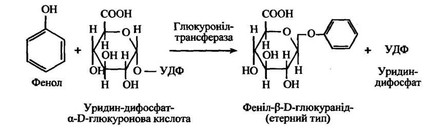
Fig. 10.40. Reaction of phenyl-β-D-glucuronide formation
Mammals such as cats are characterized by an exceptional pattern of glucuronide conjugate formation with phenol due to the lack of appropriate transglucuronidases. Therefore, these animals are particularly sensitive to the Toxic effects of phenols. However, cats excrete thyroxine, bilirubin, and iopanoic acid glucuronides into the Bile, as well as glucuronide conjugates of hydroxylated acetamidofluorene metabolites in the urine. At the same time, fish possess the necessary glucuronyltransferases, but lack the UDPGA coenzyme.
Glucuronide conjugates are classified into N-glucuronides, O-glucuronides, and S-glucuronides. In turn, O-glucuronides formed from phenols, alcohols (including steroids), and carboxylic acids are divided into four types: 1) ether type (derived from phenols and alcohols); 2) ester type (from carboxylic acids); 3) enol type (from pseudo-acids, e.g., 4-hydroxycoumarin); 4) hydroxylamine type (from compounds hydroxylated at the amino group, e.g., 2-acetylaminofluorene).
Several types of N-glucuronides are also known. For instance, the nitrogen atom to which the glucuronide moiety attaches can be part of a sulfonamide group, amino group, carbamide group, etc.
During the conjugation of thiol compounds (thiophenol, 2-mercaptobenzene, etc.) with glucuronic acid, S-glucuronides are formed.
Glycosides (glucosides and glucuronides) are generally more polar than their parent compounds and are eliminated from the site of synthesis via excretion in animals, and by accumulation in the vacuoles of leaf cells and other Tissues in plants.
An important type of conjugates is sulfuric acid esters, or ethereal sulfates. They are formed in mammals, birds, reptiles, and amphibians, but not in fish. Among invertebrates, ethereal sulfates are synthesized in Arthropods, insects, and possibly some species of mollusks.
Ethereal sulfates are subdivided into several types: 1) arylsulfates, which are esters of Phenolic Compounds; 2) alkyl sulfates, esters of primary aliphatic alcohols; 3) sulfamates, esters of sulfuric acid and amines containing a sulfonamide group; 4) steroid sulfates, esters of the hydroxyl groups of the steroid side chain, cyclic side-chain esters, and phenolic steroid esters; 5) carbohydrate sulfamates, esters of carbohydrate hydroxyl groups.
Among these five groups, arylsulfates and alkyl sulfates are the most common types of ethereal sulfates.
Ethereal sulfates are synthesized by transferring a sulfate group from adenosine-3'-phosphate-5'-phosphosulfate (PAPS) to phenols, alcohols, or amines via corresponding sulfotransferases. PAPS is formed from adenosine-5'-triphosphate (ATP) and adenosine-5'-phosphosulfate (APS) (Fig. 10.41).
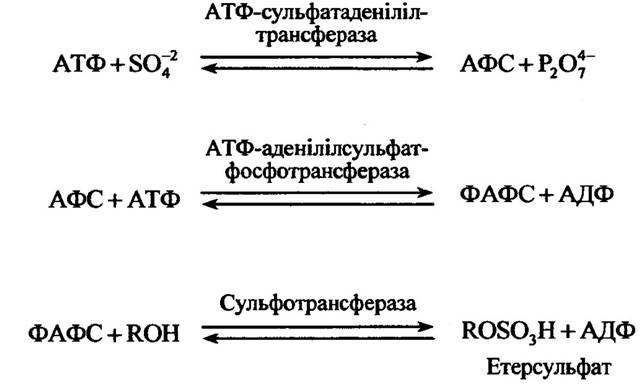
Fig. 10.41. Reactions of ethereal sulfate formation
In many animals and certain microorganisms, N-, O-, and S-methyl conjugates are formed during the transfer of a methyl group from the universal methyl donor S-adenosylmethionine via Transmethylation processes. S-adenosylmethionine is synthesized from Methionine in a reaction with ATP. The reaction leading to the formation of S-adenosylmethionine is illustrated in Fig. 10.42.
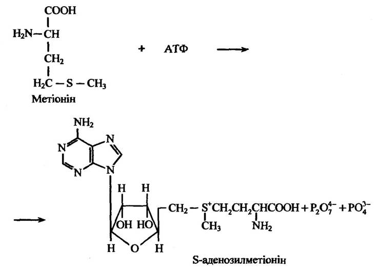
Fig. 10.42. Reaction of S-adenosylmethionine formation
Analogously, S-adenosylethionine is synthesized from the ethyl analogue of methionine (ethionine) and participates in transethynylation reactions.
In most mammals, some birds, amphibians, and insects, N-methyl conjugates are formed with both endogenous and xenobiotic compounds. For example, phenylethanolamine N-methyltransferase catalyzes the synthesis of adrenaline from noradrenaline (Fig. 10.43), as well as the N-methylation of other phenylethanolamine derivatives.
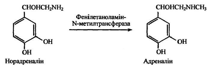
Fig. 10.43. Reaction of adrenaline formation via noradrenaline methylation
Non-specific N-methyltransferases methylate endogenous and exogenous Primary and secondary amines (serotonin, tryptamine, normetanephrine, normepyrine, pyridine, etc.). The methylation reactions of serotonin and pyridine are shown in Fig. 10.44.
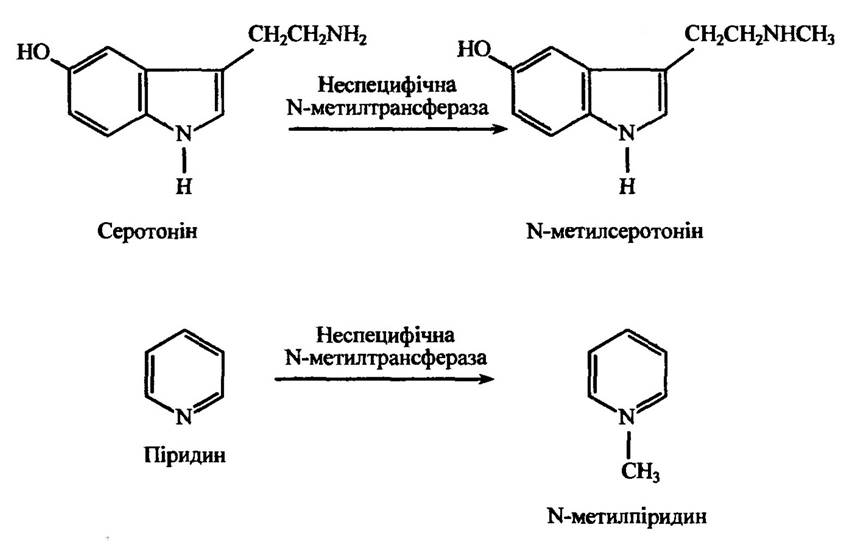
Fig. 10.44. Reactions of N-methylserotonin and N-methylpyridine formation
via the methylation of serotonin and pyridine, respectively
Specific N-methyltransferases are also known to methylate endogenous compounds such as nicotinamide, purine and pyrimidine polynucleotides, phosphatidylaminoethanol, etc. The methylation reaction of histamine by imidazole N-methyltransferase is illustrated in Fig. 10.45.
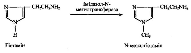
Fig. 10.45. Formation reaction of N-methylhistamine via histamine methylation
Specific N-methyltransferases of endogenous compounds are also known, which N-methylate nicotinamide, guanidinoacetic acid, phosphatidylethanolamine, as well as purine and pyrimidine polynucleotides.
Catecholamine Hormones, along with a number of catecholamine xenobiotics—specifically gallic (3,4,5-trihydroxybenzoic) and caffeic (3,4-dihydroxycinnamic) acids—are methylated by catechol-O-methyltransferase (Fig. 10.46). This enzyme is found in the liver, kidneys, blood, Skin, nerve fibers, and glandular tissues. S-adenosylmethionine serves as the methyl donor.
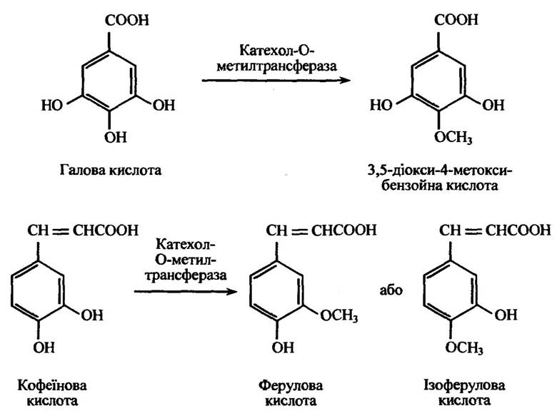
Fig. 10.46. Methylation reaction of catecholamine derivatives determined by the position of other substituents in the aromatic ring
Hydroxyindole-O-methyltransferase methylates N-acetylserotonin (Fig. 10.47), bufetin, and other hydroxyindoles. In vertebrates, this enzyme has been detected in the Pituitary Gland and the retina. Unlike catechol-O-methyltransferase, it does not methylate catechols and does not require magnesium ions for its function.
Iodophenol-O-methyltransferase methylates ortho-iodophenols, specifically 3,5-diiodo-4-hydroxybenzoic acid (Fig. 10.48), but does not act on thyroxine or triiodothyronine.
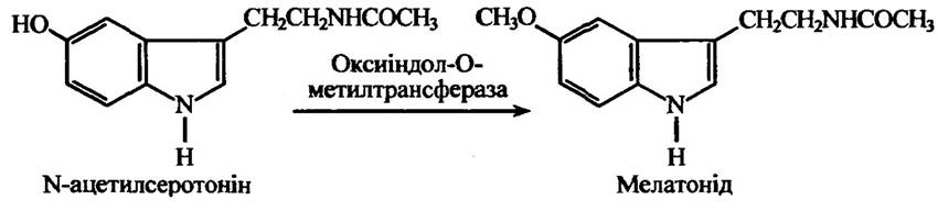
Fig. 10.47. N-acetylserotonin methylation reaction catalyzed by hydroxyindole-O-methyltransferase
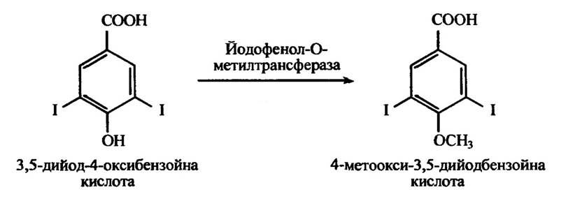
Fig. 10.48. 3,5-diiodo-4-hydroxybenzoic acid methylation reaction catalyzed by iodophenol-O-methyltransferase
In rats, dogs, and presumably certain other mammals, morphine is methylated to form codeine via conjugation, a reaction that does not require S-adenosylmethionine as a methyl group donor.
Methyl groups can also be transferred to thiol groups of xenobiotics (methyl mercaptans, ethyl mercaptans, mercaptoacetic acid, mercaptoethanol, dimercaptopropanol, thieracils, etc.) through S-methylation reactions occurring in the liver, kidneys, and lungs. Endogenous thiol compounds such as Cysteine, homocysteine, and Glutathione are not methylated by this enzyme. The S-methylation reaction is illustrated in Fig. 10.49.

Fig. 10.49. Ethanethiol methylation reaction by S-methyltransferase
Aromatic amino compounds and sulfonamides are capable of undergoing Acetylation. There are certain species-specific differences in these reactions among mammals. For instance, dogs and foxes excrete a significant amount of aromatic amines as acetylated derivatives due to the presence of an arylamine acetyltransferase inhibitor in their liver and kidneys, or because of high aromatic deacylase activity. At the same time, aliphatic amino groups in these animals are readily acetylated.
In adult birds, aromatic amines are acetylated, whereas in chicks this process does not occur due to high levels of aromatic deacylase in the kidneys. Some amphibians and fish can also acetylate aromatic amines, whereas this type of conjugation is absent in reptiles. It is known that certain insect species (such as locusts and moths) carry out both the acetylation and deacetylation of aromatic amines. Similar acetylation and deacetylation processes take place in plants.
Foreign amines can be acetylated via intermediate compounds with coenzyme A (CoA-SH), resulting in the formation of amide conjugates (Fig. 10.50).

Fig. 10.50. Amide acetylation reaction by acetyl-coenzyme A
Aromatic amines, sulfonamides, and certain foreign aromatic Amino Acids are capable of being acetylated (Fig. 10.51).
Fig. 10.51. Acetylation reaction of aniline, S-phenylcysteine, and isocyanide
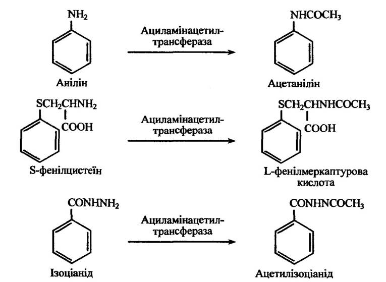
Fig. 10.51. Acetylation reactions of aniline, S-phenylcysteine, and isocyanide
Acetylation typically takes place in the liver, but it can also occur in the reticuloendothelial Cells of the spleen and lungs, as well as in the mucosa of the gastrointestinal tract.
A characteristic reaction of aromatic carboxylic acids is conjugation with glycine (yielding so-called hyphuric acids) and Other Amino Acids. Substituted acetic acids (phenylacetic and indoleacetic), acrylic acids (cinnamic), and certain natural steroidal acids (cholic acid) also form glycine conjugates. Conversely, aliphatic carboxylic acids do not undergo this reaction.
The General scheme of peptide conjugation reactions with glycine is illustrated in Fig. 10.52.
The formation of glycine conjugates with benzoic acid involves the following reactions (Fig. 10.53).

Fig. 10.52. Peptide conjugation reactions with glycine
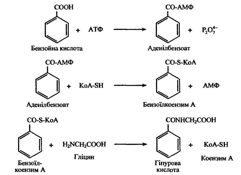
Fig. 10.53. Reactions of glycine conjugate formation with benzoic acid
Glycine is The amino acid most commonly involved in peptide conjugation in mammals; however, in humans and certain primates, conjugation is mediated by phenylacetic acid, where substituted phenylacetic acids form glycine conjugates. Furthermore, in humans and other primates, indole-3-acetic acid forms a glutamine conjugate, and p-aminosalicylic acid conjugates with glutamine.
In other animals, peptide conjugation involves different amino acids. For instance, Ornithine participates in peptide conjugation in reptiles and some birds, whereas Arginine and glutamine are utilized in certain arachnids and myriapods. In some primates, conjugates are also formed with glutamine. In cats, quinaldic acid (quinoline-2-carboxylic acid) is excreted in the urine as quinaldylglycyltaurine and quinaldylglycylglycine conjugates (Fig. 10.54).
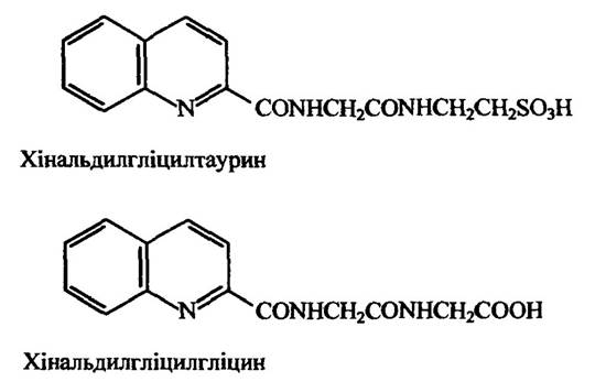
Fig. 10.54. Conjugation products of quinaldic acid
The choice of the amino acid involved in conjugation is determined by its metabolic role in the respective species.
Aromatic and aliphatic xenobiotics can also conjugate with glutathione (G-SH) to yield S-alkylglutathiones, S-arylglutathiones, cysteine derivatives, and ultimately mercapturic acids. Among the various types of such conjugation, the most prevalent are glutathione conjugates of aromatic hydrocarbons, as well as those where compounds conjugate with glutathione via the displacement of halogen atoms, nitroamido, and sulfamido groups.
In most mammals and insects, conjugation with glutathione occurs first, followed by the conversion of this conjugate into the corresponding cysteine derivative and subsequent acetylation to form premercapturic acid. However, in some mammals, such as guinea pigs, mercapturic derivatives are almost entirely absent due to a deficiency in the enzyme that acetylates arylcysteine. Similarly, locusts excrete non-acetylated cysteine derivatives.
During the formation of glutathione conjugates from aromatic hydrocarbons such as benzene, naphthalene, and anthracene, these compounds are converted into mercapturic acids in which a hydrogen atom is replaced by an L-acetylcysteine moiety. These conjugates are excreted in the urine as premercapturic acids. Treatment with mineral acids converts them into mercapturic acid (with the elimination of a water molecule) and phenols (with the elimination of N-acetylcysteine). The reaction for the Formation of the benzene-glutathione conjugate is shown in Fig. 10.55.
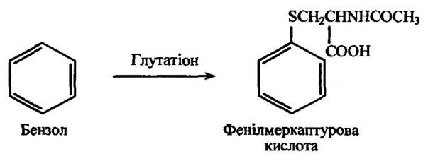
Fig. 10.55. Glutathione-mediated conjugation reaction of benzene
A number of compounds containing a labile chlorine atom—such as benzyl chloride, 4-chloronitrobenzene, 2,4-dichloronitrobenzene, and 1- and 2-menaphthyl chlorides—form mercapturic acids in which the L-acetylcysteine chain replaces chlorine. This reaction with benzyl chloride is illustrated in Fig. 10.56.
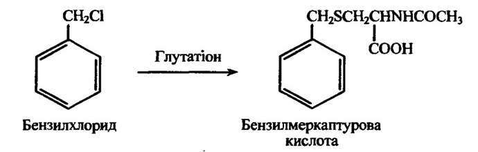
Fig. 10.56. Glutathione-mediated conjugation reaction of benzyl chloride
Various haloalkanes and nitroalkanes also form mercapturic acids through the displacement of halogen atoms or nitro groups, presumably via a mechanism analogous to the synthesis of benzylmercapturic acid. The metabolites of bromoalkanes can include alkylmercapturic or hydroxyalkylmercapturic acids (Fig. 10.57).
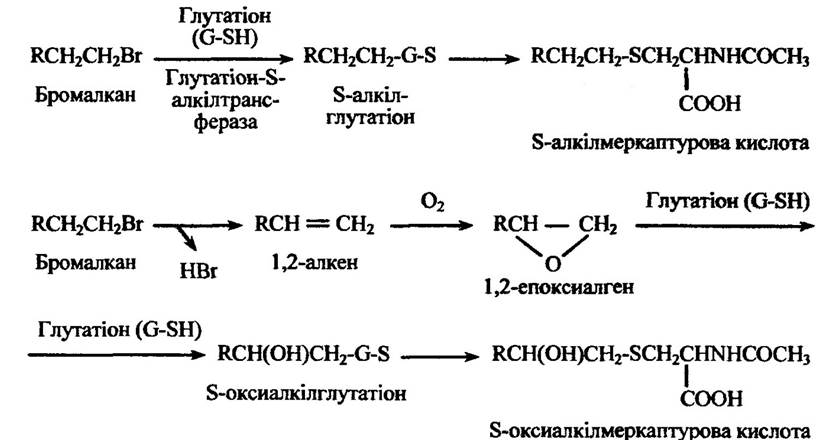
Fig. 10.57. Conjugation reactions of bromoalkane yielding alkylmercapturic
or hydroxyalkylmercapturic acids
In vertebrates (including humans), invertebrates, plants, and bacteria, the detoxification of cyanides can proceed via their sulfur-mediated conjugation (Fig. 10.58) to form thiocyanate (rhodanides). The enzyme rhodanase, which catalyzes this conjugation in vertebrates, is localized in all tissues (except blood) and is particularly abundant in the liver and kidneys. In this reaction, thiosulfate, cysteine, and glutathione do not serve as sulfur donors.

Fig. 10.58. Cyanide conjugation reactions mediated by rhodanase
Cyanide can also be detoxified through a reaction with cystine, yielding 2-aminothiazolidine-4-carboxylic acid (Fig. 10.59).
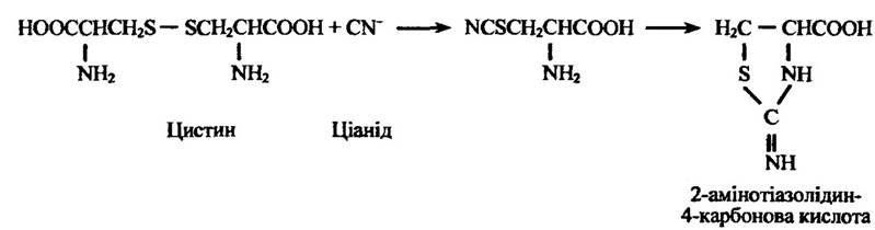
Fig. 10.59. Diagram of cystine conjugation with cyanide
If a xenobiotic molecule contains two or more functional groups, typically only one group undergoes conjugation. However, double conjugates may also form. For instance, acetamidophenols and hydroxyanisoles are capable of further conjugation with glucuronic acid or sulfate.
The listed conjugation reactions by no means exhaust all metabolic possibilities. For example, There are also pathways involving phosphate, glycinetaurine, and formyl conjugation, among others. Pesticides such as 2,4-dichlorophenoxyacetic acid, 3,4-dichloroaniline, and pentachlorophenol can conjugate with plant lignin. Heavy Metal Ions conjugate with metallothioneins.
It should be noted that xenobiotic conjugation as a mechanism to neutralize their adverse effects is characteristic only of a specific organism. Xenobiotics entering an ecosystem continue to circulate within it in a transformed form.
Last update: 06/08/2026
Editorial and Educational Adaptation: This material has been compiled based on the primary/original source text. The project team performed an editorial review, corrected technical inaccuracies, structured sections, and adapted the content for an educational format.
What was processed:
- elimination of formatting defects (OCR errors, structural breaks, corrupted characters);
- editorial organization of content;
- standardization of terminology in accordance with academic sources;
- verification of factual statements against the original source text.
All mentions of the author, publication year, and origin of the primary text have been preserved in accordance with the source.