Vertebrate Zoology - V. M. Konstantinov 2011
Chordates
Gnathostomes
Class Bony Fishes - Subclass Ray-finned Fishes - Superorder Teleost Fishes
Features of bony fish Organization
Teleost fish—the most numerous and widespread superorder of modern Fishes—inhabit all seas and oceans across various Water depths. Many species thrive in freshwater environments such as rivers, lakes, and ponds.
Due to the wide variety of habitats and lifestyles, the external Morphology of these fishes varies greatly. At the same time, they share A number of common structural features. Their Skeleton is almost entirely ossified, with Cartilage persisting only in small patches between the replacing bones. The Skeleton of the paired fins is simplified; typically, the pectoral fins lack basalia, and the bony radials articulate directly with the girdle. The pelvic fins lack both basalia and radials, with the fin skeleton consisting solely of bony rays.
The pectoral fins are positioned vertically relative to the body. The body is covered in bony scales, which form thin plates overlapping one another like roof tiles. The Mouth is terminal (located at the anterior end of the HEAD). There is no cloaca. The caudal fin is homocercal.
The Structural Features of the subclass Actinopterygii are examined using teleost fishes as an example, as they represent their most numerous and typical group.
Integument. The body is covered with bony scales originating from the dermis (corium) and forming thin, translucent plates with either smooth (cycloid scales) or serrated (ctenoid scales) outer margins. Externally, the scaly cover is coated with a microscopic layer of epidermis rich in unicellular glands that secrete mucus onto the body surface.
Class="center">
Fig. 38. Longitudinal section through the lateral line canal of a teleost fish:
1 — lateral nerve; 2 — lateral line Organs; 3 — canal; 4 — external opening of the canal
Scale size increases as the fish grows, a process that continues virtually throughout the animal's lifetime. The growth rate of scales varies across different seasons of the year. The age of fish can be determined by counting the dark and light growth bands on their scales (see details below). Some species possess naked, mucous Skin devoid of scales.
The skin contains the lateral line, which is a canal running along the sides of the body and communicating with the external environment through a series of openings piercing the scales (Fig. 38).
Skeleton. The Axial Skeleton, much like that of cartilaginous fishes, comprises THE Vertebral Column and the chondrocranium. The vertebral column consists of bony amphicoelous vertebrae and is divided into trunk and caudal regions. The vertebrae bear neural and haemal arches. The neural arches meet above to form the vertebral (spinal) canal, with neural spines projecting upward. The trunk region haemal arches do not close; instead, they articulate with Ribs that enclose the body cavity dorsally and laterally. In the caudal region, spinous processes are present on both the neural and haemal arches. The enclosed haemal arches form the haemal canal.
The braincase is mostly ossified, being formed of both dermal and chondral (endochondral) bones (Fig. 39).

Fig. 39. Diagram of the Skull Structure of a teleost fish. The operculum and circumorbital ring have been removed. Designated bones:
1 — basioccipital; 2 — exoccipital; 3 — supraoccipital; 4 — otic bones; 5 — basisphenoid; 6 — pterosphenoid; 7 — orbitosphenoid; 8 — mesethmoid; 9 — lateral ethmoid; 10 — parietal; 11 — frontal; 12 — nasal; 13 — parasphenoid; 14 — Vomer; 15 — palatine; 16 — quadrate; 17 — pterygoids; 18 — premaxilla; 19 — Maxilla; 20 — articular; 21 — dentary; 22 — anguloarticular; 23 — hyomandibula; 24 — symplectic; 25–29 — branchial arches; 30 — hyoid; 31 — copula
Chondral ossifications reflect the architectural style of the chondrocranium in cartilaginous fishes. The occipital region includes four occipital bones: the basioccipital, two exoccipitals, and the supraoccipital. They enclose the foramen magnum. Five otic bones develop in the region of each paired otic capsule. The orbital region is framed by the unpaired basisphenoid, paired pterosphenoids, and orbitosphenoids. In the region of the paired olfactory capsules, an unpaired median mesethmoid and paired lateral ethmoids develop.
Thus, chondral ossifications form the posterior portion, sides, and partially the floor of the braincase.
Dermal, or roofing, bones form the roof, and partly the sides and floor, of the braincase. The skull roof is composed of sequentially arranged paired nasal, frontal, and parietal bones.
The floor of the skull consists of the unpaired parasphenoid and the unpaired vomer lying anterior to it. Small dermal ossicles form a circumorbital ring around the Orbit.
Major changes occur in the visceral skeleton, which also contains both chondral and dermal ossifications.
The dorsal section of the mandibular arch, homologous to the palatoquadrate cartilage, has lost its upper jaw function in bony fishes and has become incorporated into the floor of the skull complex. Anteriorly, it is replaced by the Palatine bone (of mixed origin); medially, by three pterygoid bones (two dermal and one chondral); and posteriorly, by the chondral quadrate bone. The upper jaw function is fulfilled by paired bones of dermal origin—the maxilla and premaxilla. These represent novelties in the visceral skeleton.
The lower jaw is represented by a large dermal dentary bone, which overlies Meckel's cartilage. The angular bone, also of dermal origin, forms the posteroventral corner of the jaw, while the sole chondral bone—the articular—connects with the quadrate.
The hyoid arch consists of the same elements found in cartilaginous fishes: paired suspensoria (hyomandibulae), hyoids, and an unpaired copula. All these bones are of chondral origin. A series of elongated bones—the branchiostegal rays—attach to the posterior margin of the lower section of the hyoid arch.
The bony branchial arches share a similar structure with those of cartilaginous fishes, though the last (fifth) arch is heavily reduced. A novel evolutionary acquisition of bony fishes is the operculum, represented by four flat dermal bones.
Limbs and their girdles. The pectoral fin skeleton lacks basalia and consists of bony radials connecting directly to the girdle, along with dermal fin rays. The Pectoral Girdle comprises small, chondral-origin coracoids and scapulae. Dermal BONES OF THE secondary girdle attach to these primary chondral elements. The primary component is a large, crescent-shaped bone known as the cleithrum. Through smaller bones, it articulates with the neurocranium.
The Pelvic Girdle lies deep within the musculature and is represented by an unpaired, elongated plate. The pelvic fin skeleton consists solely of dermal bony rays.
Swim bladder. The swim bladder is characteristic of most teleost fishes. Developmentally, it originates as an outpocketing of the dorsal wall of the alimentary canal. In many species, the connection with the Esophagus is lost during development (physoclistous fishes), whereas in others it persists throughout life (physostomous fishes). The swim bladder primarily serves a hydrostatic function, achieved by altering the gas volume within the bladder and, consequently, changing the fish's body density. In physostomous fishes, volume changes are accomplished by compressing or expanding the bladder when gulping air; in physoclistous fishes, this occurs through the absorption or secretion of gases via a specialized network of capillaries in the gas gland (the rete mirabile). The gas filling the swim bladder is predominantly nitrogen.
In some fishes, the swim bladder is connected to the Inner ear—the membranous labyrinth—via a chain of small bones known as the Weberian apparatus. Through this mechanism, volume Changes in the bladder caused by shifts in the fish's position within the water column are transmitted to the semicircular canals of the inner ear, which function as the Organ of Equilibrium. Additionally, the Weberian apparatus transmits sounds that are picked up by the body surface, resonated by the swim bladder, and conveyed to the auditory organ (the membranous labyrinth).
Overall, the Evolution of the swim bladder was likely driven by the increased body weight resulting from The Development of a bony skeleton.
Digestive System. In most fish species, the bones surrounding the Oral Cavity bear numerous single-cusped, conical Teeth. These are located not only on the jawbones—dentaries, maxillaries, and premaxillaries—but also on the palatines, vomer, and parasphenoid.
The oral cavity is not sharply demarcated from the Pharynx, which leads into a short esophagus. The Stomach varies greatly in shape and size, and in some species is relatively weakly developed. The intestine is morphologically less differentiated than that of cartilaginous fishes. There is no spiral valve. At the very beginning of the intestine, many species possess blind outgrowths known as pyloric caeca. These increase the digestive surface area of the intestine and possibly slow down the passage of food, similar to the function of the spiral valve in cartilaginous fishes. The number of pyloric caeca varies among species: for example, the perch has 3, the salmon about 40, and the mackerel around 200.
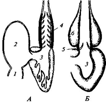
Fig. 40. STRUCTURE OF THE Heart in a shark (A) and a teleost fish (B):
1 — sinus venosus; 2 — atrium; 3 — ventricle; 4 — conus arteriosus; 5 — rudiment of the conus arteriosus; 6 — bulbus arteriosus
The Liver is multi-lobed and equipped with a Gallbladder. The Bile duct empties into the anterior, loop-like section of the intestine. The Pancreas is poorly developed, appearing as very small lobules scattered throughout the mesentery.
Respiratory system. Teleost fishes are primarily branchial-gilled. Unlike cartilaginous fishes, they lack interbranchial septa, and the gill filaments sit directly on the respective branchial arches. There are four pairs of gill filaments. Additionally, on the inner surface of the operculum, There is a pseudobranch—a single row of vestigial filaments. Respiration is driven by the coordinated Movements of the opercula and mouth, which pump water into the gill cavities and force it out. Fast-swimming fish typically rely on ram ventilation of the gills. In scaleless fishes, gill respiration is supplemented by cutaneous respiration.
Circulatory system. In most species, the ventral aorta features a Swelling at its base known as the bulbus arteriosus. Outwardly it resembles the conus arteriosus, but it consists of smooth Muscle rather than striated muscle (Fig. 40) and is incapable of robust pulsation. The number of afferent and efferent branchial Arteries (aortic arches) is four. Carotid arteries branch forward from the aortic roots to supply the Brain with Blood. The aortic roots are joined by an anastomosis, forming the cephalic arterial circle characteristic of bony fishes. The Venous system of most species is notable for the continuity of the right cardinal vein; only the left cardinal vein forms a renal portal system within the corresponding Kidney (Table 3).
Table 3
Blood parameters in various fish groups
(from N. P. Naumov and N. N. Kartashev, 1979)
Fish groups |
Blood volume, % of body mass |
Hemoglobin content, % |
Oxygen capacity of blood, % per unit volume |
Sharks |
3,7-6,8 |
3,7-6,5 |
4,5-8,7 |
Rays |
1,5-7,2 |
0,8-4,5 |
1,1-6,0 |
Bony fishes |
1,1-7,3 |
1,1-17,4 |
1,5-23,0 |
Blood volume, hemoglobin concentration, and oxygen-carrying capacity vary widely among different fish species depending on their level of mobility.
Nervous System and Sense Organs. In several respects, the brain of teleost fishes has a more primitive structure compared to that of cartilaginous fishes. Its overall relative size is smaller, with a particularly underdeveloped Forebrain; in most species, the roof of the Telencephalon is epithelial and lacks Nervous Tissue. The olfactory lobes are visible anteriorly. The Lateral ventricles are not separated even by an incomplete septum (unlike in cartilaginous fishes). Conversely, the Diencephalon, mesencephalon, and Cerebellum are relatively enlarged. There are 10 pairs of Cranial Nerves.
Organ of Vision. The visual organ has a typical piscine structure featuring a flat cornea and a spherical lens. Focusing is performed exclusively by the lens—at rest, it provides near vision, while for distance vision, a specialized muscle shifts it slightly deeper into the Eyeball; thus, accommodation in fish is relatively primitive. Teleost fishes possess Color Vision. Their retina contains functional rods and cones operating side by side, which likely explains the striking variety of body coloration seen in many teleost fishes. Deep-sea forms, however, much like sharks, possess exclusively rod-based (monochromatic) vision.
Organ of Hearing. The auditory organ in teleost fishes is represented by the inner ear, or membranous labyrinth, which perceives sound across a broad frequency range of 30 Hz – 12 kHz. Assisting in sound perception are the aforementioned swim bladder and Weberian apparatus. Movable opercula, skull bones, and fin movements contribute to sound production. As research has shown, fish are remarkably "talkative." Acoustic signaling facilitates communication between individuals of the same and different species during foraging, mating, predator warnings, and territory defense. The sounds produced by fish are diverse: clicks, whispers, creaks, pops, trills, groans, etc.
Another sensory organ located within the inner ear is The Organ of equilibrium. The sense of balance is maintained by three semicircular canals filled with fluid containing suspended mineral particles (otoliths). The sense of balance is one of the most ancient Sensory systems in vertebrates.
Olfactory organs. In most teleost fishes, the olfactory organs consist of paired nasal sacs located in the anterodorsal region of the head. The cavity of each olfactory sac communicates with the aquatic environment via two openings (anterior and posterior). Water is driven through them, allowing chemical reception to take place within the folds of the olfactory epithelium. Unlike in some other groups, the olfactory sac in teleost fishes does not communicate with the oral cavity.
Histological and physiological data attest to the exceptional acuteness of their SENSE OF SMELL. For instance, in the minnow, there are approximately 95,000 receptor Cells per 1 mm2 of olfactory epithelium. Experiments conducted in the late 1970s established that eels react unfailingly to phenylethyl alcohol at dilutions up to 2.8 ∙ 1018, and trout to 9.9 ∙ 109.
Gustatory organs. The Organs of taste are represented by taste buds composed of Cell clusters enveloped by nerve endings. Taste buds are not only found within the oral cavity but are also scattered across numerous Regions of the body within the outer layer of the skin.
Lateral line organs. The lateral line organs are long canals embedded in the skin along the sides of a fish's body. On the front of the head, they form a complex network of channels. Sensory cells lie at the bottom of these canals, communicating with the surrounding environment through multiple Pores in the scales and head bones. These visible openings are commonly referred to as the lateral line organs.
Through the lateral line organs, fish perceive wave motions propagating through the water, which inform them about nearby objects.
Excretory organs. The excretory organs are represented by long, paired, ribbon-like mesonephric Kidneys. They lie along the sides of the vertebral column above the swim bladder. Ureter ducts, homologous to the Wolffian ducts, run along the inner margins of the kidneys. Before exiting the body, the Ureters merge into a single duct that opens at the tip of the urogenital papilla. Some species possess a Urinary Bladder. Urea, dissolved in water, is excreted through the kidneys as the end product of Nitrogen METABOLISM.
The primary final product of nitrogen metabolism is ammonia. It is highly toxic, and its accumulation in the blood at high concentrations is detrimental and harmful to the Organism. Due to the high solubility of ammonia in water, extrarenal excretion of Protein metabolism products occurs via the gills and, to some extent, through the skin.
The reproductive organs have a different structure than those of cartilaginous fish. Females lack Müllerian ducts, which serve as oviducts in cartilaginous fish, while in males the Testes are not connected to the kidneys, and the Wolffian ducts function solely as ureters. Paired testes and Ovaries (the perch has a single, unpaired Ovary) contain internal cavities and specialized ducts that open onto the urogenital papilla separately from the urinary opening.
The eggs are small, with a thin gelatinous envelope; Fertilization is generally external.
Taxonomic Overview
The superorder Teleostei includes more than 30 orders. Let us examine some of them.
Last update: 13/08/2026
Editorial and Educational Adaptation: This material has been compiled based on the primary/original source text. The project team performed an editorial review, corrected technical inaccuracies, structured sections, and adapted the content for an educational format.
What was processed:
- elimination of formatting defects (OCR errors, structural breaks, corrupted characters);
- editorial organization of content;
- standardization of terminology in accordance with academic sources;
- verification of factual statements against the original source text.
All mentions of the author, publication year, and origin of the primary text have been preserved in accordance with the source.