Human Anatomy and Physiology - N. I. Fedyukovich 2003
Cells and Tissues
Tissues
Connective Tissue
Based on their properties, Connective Tissues comprise a significant group of tissues, including proper connective tissues (loose fibrous, and dense fibrous—both irregular and regular); specialized connective tissues (adipose, reticular); skeletal hard tissues (bone and Cartilage), and fluid tissues (Blood, lymph). Connective Tissue performs supportive, protective (mechanical), morphogenetic, plastic, and trophic Functions. It consists of numerous Cells and an Extracellular matrix containing various fibers (Collagen, elastic, and reticular).
Loose Fibrous connective tissue contains various cellular elements (fibroblasts, macrophages, plasma cells, mast cells, etc.). Depending on the Structure and function of the organ, these fibers are oriented differently within the ground substance. This tissue is predominantly located along the course of Blood Vessels.
Dense Fibrous Connective tissue is categorized as either regular or irregular. In regular Cytology/practical/45.html">Dense connective tissue, fibers run parallel and are bundled together, forming ligaments, tendons, membranes, and fascias. Irregular dense connective tissue is characterized by an interwoven network of fibers and a relatively sparse cellular component.
Adipose tissue forms beneath the Skin—particularly around the Peritoneum and omentum—and lacks its own characteristic ground substance. Each Cell features a central fat droplet, with The Nucleus and Cytoplasm pushed to the periphery. Adipose tissue serves as an energy depot, cushions Internal Organs against mechanical Shock, and helps retain body heat.
Skeletal Tissues include cartilage and bone. Cartilage tissue consists of cartilage cells (chondrocytes), usually grouped in clusters of two or three, embedded in a gel-like extracellular matrix. There are three MAIN TYPES OF cartilage: hyaline, fibrous, and elastic. Hyaline cartilage forms the articular and costal cartilages, contributes to the thyroid and cricoid cartilages of the Larynx, and lines the respiratory tract. Fibrocartilage is found in the intervertebral and intra-articular discs, the menisci, and covers the articular surfaces of the temporomandibular and sternoclavicular joints. Elastic cartilage builds the epiglottis, arytenoid, corniculate, and cuneiform cartilages, the auricle, as well as the cartilaginous PARTS OF THE auditory tube and external acoustic meatus.
Blood, lymph, and interstitial fluid constitute the internal environment of the Organism. Blood supplies tissues with nutrients and oxygen, removes Metabolic waste products and carbon dioxide, produces Antibodies, and transports Hormones that regulate The activity of various body systems. Although blood circulates within blood vessels and is separated from other tissues by the vascular wall, formed elements and plasma solutes can migrate into the connective tissue surrounding the blood vessels. This process enables blood to maintain the constancy of the internal environment's composition.
Depending on The Nature of the substances transported, the primary FUNCTIONS OF BLOOD include respiratory, excretory, nutritive, homeostatic, regulatory, protective, and thermoregulatory functions.
Through its respiratory function, blood transports oxygen from the Lungs to organs and tissues, and carbon dioxide from peripheral tissues back to the lungs. The excretory function involves transporting metabolic wastes (such as uric acid and bilirubin) to excretory organs (such as Kidneys, intestines, and skin) for subsequent elimination as substances harmful to the organism. The nutritive function relies on delivering Digestion-derived nutrients (like glucose and Amino Acids) to organs and tissues. The homeostatic function ensures the even distribution of blood among organs and tissues, alongside the maintenance of constant osmotic pressure and pH levels via Plasma Proteins and other mechanisms. The regulatory function entails carrying hormones—synthesized by Endocrine glands—to specific target organs to transmit systemic signals. The protective function involves the neutralization of microorganisms and their toxins by Blood Cells, antibody production, the removal of tissue breakdown products, and hemostasis via blood clot formation. The thermoregulatory function is carried out by transferring heat outward from deep-seated organs to cutaneous vessels, as well as by evenly distributing heat throughout the body owing to the high heat capacity and thermal conductivity of blood.
In humans, blood mass accounts for 6–8% of body weight and normally equals approximately 4.5–5.0 L. At rest, only 40–50% of the total blood volume circulates, while the remainder is sequestered in blood depots (Liver, Spleen, skin). The Pulmonary Circulation contains 20–25% of the blood volume, whereas the systemic circulation holds 75–85%. The Arterial System contains 15–20% of the blood, the Venous system 70–75%, and capillaries 5–7%.
Blood consists of cellular (formed) elements (45%) and a liquid portion known as plasma (55%). Once the formed elements are removed, the remaining plasma contains Water-dissolved salts, proteins, CARBOHYDRATES, biologically active compounds, as well as carbon dioxide and oxygen. Plasma comprises approximately 90% water, 7–8% protein, 1.1% other organic substances, and 0.9% Inorganic Components. It maintains the constancy of intravascular fluid volume and acid-base balance (ABB), while also participating in The transport of active substances and metabolic products. Plasma proteins are divided into two main groups:
albumins and globulins. The first group accounts for approximately 60% of plasma proteins. Globulins are represented by alpha1-, alpha2-, beta2-, and gamma-globulin fractions. Fibrinogen is also included within the globulin fraction. Plasma proteins participate in processes such as the Formation of tissue fluid, lymph, and urine, as well as water absorption. The nutritive function of plasma is related to the presence of Lipids, the concentration of which depends on dietary habits.
Blood serum differs from plasma in that it lacks fibrinogen and does not clot. Serum is prepared from Blood Plasma by removing fibrin. Blood is placed in a cylindrical vessel; after a certain time, it clots and turns into a gelatinous mass from which a light-yellow liquid—blood serum—is extracted.
Blood is a colloidal-polymer solution in which water acts as the solvent, while salts, low-molecular-weight Organic compounds, proteins, and their complexes serve as the solutes.
BLOOD OSMOTIC PRESSURE is the driving force of solvent movement across a semipermeable membrane from a less concentrated solution into a more concentrated one. Blood osmotic pressure remains relatively constant for METABOLISM and equals 7.3 atm (5600 mmHg, or 745 kPa). It depends on the concentration of dissociated ions and salts, as well as the volume of Body Fluids. The salt concentration in blood is 0.9%, which primarily dictates blood osmotic pressure.
Osmotic pressure is determined by maintaining the concentration of various substances dissolved in body fluids at the required physiological level.
Thus, osmotic pressure ensures that water is distributed evenly between Cells and Tissues. Solutions with an osmotic pressure higher than that of the intracellular contents (hypertonic solutions) cause cell shrinkage due to water shifting out of the cells into the solution. Solutions with a lower osmotic pressure than the intracellular contents (hypotonic solutions) increase cell volume as water moves from the solution into the cells. Solutions whose osmotic pressure matches that of the intracellular contents and which induce no morphological changes in cells are termed isotonic.
The Regulation of Osmotic pressure is mediated neurohumorally. Additionally, specialized osmoreceptors located in blood vessel walls, tissues, and the Hypothalamus respond to fluctuations in osmotic pressure. Their stimulation leads to alterations in the activity of excretory organs (such as the kidneys and Sweat Glands).
Blood maintains a constant pH reaction. The reaction of the medium is determined by the hydrogen ion concentration, expressed as the pH value, which is of great significance since the vast majority of biochemical reactions can only proceed normally within specific pH ranges. Human Blood has a weakly alkaline reaction: the pH of venous blood is 7.36, and that of arterial blood is 7.4. Life is sustainable within a rather narrow window of pH shifts—from 7.0 to 7.8. Despite the continuous influx of acidic and alkaline metabolic products into the bloodstream, blood pH is maintained at a relatively constant level. This constancy is upheld by physicochemical, biochemical, and physiological mechanisms.
Several Blood Buffer Systems are known (carbonate, plasma proteins, phosphate, and Hemoglobin), which bind hydroxyl (OH-) and hydrogen (H+) ions, thereby keeping the blood reaction at a constant level. Concurrently, excess acidic and alkaline metabolic products generated in the body are excreted by The Kidneys in urine, while Carbon dioxide is eliminated by the lungs.
The formed elements of blood include erythrocytes, leukocytes, and platelets.
Erythrocytes (red blood cells) are biconcave discs that lack a nucleus. Their average diameter is 7–8 µm, which is roughly equal to the internal diameter of a blood capillary. The shape of an erythrocyte enhances its gas exchange capacity, facilitating the diffusion of gases from the surface to the entire cell volume. Erythrocytes exhibit remarkable elasticity, allowing them to easily traverse capillaries possessing a diameter half that of The Cell itself. The total surface area of all erythrocytes in an adult human is approximately 3800 m2, meaning it exceeds the body surface area by 1,500 times.
The blood of men contains about 5×10¹² cells/L of erythrocytes, whereas women's blood contains 4.5×10¹² cells/L. During intense physical exertion, the erythrocyte count in the blood may rise to 6×10¹² cells/L. This increase is associated with the mobilization of sequestered blood entering the general circulation.
The primary characteristic of erythrocytes is the presence of hemoglobin, which binds oxygen (transforming into oxyhemoglobin) and releases it to peripheral tissues. Hemoglobin that has unloaded its oxygen is termed reduced hemoglobin and exhibits the characteristic color of venous blood. Having released oxygen, the blood gradually absorbs the end product of metabolism—CO2 (carbon dioxide). The reaction of hemoglobin binding to CO2 is more complex than oxygen binding, which is explained by The Role of CO2 in regulating the body's acid-base balance. Hemoglobin bound to carbon dioxide is called carbohemoglobin. Under the Influence of the enzyme Carbonic anhydrase found within erythrocytes, carbonic acid dissociates into CO2 and H2O. Carbon dioxide is eliminated by the lungs, preventing any alteration in blood reaction. Hemoglobin binds particularly easily to carbon monoxide (CO) owing to its high chemical affinity for hemoglobin (300 times higher than that for O2). Hemoglobin blocked by carbon monoxide can no longer function as an oxygen carrier and is referred to as carboxyhemoglobin. Consequently, the body develops oxygen deprivation accompanied by vomiting, headache, and loss of consciousness.
Hemoglobin consists of the protein globin and a prosthetic heme group, which attaches to the four polypeptide chains of globin and imparts the red color to blood. Normally, blood contains about 140 g/L of hemoglobin: 135–155 g/L in men and 120–140 g/L in women.
A decrease in The amount of erythrocyte hemoglobin in the blood is termed anemia. It occurs in cases of Hemorrhage, intoxication, or deficiencies in vitamin B12, Folic acid, and other factors.
The lifespan of red blood cells (erythrocytes) is about 3–4 months. The process of erythrocyte destruction, during which hemoglobin is released into the plasma, is called hemolysis.
When blood is left to stand in a vertical test tube, erythrocytes settle downward. This occurs because the specific gravity of erythrocytes is higher than that of plasma (1.096 versus 1.027).
The ERYTHROCYTE SEDIMENTATION RATE (ESR) is expressed in millimeters of plasma Column height above the erythrocytes per unit of time (usually 1 hour). This reaction characterizes certain PHYSICOCHEMICAL PROPERTIES OF blood. The normal ESR is 5–7 mm/h for men and 8–12 mm/h for women. The Mechanism of erythrocyte sedimentation depends on many factors, such as the erythrocyte count, their morphological features, surface charge, aggregation capacity, plasma protein composition, and others. An elevated ESR is typical for pregnant women—up to 30 mm/h, patients with infectious and inflammatory processes, as well as malignant tumors—up to 50 mm/h or more.
Leukocytes are white blood cells. They are larger than erythrocytes and contain a nucleus. The lifespan of leukocytes is several days. The normal leukocyte count in human blood is 4–9109/L and fluctuates throughout the day, reaching its lowest level in the morning on an empty Stomach.
An increase in the leukocyte count in the blood is called leukocytosis, and a decrease is called leukopenia. Both physiological and reactive leukocytosis are distinguished. The former is most commonly observed after meals, during Pregnancy, under physical exertion, pain, emotional stress, and other conditions. The second type is characteristic of inflammatory processes and infectious diseases. Leukopenia is observed in certain infectious diseases, exposure to ionizing radiation, medication use, and others.
All types of leukocytes possess amoeboid motility and, in the presence of appropriate chemical stimuli, pass through the capillary endothelium (diapedesis) and migrate toward the stimulus: microbes, foreign bodies, or antigen-antibody complexes.
Based on the presence of granules in their cytoplasm, leukocytes are divided into granular (granulocytes) and agranular (agranulocytes).
Cells whose granules stain with acidic Dyes (such as eosin) are called eosinophils; those staining with basic dyes (such as methylene blue) are called basophils; and those staining with neutral dyes are called neutrophils. The first stain pink, the second blue, and the third pink-purple.
Granulocytes account for 72% of the total leukocyte count, comprising 70% neutrophils, 1.5% eosinophils, and 0.5% basophils. Neutrophils are capable of migrating through intercellular spaces to infected areas of the body, engulfing and digesting pathogenic Bacteria. The number of eosinophils increases during allergic reactions, Bronchial Asthma, and hay fever; they exhibit antihistamine activity. Basophils produce heparin and histamine.
Agranulocytes are leukocytes that consist of an oval-shaped nucleus and non-granular cytoplasm. They include monocytes and lymphocytes. Monocytes have a Kidney-shaped nucleus and are formed in the Bone Marrow. They actively infiltrate sites of inflammation and engulf (phagocytize) bacteria. Lymphocytes are produced in the Thymus gland from stem lymphoid Cells of the bone marrow and spleen. Lymphocytes produce antibodies and participate in cellular immune responses. There are T- and B-lymphocytes. T-lymphocytes independently destroy microorganisms, Viruses, and transplanted tissue cells using Enzymes, and are known as killer cells. B-lymphocytes, upon encountering a foreign substance, neutralize and bind these substances using specific antibodies, preparing them for phagocytosis. A condition in which the lymphocyte count exceeds the normal baseline is called lymphocytosis, while a decrease is termed lymphopenia.
Lymphocytes are the core component of The Immune System; they participate in cell growth, tissue regeneration, and the Regulation of the GENETIC APPARATUS OF other cells.
The ratio of Different types of leukocytes in the blood is called the leukocyte differential count (Table 1).
Class="center">Table 1 Leukocyte differential count
|
Leukocytes, 10% |
Eosinophils, % |
Basophils, % |
Neutrophils, % |
Lymphocytes, % |
Monocytes, % |
||
|
myelocytes |
band |
segmented |
|||||
|
4.0-9.0 |
1-4 |
0-0.5 |
0-1 |
2-5 |
55-68 |
25-30 |
6-8 |
The count of specific types of leukocytes increases in A number of diseases. For example, lymphocyte levels rise in whooping cough and typhoid fever, monocyte levels in malaria, and neutrophil levels in Pneumonia and other infectious diseases. Eosinophil counts increase in allergic conditions (bronchial asthma, scarlet fever, etc.). Characteristic Changes in the leukocyte differential count enable an accurate Diagnosis.
Platelets (thrombocytes) are colorless, spherical, anucleate cell fragments with a diameter of 2–5 µm. They are formed from large bone marrow cells called megakaryocytes. The lifespan of platelets ranges from 5 to 11 days. They play a crucial role in blood clotting. A significant portion of them is stored in the spleen, liver, and lungs, and is released into the bloodstream as needed. During physical exertion, food intake, and pregnancy, the platelet count in the blood increases. The normal platelet count is approximately 250×109/L.
Blood Groups are immunogenetic and individual blood characteristics that classify people based on the similarity of specific Antigens—aglutinogens—on erythrocytes and antibodies—agglutinins—present in blood plasma.
The blood group is determined by the presence or absence of specific mucopolysaccharides—agglutinogens A and B—in the donor Erythrocyte membranes, and agglutinins α and β in the recipient's blood plasma (Table 2).
Table 2 Dependence of the blood group on the presence of erythrocyte agglutinogens and plasma agglutinins
|
Blood groups |
Agglutinogens in erythrocytes |
Agglutinins in serum |
|
0(I) |
— |
α, β |
|
A (II) |
A |
β |
|
B (III) |
B |
α |
|
AB(IV) |
A, B |
— |
Accordingly, four blood groups are distinguished: 0 (I), A (II), B (III), and AB (IV). When similar erythrocyte agglutinogens are combined with plasma agglutinins, an agglutination (clumping) reaction of erythrocytes occurs, which underlies blood group incompatibility. This principle must be strictly followed during blood transfusions.
The science of blood groups has become significantly more complex with the discovery of new agglutinogens. For example, group A has several subgroups; furthermore, new agglutinogens such as M, N, S, P, and others have been identified. These factors occasionally cause complications during repeat blood transfusions.
People with blood group I are considered universal Donors. However, it has been found that this universality is not absolute. This is due to the fact that individuals with blood group I often exhibit high titers of immune anti-A and anti-B agglutinins. Transfusing such blood can lead to severe complications and potentially fatal outcomes. These findings have served as the basis for transfusing exclusively same-group blood (Fig. 4).
Transfusion of incompatible blood leads to The Development of transfusion reactions (thrombosis, followed by erythrocyte hemolysis, kidney damage, etc.).
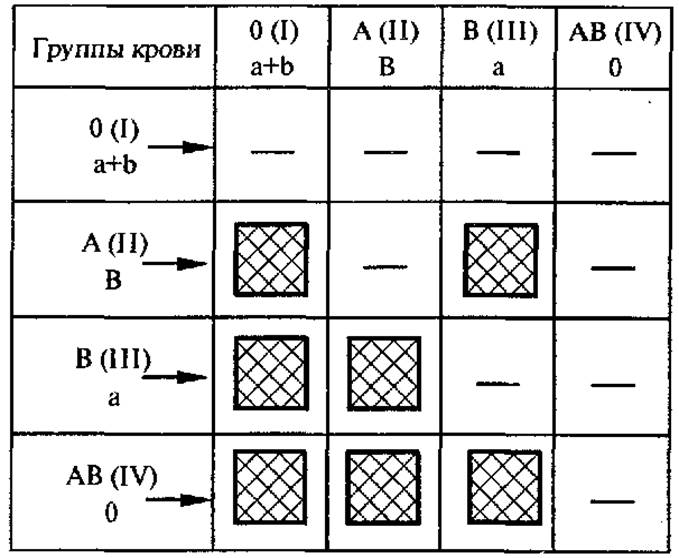
Fig. 4. Blood type compatibility:
dash — compatible; square — incompatible
Besides the main agglutinogens A and B, erythrocytes may contain others, notably the so-called Rhesus factor (Rh factor), which was first discovered in the blood of the rhesus macaque monkey. Based on the presence or absence of the Rh factor, organisms are classified as Rh-positive (about 85% of people) and Rh-negative (about 15% of people). In medical practice, the Rh factor is of great significance. Thus, in Rh-negative individuals, blood transfusions or subsequent pregnancies induce The formation of Rh antibodies. Transfusion of Rh-positive blood into individuals with Rh antibodies leads to severe hemolytic reactions accompanied by the destruction of the transfused erythrocytes.
The development of Rh-incompatibility during pregnancy is based on The entry of Rh-positive fetal erythrocytes through the Placenta into the body of an Rh-negative woman, leading to the formation of specific antibodies (Fig. 5).
In such cases, the first child, having inherited the Rh-positive trait, is born normal. However, during a second pregnancy, maternal antibodies penetrating the fetal bloodstream cause erythrocyte destruction, accumulation of bilirubin in the newborn's blood, and the onset of hemolytic jaundice with damage to the child's internal organs.
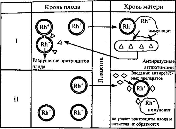
Fig. 5. Development of Rh incompatibility and its Prevention:
I — Rh incompatibility; II — prevention of Rh incompatibility
Blood clotting is a protective reaction that prevents blood loss and the entry of pathogens into the body. It is a multi-step process involving 12 factors present in blood plasma, as well as substances released from damaged tissues and platelets. Blood clotting is divided into three stages. In The First stage, blood flowing from a wound mixes with substances from damaged tissues and destroyed platelets and comes into contact with air. Then, the released thromboplastin precursor is converted into active thromboplastin under The Influence of plasma factors and Calcium Ions (Са2+). In the second stage, with the participation of thromboplastin, plasma factors, and calcium ions, the inactive plasma protein prothrombin is converted into Thrombin. In the Third Stage, thrombin (a proteolytic enzyme) cleaves the plasma protein fibrinogen molecule into smaller fragments and creates a network of fibrin threads (an insoluble protein) that precipitates out. Blood formed elements are trapped within the fibrin network, forming a clot that prevents blood loss and the penetration of microorganisms into the wound. After the removal of fibrin from plasma, the remaining fluid is serum.
Blood serves as a therapeutic agent. Blood transfusions and blood products are widely used in clinical practice. Donation is widespread to ensure an adequate blood supply. People who donate blood for medical purposes are called donors. For active donors, a single blood donation volume is 250— 450 ml. Typically, this results in a decrease in hemoglobin and erythrocyte counts proportional to the amount of blood withdrawn. The recovery rate of the donor's blood depends on many factors, including the volume of blood donated, age, gender, Nutrition, etc.
Last update: 08/08/2026
Editorial and Educational Adaptation: This material has been compiled based on the primary/original source text. The project team performed an editorial review, corrected technical inaccuracies, structured sections, and adapted the content for an educational format.
What was processed:
- elimination of formatting defects (OCR errors, structural breaks, corrupted characters);
- editorial organization of content;
- standardization of terminology in accordance with academic sources;
- verification of factual statements against the original source text.
All mentions of the author, publication year, and origin of the primary text have been preserved in accordance with the source.