Peptide Self-Regulation in Living Systems (Facts and Hypotheses) - Shataeva, L. K. 2003
Peptide-Cell Membrane Interactions
Structure of the Phospholipid Bilayer of the Membrane
It has long been evident that viewing the Functions of living Cell membranes solely through the lens of The chemical properties of their constituent components is insufficient; one must account not only for their physicochemical and electrochemical characteristics, but also for the Spatial Organization of these components, namely molecular Asymmetry and the liquid-crystalline state of Lipids. Lipids differ from the other three Major Groups of substances that make up living organisms (Proteins, CARBOHYDRATES, and NUCLEIC ACIDS) in that they are insoluble in aqueous environments. These pale yellow, pasty substances dissolve readily in "lipid Solvents" such as chloroform, diethyl ether, and benzene. Lipids are classified into several groups, of which Phospholipids and sterols are incorporated into Plasma Membranes, whereas triglycerides (fats) are typically located in the intercellular space.
Phospholipids are glycerol esters that typically feature different substituents at the 1, 2, and 3 carbon atoms, with long-chain hydrophobic substituents—alkyls or alkenyls—at positions 1 and 2. At position 3, hydrophilic groups such as Choline, ethanolamine, Serine, glycerol, Inositol, and phosphorylated inositol forms are attached to the lipid via a phosphodiester bond. This amphiphilic nature of phospholipids drives their tendency to form micelles In aqueous solutions (with a critical micelle concentration, or CMC, on the order of 10-10 M) into a thermodynamically favorable micellar geometry: a lipid bilayer in which polar heads form two hydrophilic surfaces spaced 75—80 Å apart, thereby shielding the hydrophobic regions (Preparative BIOCHEMISTRY OF LIPIDS, 1981; Lishko & Shevchenko, 1987). Fig. 9 illustrates the core Structure/83.html">Structural elements of the most common phospholipids and a schematic of a bilayer lipid membrane with an embedded polypeptide chain (Deenen, 1981; Kagawa, 1985).
Currently, more than 40 variants of polar groups and roughly 200 combinations of hydrophobic chains have been identified in phospholipids from various sources. In formed bilayers, saturated hydrocarbon chains typically adopt a zigzag conformation and run parallel to one another. The axis of the first acyl chain coincides with the axis of the glycerol residue; the second acyl chain, following a bend at the —CO—CH2— segment, aligns parallel to the first chain.
The polar groups of phospholipids exposed to the aqueous environment are oriented According to the sign of their net charge: negatively charged phosphatidylserine groups are directed perpendicularly to the bilayer plane, whereas positively charged phosphatidylcholine and phosphatidylethanolamine groups are oriented parallel to The Lipid Bilayer surface (Brown & Wolken, 1982).
Phosphatidylcholine (lecithin) is abundant in the Cells of higher organisms (accounting for 30–60% of total phospholipids), whereas its content in bacterial membranes does not exceed 6%.
Studies of both natural and synthetic phospholipids have demonstrated that the ionization constants of the ionogenic groups forming the polar HEAD span a wide range, from pKa = 2.72 (phosphate group) to pKa = 11.6 (choline) (Introduction/36.html">Biological Membranes, 1990; Tomoaia-Cotisel, 1999). Choline is one of the strongest organic bases, second in basicity only to the Arginine side chain, allowing the polar head of phosphatidylcholine (PC) to maintain a zwitterionic structure across a broad pH range. Conversely, phosphatidylserine (PS) bears two permanently ionized acidic groups. Thus, both surfaces of the Lipid Bilayer Membrane feature polar groups whose ampholytic properties are comparable to those of Amino Acids and Peptides. Furthermore, although the hydrocarbon chains of phospholipids situated between the two ampholytic surfaces of the lipid bilayer are entirely hydrophobic, the overall structure remains fairly permeable to Water molecules. Both Artificial and natural bilayer membranes exhibit a water permeability of approximately 2 mL/(cm2∙s) (Kagawa, 1985).
Class="center">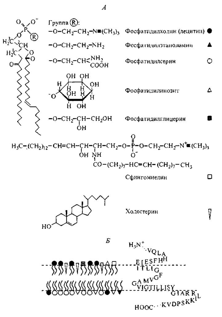
Fig. 9. Principal phospholipids incorporated into the bilayer structures of plasma membranes (A), and a schematic of the lipid bilayer structure in an erythrocyte membrane (B) with an embedded Amino Acid Sequence from a segment of the Glycophorin chain (Preparative Biochemistry of Lipids, 1981; Kagawa, 1985; Lishko & Shevchenko, 1987).
It was previously assumed that The properties of biological membranes were largely dictated by The structure of the lipid bilayer itself, such that the overall closed boundary of a living cell resembled a soap bubble. Over the past 20 years, detailed investigations into the structure and mobility of biological membrane components have substantially shifted our understanding of cell membrane Structure and function. The Functional Significance of the lipid bilayer has proven to be far broader than merely serving as a hydrophobic barrier between The Cell interior and the external environment.
The dynamic STRUCTURE OF THE lipid bilayer has been most thoroughly characterized using artificial bilayer vesicles. These studies have demonstrated that a phospholipid molecule as a whole can rotate around its longitudinal axis and exhibits considerable mobility within the layer, with lateral diffusion coefficients of 10-7–10-9 cm2/s. Polar heads form short-lived (10-6–10-7 s) clusters of 20–30 molecules On the surface, which can give rise to transient defects within the bilayer structure. Water molecules diffuse across the lipid bilayer by entering these free volumes situated between the hydrophobic tails of the lipids. Within the bilayer, phospholipid molecules can undergo transbilayer migration (flip-flop). However, this occurs relatively infrequently in artificial bilayer membranes due to the energetic penalty of transferring a polar head across the Hydrophobic core (Deenen, 1981). Only selective interactions with integral proteins of natural membranes can facilitate the rapid translocation of a phospholipid from one layer to the other. For instance, a protein has been isolated from bovine Liver that selectively interacts with PC and transports it from the outer leaflet of the membrane to the inner leaflet, or from artificial vesicles into The Plasma Membrane. Following the Hydrolysis of this complex, a
peptide fragment that selectively interacts with the phospholipid was isolated (Etemadi, 1980):

It is hypothesized that the hydrophobic block (underlined) may serve as a region capable of binding to the hydrophobic chain of a phospholipid. Notably, 4 out of these 6 amino acid residues feature an aromatic side group.
The distribution of various phospholipids between the outer and inner leaflets follows specific patterns characteristic of each tissue. In particular, PS is unique because it is exclusively localized on the inner surface of The erythrocyte membrane. When exogenous labeled PS is added to erythrocytes, it is immediately translocated to the inner membrane surface, as Membrane Proteins maintain an asymmetrical charge distribution across the two leaflets of the plasma membrane. If endogenous PS appears on the outer surface due to local membrane damage, the erythrocyte fails to maintain Homeostasis and is eliminated from the bloodstream by the reticuloendothelial system (Connor, Schroit, 1988).
The Lipid Composition of Cytoplasmic membranes exhibits distinct tissue Specificity. Table 10 presents variations in the phospholipid compositions of cytoplasmic membranes across certain cell types. At the same time, species-related differences in phospholipid composition are significantly smaller than tissue-related differences (McMurray, 1973).
More than 60 years ago, J. Bernal (1969) suggested viewing individual intracellular structures as liquid crystals. The most evident example of a profound analogy between liquid crystals and biological structures is the lipid bilayer of the plasma membrane (Brown, Wolken, 1982).
The phase state of phospholipids is Temperature-dependent: upon heating, an endothermic phase transition from the gel to the liquid-crystalline state is observed. Apparently, phospholipids, having a limited number of molecular Conformations, self-assemble into bilayer membranes and compensate for The Diversity of conformational states through phase transitions within liquid-crystalline structures. These transitions are associated with an increase in the conformational freedom of the hydrocarbon moieties of lipid molecules. For example, measurements of the temperature dependence of heat capacity and enthalpy in PC vesicles revealed two phase transitions—at 34 and 41 °C. Concurrently, the distance between fatty acid chains increased from 0.48 to 0.53 nm (Volkenshtein, 1981). Depending on the compositional makeup of the lipid bilayer, several quasi-equilibrium liquid-crystalline phases may coexist within it.
Table 10 Lipid composition of plasma membranes, % (after: Brown, Wolken, 1982; Kagawa, 1985)
Lipid component |
Erythrocytes |
Myelin |
Retinal rod cells |
Hepatocytes |
Phosphatidylethanolamine |
20 |
14 |
38.5 |
20 |
Phosphatidylserine |
11 |
7 |
9.2 |
4 |
Phosphatidylcholine |
23 |
11 |
44.5 |
43 |
Phosphatidylinositol |
2 |
0 |
0 |
7 |
Sphingomyelin |
18 |
6 |
1.3 |
20 |
25 |
25 |
0 |
6 |
|
Cerebroside |
0 |
21 |
6.5 |
— |
Under physiological conditions (above the phase transition temperature), the phospholipid bilayer adopts a liquid-crystalline state, meaning it simultaneously exhibits fluidity and an ordered arrangement of elements. This fluidity, combined with a sufficiently high surface tension at the aqueous interface, results in the self-closure of the bilayer. In fact, plasma membranes are never formed de novo: they bud off and assemble from preexisting membranes by the incorporation of additional structural components (Brown, Wolken, 1982). However, the principles governing the self-assembly of plasma membrane phospholipid layers remain insufficiently understood, although the de novo formation process of Endoplasmic reticulum membranes has been investigated in detail using Electron Microscopy (Biryuzova, 1993). These membranes are synthesized via Replication on the outer surface of the double membrane of the Cell Nucleus, which temporarily assumes a bean-like shape. Self-assembly of the membranes occurs on its concave surface, and as The Nucleus flattens back into a spherical shape, they slide off and smooth out the surface relief. This example demonstrates that one-dimensional sequences in polypeptide chains and DNA Helical structures are not the only templates capable of directing the synthesis of complementary one-dimensional structures. The two-dimensional surface of a phospholipid bilayer likewise serves as a template for the assembly of a complementary phospholipid layer.
The fluidity of bilayers depends on the degree of unsaturation of the hydrophobic chains and the presence of additional substances in the membrane. Notably, cholesterol—one of the major Lipid Components of all eukaryotic plasma membranes—significantly affects bilayer fluidity (Deinum et al., 1988). Erythrocyte membranes contain up to 30% cholesterol, as it readily interacts with phospholipids to form 1:1 complexes. Its flat steroid nucleus easily penetrates the Membrane Structure and enhances the orientational order of hydrocarbon chains in the liquid-crystalline phase: the steroid rings span 9–12 carbon units of the lipid. Sphingomyelins exhibit the highest affinity for cholesterol. Nevertheless, the selectivity of intermolecular interactions among phospholipids is a distinct issue in clinical biochemistry and will not be addressed in this section (Nikiforova, 1981).
The asymmetric arrangement of positively and negatively charged phospholipid groups on opposite sides of the plasma membrane gives rise to a substantial Electrochemical Potential gradient between the outer and inner leaflets of this structure. Until recently, comparing the electrostatic potentials of phospholipid monolayers with that of a bilayer separating two aqueous phases was restricted to artificial symmetric membranes, where the dipole moments of the lipid molecules mutually compensate each other. The electrostatic capacitance of such a lipid bilayer depends on the chain length of the fatty acid residues of the phospholipids used and reaches 0.7–0.8 µF/cm2 (Boguslavsky, 1978), whereas the electrical resistance of the bilayer ranges from 103 to 106 Ω ∙ cm2 (Volkenshtein, 1981).
The features distinguishing cellular membranes and their models from traditional objects of electrochemical research begin with their dimensions: they are nanosystems in the literal sense of the word, given that the thickness of the lipid bilayer is on the order of 7.0–7.5 nm, with the polar outer layer being 1.4 nm thick, while the remaining membrane portion, constructed from hydrocarbon chains, acts as an insulator. The well-known challenges in studying electrochemical processes at the insulator–electrolyte solution interface are compounded when transitioning to these ultrathin bilayer membranes, as their polar region is comparable in thickness to the interfacial solvent layer.
The structural heterogeneity of the cell boundary zone is determined not only by the distribution of phospholipid charges on the outer membrane surface and associated low-molecular-weight counterions, but also by the structure of the adjacent water molecule layers and the arrangement of peptide molecules near this surface. Specifically, apolipoproteins contain chain regions with an asymmetric distribution of hydrophilic groups capable of organizing into helical structures ("Edmundson wheels", see Fig. 4). During Isolation and Purification, these regions typically lose their helical conformation, but upon The addition of phospholipids, the proportion of helical segments is restored. This process is facilitated by the interaction of peptide side groups with the polar (ionized) phospholipid head groups. Here, the peptide positions itself on the phospholipid monolayer such that the axis of the α-Helix is oriented parallel to the monolayer surface (Polyakov, Panin, 2000).
The order inherent in the liquid-crystalline state of phospholipids dictates two key Properties of the cytoplasmic membrane: spontaneous polarization and its modulation in response to environmental fluctuations (temperature, external electric field, mechanical pressure).
The native asymmetry in the distribution of positively and negatively charged groups across the plasma membrane is an intrinsic feature of all natural membranes. As a result, the inner Regions of the lipid bilayer, which possess a low dielectric constant, are effectively positioned between the plates of a capacitor; that is, they are subjected to a potential difference between the polar outer and inner surfaces of the membrane. Driven by this intra-membrane electrostatic field, the hydrophobic portions of the phospholipids undergo polarization. This may involve both molecular (electronic) polarization and orientational polarization, which dictates the packing of aliphatic chains within the membrane and their liquid-crystalline state. Any alteration in this bilayer state induced by an external electrostatic field primarily results in a shift in the Membrane Potential.
A light wave can serve as an effective external electrical stimulus. The optical frequency range is such that it excites oscillations exclusively within the electron shells, leaving the molecular conformations within the bilayer structure unaffected. Thus, the hydrophobic interlayer of the membrane acts as a regular dielectric medium capable of functioning as a waveguide for electromagnetic waves, specifically photons.
Investigations into the structure of chloroplast lipid layers have demonstrated that during Photosynthesis, they function analogously to a lens, focusing light onto the active layer of chlorophyll pigment (Brown & Wolken, 1982). The anisotropy of the membrane's liquid-crystalline structure determines its sensitivity to the direction of plane-polarized light (Jaffe, 1981). Dense Intercellular junctions facilitate the transmission of such electromagnetic signals from Cell to Cell, enabling the entire tissue (or organ) to become "informed" almost simultaneously.
External electrostatic fields also influence The cell membrane, primarily by affecting the lateral distribution of charges on its surface, which significantly alters the polarization of The Cell as a whole. This phenomenon has been studied experimentally during the Cytology/cytology/16.html">Early stages of frog embryo development (Hinkel et al., 1981). It was found that the processes of developing Nerve Cells—neurites—grow more rapidly toward the cathode than toward the anode. Similarly, dorsal ROOT ganglia from chick embryos exhibited preferential growth toward the cathode. The direction of a constant external electric field also influenced The Development of frog embryo myoblasts. Initially spherical in shape, myoblasts grew perpendicularly to the applied field direction, causing the Cells of the resulting Muscle tissue to elongate transverse to the electric field lines. This phenomenon was observed only at weak external fields of 70 mV/mm or lower, corresponding to approximately 1 mV per diameter of the growing cell. It is suggested that this can be explained by the lateral Electrophoresis of Membrane Receptors—specific for binding growth factors—across the cell surface toward the cathode (Jaffe, 1981).
The liquid-crystalline Nature of the lipid bilayer dictates its high sensitivity not only to fluctuations in External temperature and electrical fields but also to external mechanical stress. A two-dimensional elastomer model has been developed for plasma membranes, consisting of a loose network of flexible hydrocarbon chains (Evans & Skalak, 1982).
In the general case, the deformation of a two-dimensional system cannot be regarded as isotropic. However, if the radii of curvature of the membrane surface are much larger than the distance between the layers of aliphatic chains in the bilayer membrane (a condition always met in Eukaryotic cells), the surface deformation of the entire membrane will closely parallel that of each individual layer, and The change in membrane Entropy will be inversely proportional to its stretch. This model is analogous to the textbook example of entropy reduction during the mechanical stretching of a rubber band. The primary contribution to the change in Free energy of this system is made by the work required to order the network elements, provided the material density remains constant. For such elastomers, tensile deformation leads to minor changes in internal free energy G, as its primary variations are directly linked to the increment in configurational entropy S:
dG = -TdS,
where T is temperature. As noted previously, entropy is defined by the Boltzmann equation via the energy distribution across the possible configurations of the molecular network, which include the configurations of the various liquid-crystalline states of the bilayer.
An interesting characteristic of entropic elastomers is that their shear modulus increases with temperature. In other words, the work required to stretch an elastomeric surface works against the thermal disordering processes that occur in systems striving for equilibrium. Consequently, the reversible heat exchange during the stretching of an entropic elastomer is accompanied, on average, by a decrease in the degrees of freedom of the molecular chains, a reduction in entropy, and a corresponding increase in structural order. As a result, the membrane's sensitivity to mechanical pressure—including variable mechanical stresses from sound waves—is enhanced. Furthermore, the ferroelectric properties of the liquid-crystalline structures within the lipid bilayer enable The conversion of these mechanical stresses into an electrical signal.
It can be hypothesized that the thermodynamic properties of the lipid bilayer thus contribute to the GENERATION AND PROPAGATION of environmental information signals, thereby participating in the self-regulation of living cells and the stabilization of homeostasis.
The combination of the aforementioned chemical, dynamic, and structural Characteristics of Lipid bilayer membranes determines their functional properties: electrical excitability; the coupling of mechanical properties and polarizability; and compatibility with oriented blocks of polypeptide chains.
The latter property of the phospholipid bilayer is of fundamental importance and warrants separate consideration.
Last update: 06/08/2026
Editorial and Educational Adaptation: This material has been compiled based on the primary/original source text. The project team performed an editorial review, corrected technical inaccuracies, structured sections, and adapted the content for an educational format.
What was processed:
- elimination of formatting defects (OCR errors, structural breaks, corrupted characters);
- editorial organization of content;
- standardization of terminology in accordance with academic sources;
- verification of factual statements against the original source text.
All mentions of the author, publication year, and origin of the primary text have been preserved in accordance with the source.