Chordate Zoology - textbook - Y. V. Tsaryk - 2013
Chapter 4. DIVISION GNATHOSTOMATA, or ECTOBRANCHIATA. SUPERCLASS PISCES
4.1. CLASS CHONDRICHTHYES
4.1.1. Organizational Features
Cartilaginous Fishes exhibit A number of specific characteristics:
✵ the Skin is naked or covered with placoid scales;
✵ the Skeleton is cartilaginous, with the Axial Skeleton composed of cartilaginous amphicoelous vertebrae (it can also be represented by the notochord), and well-developed vertebral arches;
✵ the Skull is amphistylic, hyostylic, and in Holocephali, autostylic;
✵ the caudal fin is heterocercal;
✵ there is no common operculum, and the gills open externally through individual slits;
✵ a swim bladder is absent;
✵ a spiral valve is developed in the intestine;
✵ The Heart features a conus arteriosus between the ventricle and the ventral aorta;
✵ Fertilization is strictly internal, and males possess specialized outgrowths of the pelvic fins called claspers (pterygopodia);
✵ oviparous, ovoviviparous, and viviparous forms occur.
Appearance and integument. Cartilaginous fishes differ from jawless fishes in having mobile paired fins, which allow for more agile swimming. This is one of the ancient groups of marine predators. We will examine the Structural Features of cartilaginous fishes using sharks as an example, whose Morphology has remained virtually unchanged for over 250 million years.
Unpaired fins are represented by the dorsal and caudal fins. The anterior dorsal fin (pinna dorsalis anterior) is located in the middle of the trunk, while the posterior dorsal fin (pinna dorsalis posterior) is situated in the caudal region.
Some species have a free, sharp spine on the anterior margin of their fins.
The caudal fin (pinna caudalis) is heterocercal. Paired fins are represented by the pectoral (pinnae pectorales) and pelvic (pinnae ventrales) fins.
The pelvic fins meet at their inner, or medial, margins. In males, as previously mentioned, a special fleshy outgrowth—the copulatory organ, or clasper (pterygopodium)—is developed at the posterior end of each pelvic fin.
Forward propulsion is achieved by the lateral bending of the caudal region to the left and right. The pectoral and pelvic fins act as hydrofoils, directing the body upward, downward, and sideways.
On the ventral side of the body, between the posterior margins of the pelvic fin bases, cartilaginous fishes have a wide longitudinal slit—the cloaca. Adjacent to the posterior margin of the cloaca are two abdominal pores that connect to the coelom.
In sharks, a r o s t r u m (rostrum) is present anteriorly, on the ventral side of which are two nasal pits forming the incurrent and excurrent openings of the olfactory cavities. The eyes (oculus) are partially covered by skin folds—eyelids (palpebra). At the anterior and posterior margins of the eyes, the white fibrous tunic—the sclera (sclera)—is visible, while the rest of the eye is covered by a transparent cornea (cornea), beneath which the iris (iris) is visible.
Behind the eyes is a slit—the spiracle (spiraculum), which connects the pharyngeal cavity with the external environment.
Five (in some, six or seven) transverse gill slits are located anterior to the pectoral fins (in rays, they are located lower, on the ventral side). Each gill slit is covered by a skin fold.
The Mouth is crescent-shaped. The Teeth vary in shape, most commonly being triangular, and are arranged in several rows. On the ventral surface of the rostrum, between the mouth and the first gill slit, and on the DORSAL SIDE OF the HEAD above the eyes, rows of openings are visible—pores leading to the tubules of the lateral line system.
The skin of cartilaginous fishes is composed of a multilayered epidermis and a corium. The epidermis contains numerous unicellular glands whose mucous secretion covers the shark's body in a thin layer. The Cells of the lower epidermal layers contain pigments. Special pigment cells are also present in the corium. Some rays are able to change their coloration depending on the color of the substrate they lie on. This is achieved through the contraction and migration of pigment cells deeper into the body, or, conversely, by the expansion of the entire pigment Cell.
Placoid scales develop in the skin of cartilaginous fishes (Fig. 4.1). This type of scale consists of a rounded or rhombic basal plate embedded in the corium and a spine (projection) whose tip projects outward through the epidermis.
Class="center">
Fig. 4.1. Structure of a placoid scale
On the jaws, placoid scales are enlarged, assume a different shape, and form teeth. In this case, primarily, the spine enlarges and the enamel thickness increases. A worn or broken tooth falls out, and a new one grows in its place. Tooth replacement can occur throughout the fish's life. The teeth of rays are mostly flat and blunt, adapted for crushing animals protected by shells or carapaces. In holocephalans, individual teeth fuse into a tooth plate. The teeth of all vertebrates are constructed on the placoid model; in fact, they are modified placoid scales of their ancestors. Modified scales also form fin spines in horn sharks and spiny dogfish, stings in stingrays, saw-like teeth in sawsharks and sawfishes. In some rays, scales are completely absent, or there are individual enlarged scales whose denticles are transformed into powerful thorns (thornback ray).
The skin of cartilaginous fishes provides them with sufficiently reliable mechanical protection, while simultaneously being permeable to Water and substances dissolved in it.
Skeletal and muscular systems. In modern cartilaginous fishes, bones are absent from the skeleton. The internal skeleton is entirely cartilaginous, though certain areas may contain calcium salts, which increases the strength of the elements. The skeleton is divided into the axial skeleton, the cranial skeleton (neurocranium and splanchnocranium), the skeleton of paired and unpaired fins, and the girdle skeleton of paired fins. The axial skeleton of elasmobranch fishes consists of THE Vertebral Column (columna vertebralis), which is divided into trunk and caudal regions.
The vertebrae of the axial skeleton are concave both anteriorly and posteriorly (Fig. 4.2). Such vertebrae are called biconcave, or amphicoelous. In the center of the vertebral centrum, There is a canal through which the notochord passes. It runs through the entire vertebral column, which resembles a string of beads: expanded at the junctions of the vertebrae and constricted within the vertebral centra. Above the vertebral centra are the neural arches. Intercalary plates are located between the neural arches of adjacent vertebrae, which, together with the arches, form the walls of the cartilaginous canal housing the Spinal Cord.
In the trunk region, poorly developed lower arches of the vertebrae form transverse processes to which very short cartilaginous Ribs are attached. In the caudal region, the expanded lower arches of the right and left sides are fused with each other, and small lower intercalary plates are located between them. This structure forms the hemal canal, through which the caudal artery and vein pass.

Fig. 4.2. Fragment of a shark vertebral column (A) and cross-section of a vertebra (B)
During ontogeny, cartilaginous primordia form within the Connective Tissue Sheath of the notochord, initially in each body segment: two paired upper and two paired lower primordia of the lower arches. Later, ring-shaped cartilages—vertebral centra—begin to develop around the notochord. In holocephalans, vertebral centra do not form. The connective tissue sheath of the notochord thickens, and ring-like calcifications develop within it, outwardly resembling vertebrae. The anterior pair of cartilaginous primordia develops into neural arches that rest on the notochord sheath, while the posterior pair transforms into intercalary plates. In the caudal region, hemal arches and lower intercalary plates develop. Ribs are absent.
The skull of cartilaginous fishes is divided into two regions: the neurocranium (axial) and the splanchnocranium (visceral) (Fig. 4.3). The neurocranium of elasmobranch fishes is a solid cartilaginous box that surrounds the Brain on all sides: in the cranial roof, there is an opening covered by connective tissue—the fontanelle. The anterior end of the skull is elongated into a rostrum. At the Base of the rostrum, paired nasal capsules fuse with the skull, and otic capsules grow into the posterior lateral walls.
The occipital region contains an opening through which the spinal cord passes. On the lateral surfaces of the skull, there are depressions—orbits—that protect the eyes from above and from the sides. The skull is platybasic: its base is broad, and the brain is located between the walls of the orbits.
The splanchnocranium was formed As a result of the transformation of the gill arches, which in ancestors served as support in the pharyngeal region. It consists of the mandibular arch, the hyoid arch, and the branchial arches. The mandibular arch is formed by two paired cartilages. The left and right cartilages are connected (fused) anteriorly. The function of the upper jaw is performed by the palatoquadrate Cartilage (palatoquadratum), which forms the jaw joint with the lower jaw (Meckel's cartilage - cartilago Meckeli). Teeth are located on the upper and lower jaws. In many sharks, 1-2 pairs of small labial cartilages lie anterior to the mandibular arches, which are interpreted as remnants of two reduced gill arches.
The hyoid arch is located immediately behind the jaws. It consists of two paired cartilages and one unpaired cartilage. In elasmobranch fishes, the large upper paired cartilage—the hyomandibula (hyomandibulare)—is movably attached at its upper end to the otic region of the neurocranium by means of a joint and ligaments, while its lower end is movably connected both to both elements of the mandibular arch in the region of the jaw joint and to the lower paired element of the hyoid arch—the hyoid (hyoideum). The right and left hyoids are connected to each other by a small unpaired cartilage—the copula (copula). This type of connection between the mandibular arch and the neurocranium—via the upper element of the hyoid arch—is called hyostyly.
Behind the hyoid arch are five pairs (six to seven in frilled and cow sharks) of branchial arches, separated from each other by gill slits.
Unlike elasmobranchs, the skull of holocephalans lacks a rostrum or has a poorly developed one, and the nasal capsules are fused to the braincase anteriorly. The teeth are fused into a tooth plate. The skull is of the tropibasic type (only a thin interorbital septum is located between the orbits).
The skeleton of unpaired fins is formed by rows of rod-like cartilages of various sizes—radials—embedded in the Muscles, which extend to the base of the fin. Sometimes PARTS OF THE radials fuse into plates.

Fig. 4.3. Shark skull
The caudal fin of most cartilaginous fishes is heterocercal (Fig. 4.4). The upper lobe of the fin is significantly larger than the lower lobe, and the end of the vertebral column extends into it.
The skeletal support of the caudal fin is formed by elongated upper and lower arches of the vertebrae, as well as a series of dorsospinal and ventrospinal unsegmented cartilages. The dermal lobes of the fin are supported by elastotrichia. In some rays and chimaeras, the lobes of the caudal fin are reduced or have disappeared completely.
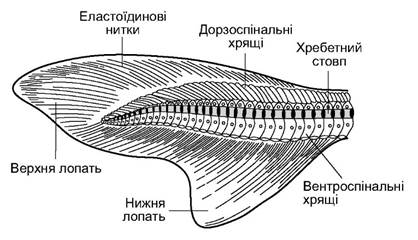
Fig. 4.4. Heterocercal caudal fin
The Pectoral Girdle (shoulder girdle) has the form of a cartilaginous arch (Fig. 4.5) embedded within the muscular wall behind the gill region. In sharks and chimaeras, it is not connected to the axial skeleton in any way, whereas in rays, due to the enlargement of the pectoral fins, the upper parts of the cartilaginous girdle arch connect to the vertebral column. On each side of the girdle's lateral surface, there is a projection serving as the attachment site for the fin skeleton, which consists of the same elements as those in the unpaired fins.
The Pelvic Girdle (or pelvic fin girdle) of cartilaginous fishes is formed by a cartilaginous plate (see Fig. 4.5) which lies within the Muscles of the abdominal wall just anterior to the cloaca. The Skeleton of the pelvic fin is attached to its lateral surfaces.
The somatic Muscular System is clearly segmented and consists of Muscle segments called myomeres. The myomeres are separated from each other by myosepta, which form a complex zigzag line resembling a sideways letter W. In other parts of the body, the musculature may differ. For example, the visceral muscular system consists of smooth muscle layers surrounding the digestive tract. In cartilaginous fishes, in the region of the jaw and gill arches, the smooth muscle fibers of the visceral muscular system are replaced by striated ones, forming highly differentiated muscle groups that control the movement of the jaw and gill arches.
A characteristic feature of the muscles of cartilaginous fishes is their relative autonomy, meaning their ability to contract even after The connection between the muscles and the Central Nervous System has been severed. Specifically, a decapitated shark can still swim, or in other cases, attack prey even after its Internal Organs have been removed. The chemical composition of the muscles of cartilaginous fishes is also unique. They have a high urea content—up to 1.50–2.84% in marine species and up to 0.7% in freshwater species (compared to up to 0.02–0.03% in bony fishes). This high concentration of urea is closely linked to osmoregulation. The muscles of cartilaginous fishes also contain high levels of heavy metal salts: Ti, Mn, Co, Ni. The Significance of this phenomenon in the life of sharks remains unexplained.
The General Anatomy of a shark is shown in Fig. 4.6.

Fig. 4.5. Limb girdles and fin skeletons of a shark: A - pectoral fin and pectoral girdle; B, C - pelvic girdle and pelvic fin skeleton of female and male; D - pelvic fins of female and male
The Digestive System begins with the mouth (stoma) (Fig. 4.6), which is a slit-like opening bounded by the upper and lower jaws. At the bottom of the buccopharyngeal cavity lies the Tongue (lingua). This is a fold of the mucous membrane supported by an unpaired element—the basihyal (copula) of the hyoid arch. This tongue lacks intrinsic muscles, so its movement is dependent on the motion of the hyoid and branchial apparatuses.
The short, muscular Esophagus transitions smoothly into a spacious, V-shaped Stomach. Behind The Stomach begins the intestine, which is divided into three sections: the Small Intestine, which includes a short duodenum where the pancreatic duct and Bile duct empty; the Large Intestine, which contains a spiral valve with 12–13 turns; and the rectum with a finger-like projection—the rectal gland. The rectal gland is an organ involved in salt balance. The rectum opens into the cloaca. A large portion of the Abdominal cavity is occupied by a three-lobed Liver, which is rich in Lipids (in the basking shark, up to 70% of the liver's mass).

Fig. 4.6. Dissected shark (male)
The Gallbladder (vesica fellea) is located on the right side of the central lobe of the liver. Its duct (ductus cysticus) joins the hepatic ducts (ductus hepaticus) and empties into the duodenum.
The Pancreas has two lobes: dorsal and ventral. The pancreatic duct also empties into the duodenum.
The Respiratory system of cartilaginous fishes is represented by gill pouches that open into the buccopharyngeal cavity via internal gill slits, and to the outside on the sides of the head via external gill slits. The gill pouches are separated from each other by interbranchial septa (septum branchiale). Each interbranchial septum originates on a gill arch, extends to the external gill slit, and ends in a fold that covers the slit (Fig. 4.7). Gill lamellae are located on the interbranchial septa. Many cartilaginous fishes possess a spiracle—a vestigial slit between the mandibular and hyoid arches. At the base of the gill lamellae, there are cartilaginous gill rays that support the interbranchial septum. Beneath the gill arch, at the base of the interbranchial septum, there are three openings of the branchial vessels: an unpaired wide vessel—the afferent branchial artery, through which venous Blood flows to the gills, and two small vessels—the efferent branchial Arteries, through which arterial blood flows away from the gills.

Fig. 4.7. Cytology/practical/72.html">Cross section of shark gills
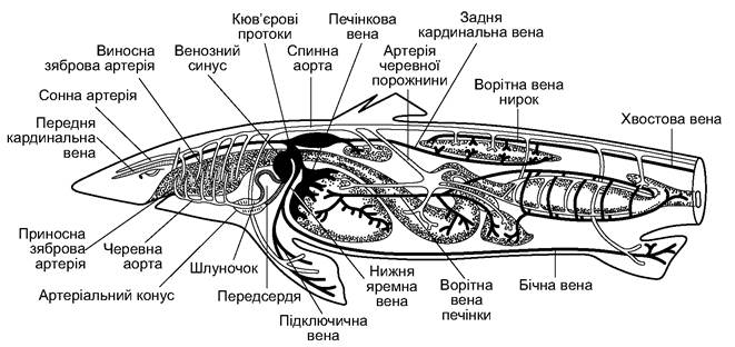
Fig. 4.8. General Topography of the Circulatory system OF a shark
The heart (cor) of cartilaginous fishes, as noted, is two-chambered, yet it consists of four compartments. The posterior compartment, where all Veins empty, is called the sinus venosus. It is connected to the atrium by a wide opening. Next, blood enters the ventricle—the central compartment of the heart with thick muscular walls. Anterior to the ventricle, in the form of a thick tube, lies the anterior compartment of the heart—the conus arteriosus, which transitions into the ventral aorta. The heart is located in a special pericardial cavity (cavum pericardiale), which is a remnant of the coelom. This cavity is separated from the abdominal cavity by a transverse septum and is lined internally by a thin parietal layer of the Peritoneum—the Pericardium (pericardium). The visceral layer tightly envelops the outside of The Heart and is called the epicardium (epicardium). Beneath it
lies a thick layer of muscle, which forms The basis of the heart wall—the myocardium (myocardium). The internal cavity of the heart is lined with a thin layer called the endocardium (endocardium).
The ventral aorta (aorta ventralis) arises from the conus arteriosus and branches into five afferent branchial arteries (arterii branchiales advehentes). Arterial blood, oxygenated in the capillaries of the gill lamellae, is collected into efferent branchial arteries (arterii branchiales revehetes). The common carotid artery (arteria carotis communis), which supplies blood to the head, branches off from the first efferent branchial artery.
The efferent branchial arteries from both sides merge to form the dorsal aorta (aorta dorsalis) located beneath the vertebral column. A series of arteries branch off from the dorsal aorta, supplying blood to all the Organs of the shark; further on, the dorsal aorta enters the hemal canal of the caudal vertebrae, where it is referred to as the caudal artery (arteria caudalis).
Venous blood returns to the heart through veins, which are wider, thin-walled vessels. The caudal vein (vena caudalis) collects blood from the tail, enters the body cavity, and divides into the right and left renal portal veins (venae portae renalis), which enter the Kidneys and break down into capillaries. The capillaries merge to form the right and left posterior cardinal veins (venae cardinalis posterior). Along the sides of the body run paired lateral veins (venae lateralis), which collect blood from the pelvic fins and body walls. In the region of the heart, each of these merges with the subclavian vein (vena lateralis), which carries blood from the pectoral fins. From the head, venous blood is collected into paired anterior cardinal veins (venae cardinalis anterior) and paired inferior jugular veins (venae jugularis inferior). The posterior and anterior cardinal veins and the inferior jugular veins on each side of the body merge into the Cuvierian ducts (ductus cuvieri), which empty into the sinus venosus of the heart.
From the stomach, intestine, and Spleen, venous blood is collected into several veins that merge before the liver to form the HEPATIC PORTAL VEIN (vena portae hepatica). Within the liver, the portal vein breaks down into capillaries, where the blood is filtered and then enters the hepatic vein (vena hepatica), which carries it to the sinus venosus.
The spleen of cartilaginous fishes is large and Functions as a hematopoietic organ.
The excretory system of cartilaginous fishes consists of paired trunk, or mesonephric, kidneys (Fig. 4.9), which are located on the dorsal side of the ABDOMINAL CAVITY AND covered by the peritoneum. The bulk of the kidneys is composed of nephrons, which consist of Malpighian corpuscles (glomeruli of capillary vessels enclosed in Bowman's capsules) from which renal tubules extend.
The renal tubules empty either into the Wolffian duct, which functions as a Ureter in females, or into a ureter separated from the Wolffian duct in males. In males, the Wolffian duct functions as a vas deferens. Metabolic waste products enter the kidneys after filtration in the Malpighian glomeruli. Notably, cartilaginous fishes are characterized by a high concentration of salts in their bodies, which allows them to maintain osmotic equilibrium with their environment (seawater). Excess salts and urea are excreted. Marine cartilaginous fishes excrete small amounts of urine: from 2 to 50 ml per 1 kg of body weight per day. For cartilaginous fishes living in brackish or fresh waters, this value increases to 250 ml per 1 kg of body weight per day (e.g., sawfish).

Fig. 4.9. Diagram of the Urogenital System of cartilaginous fishes: A - male; B - female
Reproductive System and breeding characteristics. Males have paired Testes (Testis) suspended on mesenteries on both sides of the esophagus above the liver (see Fig. 4.9). The ducts of the seminiferous tubules of the testis empty into the renal tubules of the anterior part of the Kidney, which has a modified STRUCTURE AND FUNCTIONS as an Epididymis. The ducts of the testis (vas deferens) open into the Wolffian duct, which functions as a seminal duct. A Seminal Vesicle is located in its posterior section. The seminal ducts of the right and left sides open into the cavity of the urogenital papilla, which in turn opens into the cloacal cavity. During copulation, due to the contraction of the walls of the seminal vesicle, spermatozoa enter the male's cloaca, and from there, with the help of claspers (pterygopodia), into the female's cloaca.
Females have paired Ovaries suspended on mesenteries in the same locations as the male testes. In females, the Wolffian duct functions as a ureter. They have well-developed Müllerian ducts that lie on the ventral surface of the respective (right and left) kidneys.
The anterior ends of the Müllerian ducts function as oviducts. In the region of the anterior kidney, each oviduct has an enlargement called the shell gland; in the posterior part of the oviduct, there is a prominent enlargement known as the "Uterus." The right and left oviducts open into the cloaca through separate openings on the sides of the urogenital papilla. There is no direct connection between the ovaries and the oviducts. The egg, rupturing the egg membrane, enters the body cavity and then the funnel of the oviduct, where it is fertilized. In the region of the shell glands, it is covered with an albumen coat, and on top of that, a horny shell. The latter often has appendages. Using these appendages, the egg can attach to Algae (Fig. 4.10). The eggs are large and contain a lot of yolk. Embryonic development is slow. Many sharks and rays are characterized by ovoviviparity: the fertilized egg is retained in the uterine part of the oviduct until the embryo hatches. No special connection between the maternal Organism and the embryo has been detected during this period.

Fig. 4.10. Eggs of cartilaginous fishes:
A - horn shark; B - catshark; C - sawshark; D - ray; E — chimaera
Such a connection occurs in houndsharks (genus Mustelus) and hammerhead sharks (genus Sphyrna). These fishes possess a unique Placenta (Fig. 4.11).
In almost all elasmobranchs, the newborn young are fully prepared for an independent life.
The Nervous System of modern cartilaginous fishes, as predators, is more complex compared to that of cyclostomes and ancient bony fishes. It consists of the brain, the spinal cord, and the nerves branching from them. In the trunk region, Spinal Nerves form a complex plexus that extends into the paired fins.

Fig. 4.11. Houndshark embryo with a yolk sac "placenta"
The Sensory Organs are differentiated into a system of cutaneous lateral line organs; the eyes have a structure adapted for orientation in water; the statoacoustic organs consist of three semicircular canals.
The brain includes a Forebrain with a roof (Pallium) formed of Nervous Tissue (Fig. 4.12). Externally, it appears divided into hemispheres, but internally, no such division exists yet. The Diencephalon is divided into regions and includes the infundibulum. The Midbrain is the primary visual center, the Medulla Oblongata is the center for statoacoustics, lateral line organs, and the visceral apparatus (jaw and gill arches). The Cerebellum is responsible for the motor activity of the entire body. The Cranial Nerves, numbering ten pairs, exhibit complex branching.
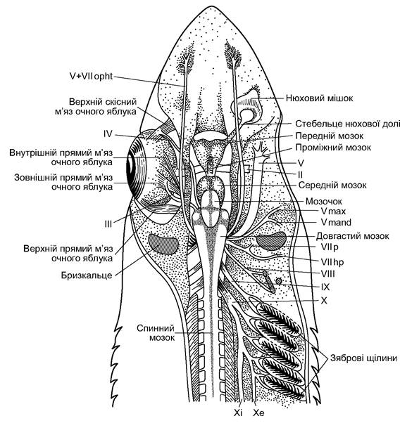
Fig. 4.12. Brain and cranial nerves of a shark. Nerves: II-X - cranial nerves; II - optic; III - oculomotor; IV - trochlear;
Vmax - maxillary branch of the Trigeminal nerve; Vmand - mandibular branch of the trigeminal nerve; V+VIIopht - ophthalmic Branches of the trigeminal and facial nerves; VIIp - palatine branch of the Facial Nerve; VIIhm - branch innervating the hyomandibula; VIII - auditory; IX - glossopharyngeal;
X - vagus; Xi - internal branch of the Vagus nerve; Xe - lateral branch of the vagus nerve
The sympathetic nervous system of cartilaginous fishes, like that of cyclostomes, is represented by a chain of ganglia that do not yet have a direct longitudinal connection (which only appeared in lungfishes and bony fishes).
As previously mentioned, cartilaginous fishes rely most heavily on Sensory systems such as the olfactory organs, the lateral line, and the organs of Vision (Fig. 4.13).

Fig. 4.13. Structure of a shark's eye
The olfactory organs and the lateral line are well developed. Thanks to the lateral line, these animals sense the mechanical displacement of water. On the head of some cartilaginous fishes, there may be clusters of ampullae of Lorenzini (capsules embedded in the skin that open to the outside via a thin canal) (Fig. 4.14).

Fig. 4.14. Diagram of the arrangement of the lateral line organs
Thanks to these ampullae, the fish sense changes in water Temperature (from 0.05°C) and, likely, variations in electric field parameters (from 0.10-0.01 mV/cm).
Some representatives of cartilaginous fishes, particularly from the order of electric rays, possess organs that generate electric fields as well as electric discharges. These electric organs are structurally modified regions of striated muscles (Fig. 4.15). Muscle fibers are transformed into electroplaques, separated by gelatinous connective tissue.
The lower surface of the electroplaque accumulates negative charges, while the upper surface accumulates positive ones. Stacks of 30-40 rows of electroplaques form columns separated from each other by Fibrous connective tissue. Each electric organ consists of 350-600 columns. It is innervated by powerful branches of the cranial nerves (the seventh, ninth, and tenth pairs). The electric organ operates on THE PRINCIPLE OF a Leyden jar, accumulating electric potential generated during METABOLISM in the electroplaques. Discharges in some rays can reach 60-300 V, and the current strength can reach 5 A. Within a few seconds, a ray can generate over a hundred such discharges.

Fig. 4.15. Electric organs of the ray Torpedo marmorata: A - dissected electric organs and brain;
B - STRUCTURE OF THE electric organ (two electroplaques)
Last update: 19/08/2026
Editorial and Educational Adaptation: This material has been compiled based on the primary/original source text. The project team performed an editorial review, corrected technical inaccuracies, structured sections, and adapted the content for an educational format.
What was processed:
- elimination of formatting defects (OCR errors, structural breaks, corrupted characters);
- editorial organization of content;
- standardization of terminology in accordance with academic sources;
- verification of factual statements against the original source text.
All mentions of the author, publication year, and origin of the primary text have been preserved in accordance with the source.