Human Anatomy and Physiology (with Age-Related Features of the Child's Body) - Sapin M.R., Sivoglazov V.I. 2002
Musculoskeletal System
The Study of Bones and Their Joints (Osteoarthrology)
General Anatomy of Bone Joints
All bone connections are divided into three main groups: continuous connections, symphyses (or hemiarthroses), and discontinuous connections (or synovial joints).
Continuous connections of bones are formed by various types of Connective Tissue. These joints are strong and elastic, yet their mobility is limited. Continuous joints are subdivided into fibrous, cartilaginous, and bony connections.
Fibrous connections include syndesmoses, sutures, and gomphoses (dentoalveolar syndesmoses).
Syndesmoses are bone connections mediated by ligaments and membranes of various shapes. Examples include the interosseous membranes of the forearm and leg, the yellow ligaments (ligamenta flava) connecting the vertebral arches, and the ligaments that reinforce joints. Sutures are connections between the edges of Skull bones via thin layers of Fibrous connective tissue. There are serrate sutures (e.g., between the parietal bones), squamous sutures (the junction of the Temporal bone's squamous part with the Parietal bone), and plane sutures (between the BONES OF THE facial skull). A gomphosis refers to The connection between a tooth ROOT and the dental alveolus, where the tooth is essentially "driven into" the socket.
Cartilaginous connections (synchondroses) are joints mediated by Cartilage—for instance, the joints between vertebral bodies and the connections between the Ribs and the Sternum.
Bony connections (synostoses) develop as synchondroses undergo ossification between the epiphyses and diaphyses of long bones, the individual bones of the cranial base, the bones forming the hip bone, and elsewhere.
Symphyses are also a type of cartilaginous connection. Within the mass of cartilage forming them, There is a small slit-like cavity containing a minimal amount of fluid. The Pubic Symphysis is a prime example of a symphysis.
Joints, or synovial joints, represent discontinuous bone connections that are robust and highly mobile. All joints possess the following mandatory anatomical elements: articular surfaces of the bones covered with articular cartilage; the articular capsule; the joint cavity; and synovial fluid (Fig. 13). The articular surfaces are covered with resilient hyaline cartilage; only the temporomandibular and sternoclavicular joints feature fibrous cartilage. The thickness of the articular cartilage ranges from 0.2 to 6.0 mm and is directly dependent on the functional load experienced by the joint: the greater the load, the thicker the cartilage. The articular capsule consists of a dense outer layer—the fibrous membrane—which attaches to the bones near the margins of the articular surfaces, where it transitions into the periosteum. The thin inner layer of the Joint Capsule is formed by the synovial membrane, which features folds and villi that increase its free surface area facing the joint cavity.
The fibrous layer of the articular capsule is thickened in certain areas, forming intracapsular ligaments. Ligaments may also be located outside the capsule, adjacent to it (extracapsular ligaments).
Ligaments reinforce the joint, guide its movements, and restrict excessive motion. They possess exceptional tensile strength. For instance, the tensile strength of the iliofemoral ligament reaches up to 350 kg, while the long plantar ligament can withstand up to 200 kg.
In a healthy living person, the joint cavity normally appears as a narrow slit containing synovial fluid. Even in large joints such as the knee or hip, the volume of this fluid does not exceed 2–3 cm3. The pressure within the joint cavity is subatmospheric.
Articular surfaces rarely match each other in shape perfectly.
To achieve congruence (from Latin congruens – corresponding), joints possess A number of auxiliary structures, such as articular discs, menisci, and labra. For example, the temporomandibular joint features an articular disc fused with the capsule along its outer margin, dividing the joint cavity into two compartments. The knee joint contains crescent-shaped medial and lateral menisci located between the articular surfaces of the Femur and Tibia. Along the rim of the acetabulum in the hip joint, there is a fibrocartilaginous acetabular labrum, which deepens the articular surface on the hip bone to better accommodate the spherical femoral HEAD.
Class="center">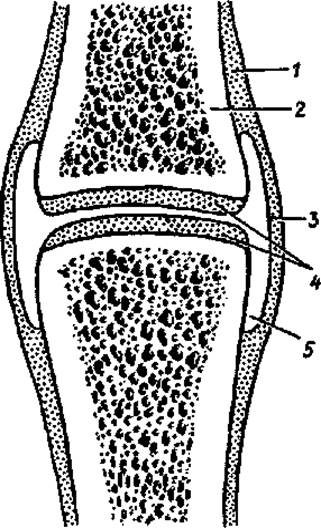
Fig. 13. Diagram of joint Structure: 1 — periosteum, 2 — bone, 3 — articular capsule, 4 — articular cartilage, 5 — joint cavity
Classification of Joints
Depending on the number of articular surfaces participating in The formation of a joint, they are classified as simple (two articular surfaces), complex (more than two articular surfaces), compound, and combined. When two or more anatomically independent joints function together, they are termed combined (e.g., both temporomandibular joints). Complex joints contain an intra-articular disc or menisci between their mating surfaces, which divide the joint cavity into two compartments.
The shape of the articulating surfaces determines the number of axes around which movement can occur. Accordingly, joints are divided into uniaxial, biaxial, and multiaxial joints (Fig. 14).
For convenience, the shape of an articular surface is often compared to a segment of a body of revolution, with each joint having one, two, or three axes of movement. For example, cylindrical and ginglymus (Hinge) joints are uniaxial. Examples of uniaxial cylindrical joints include the median atlantoaxial joint, as well as the proximal and distal radioulnar joints. In a hinge joint, the cylindrical surface features a groove or ridge perpendicular to the cylinder's axis, along with a corresponding depression or protrusion on the opposing articular surface; the interphalangeal JOINTS OF THE hand serve as classic examples. A variation of the hinge joint is the cochlear (screw) joint. The distinction between a screw joint and a hinge joint is that the groove runs in a spiral rather than perpendicularly to the joint axis. The humeroulnar joint is an example of a screw joint.
Ellipsoid, condylar, and saddle joints are biaxial. The radiocarpal joint is ellipsoid in shape. A condylar joint is structurally close to an ellipsoid one, where the articular head resembles an ellipse, but its articular surface is situated on a condyle. For instance, the knee and atlanto-occipital joints are condylar (with the former also being complex and the latter combined).
The articular surfaces of a saddle joint resemble two "saddles" with axes intersecting at a right angle. The carpometacarpal joint of the thumb is a saddle joint; it is unique to humans and allows for the opposition of the thumb to the other digits. The evolutionary adaptation of this joint into a typical saddle shape is closely linked to labor activity.
Spheroidal (ball-and-socket) and plane joints are multiaxial. In addition to movement along three axes, multiaxial joints are capable of circumduction (circular motion). The shoulder and hip joints are prime examples of multiaxial joints, with the latter being considered a cotyloid (cup-shaped) joint due to the significant depth of its socket.
Plane joints also belong to the multiaxial category. A flat articular surface represents a segment of a very large sphere. Movements in plane joints can occur around three axes, but they are of a very limited range. Examples include the intercarpal and tarsometatarsal joints.
Movements in joints are determined by the shape of their articular surfaces. Movements around the frontal axis include flexion and extension (occurring in the sagittal plane); around the sagittal axis—adduction and abduction (occurring in the frontal plane); and around the vertical (longitudinal) axis—rotation.
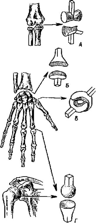
Fig. 14. Schematic representation of articular surfaces. Joints: A — hinge joint, B — ellipsoid joint, C — saddle joint, D — ball-and-socket joint
The range of motion in a joint depends on the congruence between the articulating surfaces. The greater the conformity, the more limited the mobility within the joint (e.g., the hip joint); conversely, the less the articular surfaces match one another, the greater the mobility in such a joint (e.g., the shoulder joint).
The degree of joint mobility is determined by the difference in the angular dimensions of the articular surfaces of the articulating bones. For instance, if the angular span of the glenoid cavity is 150° and that of the articular head is 230°, the arc of potential movement equals 80°. The greater the difference in curvature between the articular surfaces, the wider the potential range of motion in that joint.
Joint mobility is also influenced by the tension of the joint capsule, the ligamentous apparatus, muscular development and elasticity, as well as sex and age characteristics, The Nature of labor, and the type of sport.
Age-related and functional changes in bone connections
Joints (synovial joints) begin to form during the 6th to 11th weeks of embryonic development. During this period, the articular surfaces of the articulating bones, the joint cavity, and other joint structures begin to take shape.
Newborns already possess all the anatomical elements of a joint. However, the epiphyses of the articulating bones consist of cartilage; endochondral ossification for most of them begins after birth (during the 1st–2nd years of life) and continues until adolescence. Between the ages of 6 and 10, The structure of the synovial membrane and joint capsule becomes more complex, the number of villi and folds increases, and the vascular networks and nerve endings of the synovial membrane are formed. In the fibrous layer of the joint capsule in children aged 3 to 8, the number of Collagen fibers increases and they thicken significantly, ensuring its structural integrity. The final formation of all joint structures is completed by the age of 13–16. Under conditions of normal physiological activity, joints retain a constant range of motion for a long time and are minimally prone to Aging. With prolonged and excessive mechanical loads, as well as with age, Structural and functional changes occur in the joints: the articular cartilage thins, the fibrous membrane of the joint capsule and ligaments undergo sclerosis, and bony projections—osteophytes—form along the periphery of the articular surfaces. These anatomical alterations lead to functional impairments, manifested by restricted mobility and a reduced range of motion.
Review and Self-Assessment Questions:
1. Name the Organs belonging to the passive and active parts of The Musculoskeletal System. What is the basis for this classification?
2. Which organs (structures) are referred to as the rigid Skeleton and the soft skeleton, and why?
3. What do you know about The chemical composition of bones and their mechanical properties?
4. Describe the Classification and Structure of each type of bone.
5. Discuss the various modes of Bone Development and formation.
6. What age-related features of bone Structure and function are you familiar with?
7. What are the Types of bone connections? Provide their characteristics.
8. Describe the anatomical and biomechanical classifications of bone joints.
9. What can you tell about the age-related and functional features of Bone Articulations?
Last update: 10/08/2026
Editorial and Educational Adaptation: This material has been compiled based on the primary/original source text. The project team performed an editorial review, corrected technical inaccuracies, structured sections, and adapted the content for an educational format.
What was processed:
- elimination of formatting defects (OCR errors, structural breaks, corrupted characters);
- editorial organization of content;
- standardization of terminology in accordance with academic sources;
- verification of factual statements against the original source text.
All mentions of the author, publication year, and origin of the primary text have been preserved in accordance with the source.