Human Anatomy and Physiology (with Age-Related Features of the Child's Body) - Sapin M.R., Sivoglazov V.I. 2002
Musculoskeletal System
The Study of Bones and Their Joints (Osteoarthrology)
General Anatomy of the Skeleton
The Musculoskeletal System (the apparatus of support and movement) comprises bones, bone joints, and Muscles. Its primary function is not only to provide structural support, but also to enable the locomotion of the body and its parts in space. The musculoskeletal system is divided into passive and active components. Bones and Their Joints make up the passive component, while muscles form the active component, driving the skeletal bones into motion through their ability to contract.
The Skeleton (from the Greek sceleton, meaning dried up) is a complex of bones varying in shape and size. The human skeleton includes the BONES OF THE trunk, HEAD, upper limbs, and lower limbs (Fig. 11). The bones are interconnected by various TYPES OF JOINTS and serve Functions of support, movement, protection, and mineral storage (acting as a depot for various salts). The bony skeleton is also referred to as the hard or rigid skeleton.
Class="center">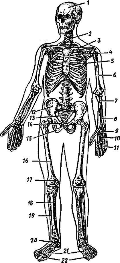
Fig. 11. Human skeleton. Anterior view:
1 — Skull, 2 — Vertebral Column, 3 — clavicle, 4 — rib, 5 — Sternum, 6 — humerus, 7 — radius, 8 — ulna, 9 — Carpal Bones, 10 — Metacarpal bones, 11 — Phalanges of the hand, 12 — ilium, 13 — sacrum, 14 — pubis, 15 — ischium, 16 — Femur, 17 — Patella, 18 — Tibia, 19 — Fibula, 20 — Tarsal Bones, 21 — Metatarsal Bones, 22 — phalanges of the FOOT
The supporting function of the skeleton lies in the fact that the bones, together with their connections, form the structural framework of the entire body, to which soft Tissues and Organs are attached. Soft tissues such as ligaments, fasciae, capsules, and organ stroma are collectively known as the soft skeleton, as they also perform mechanical functions (attaching organs to the hard skeleton, supporting the organ stroma, and protecting them).
The supporting and locomotor Functions of the skeleton are complemented by the Shock-absorbing (spring) function of articular cartilages and other structural adaptations (such as the arches of the foot), which mitigate jolts and impacts.
The protective function is manifested in The formation of bony cavities that house vital organs: the skull protects the Brain, THE VERTEBRAL COLUMN protects the Spinal Cord, and The thoracic cage protects The Heart, Lungs, and major Blood Vessels. The pelvic cavity contains the reproductive organs. Inside the bones lies the Bone Marrow, which gives rise to blood and immune system Cells.
Support and movement are made possible by the architecture of bones, which act as long and short levers that are movably connected to one another, actuated by muscles, and controlled by The Nervous system. Additionally, bones determine The pathway of blood Vessels and nerves, as well as body shape and proportions.
Bones serve as a reservoir (depot) for salts of phosphorus, calcium, iron, magnesium, copper, and Other Compounds, thereby maintaining the constancy of the mineral composition in the body's internal environment,
The skeleton consists of 206 bones (85 paired and 36 unpaired). The mass of the "living" skeleton in newborns is about 11% of body weight, and ranges from 9 to 18% in children of various ages. In adults, The ratio of skeletal mass to body weight remains at approximately 20% until advanced and old age, after which it slightly decreases.
Each bone, functioning as an organ, is composed of various types of tissue, with Bone tissue—a specialized form of Connective Tissue—playing the predominant role.
The chemical composition of bone is complex, consisting of both organic and inorganic substances. Inorganic substances make up 65–70% of the dry bone mass and are represented primarily by Calcium and phosphorus salts. In small quantities, bone contains over 30 other various elements. Organic substances, known collectively as ossein, account for 30–35% of the dry bone mass and comprise bone cells and Collagen fibers. The elasticity and resilience of a bone depend on its organic constituents, whereas its hardness is attributed to mineral salts. The combination of inorganic and organic substances in living bone grants it extraordinary strength and flexibility. In terms of hardness and elasticity, bone can be compared to copper, bronze, or cast iron. In youth and childhood, bones are more elastic and resilient because they contain a higher proportion of organic substances and fewer inorganics. In elderly and aged individuals, inorganic substances predominate, rendering the bones more brittle.
Every bone comprises dense (compact) and spongy (cancellous) bone substance. The distribution of compact and spongy substance depends on the bone's Location in the body and its specific function.
Compact bone substance is found in those bones and regions of bones that perform primarily supporting and locomotor functions, such as the shafts (diaphyses) of long bones.
In areas where a large volume must remain lightweight yet durable, spongy bone substance is formed, such as in the epiphyses of long bones.
Spongy substance is also located in short (cancellous) and flat bones. Within these bones, bony plates form intersecting trabeculae (beams) of varying thickness oriented in multiple directions. The spaces between these trabeculae (marrow cells/areolae) are filled with Cytology/practical/86.html">Red bone marrow (see "Immune System"). In long bones, bone marrow resides within the central channel known as the medullary cavity. In adults, a distinction is made between red and yellow bone marrow. Red bone marrow fills the spongy substance of flat bones and the epiphyses of long bones, whereas yellow bone marrow (fatty marrow) is located in the diaphyses of long bones.
The entire bone, with the exception of articular surfaces, is covered by a connective tissue membrane called the periosteum. The articular surfaces of the bone are covered with articular Cartilage.
Classification of Bones
Bones are classified into long (tubular), short (tubular), spongy, flat, irregular (mixed), and pneumatic bones (Fig. 12).
Tubular bones are those located in Regions of the skeleton where movements with a wide range occur (e.g., in the limbs). A tubular bone consists of an elongated middle portion—either cylindrical or three-sided—known as the body or diaphysis, and thickened ends known as the epiphyses. The epiphyses feature articular surfaces covered with articular cartilage, which serve for articulation with adjacent bones. The region of the bone located between the diaphysis and epiphysis is called the metaphysis. Tubular bones are further divided into long tubular bones (e.g., the humerus, femur, and bones of the forearm and leg) and short tubular bones (metacarpals, metatarsals, and phalanges). The diaphyses are constructed of compact bone, while the epiphyses are made of spongy bone covered by a thin layer of compact bone.
Spongy (short) bones consist of spongy substance enclosed by a thin layer of compact substance. Spongy bones typically have the shape of an irregular cube or polyhedron. Such bones are located in areas where heavy mechanical loads are combined with high mobility, such as the carpal and tarsal bones.
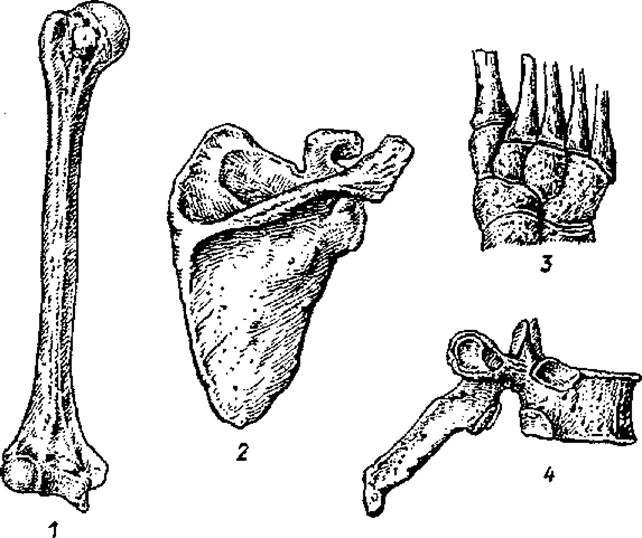
Fig. 12. Various types of bones:
1 — long (tubular) bone, 2 — flat bone, 3 — spongy (short) bones, 4 — irregular bone
Flat bones consist of two plates of compact bone tissue enclosing a layer of spongy bone tissue. These bones contribute to forming the walls of Body Cavities and limb girdles, and serve a protective function (bones of the cranial vault, sternum, Ribs).
Irregular bones have a complex shape. They consist of several parts with different structures, such as the vertebrae and the bones of the skull base.
Pneumatic bones contain a mucous membrane-lined cavity filled with air within their body. Examples include the frontal, sphenoid, ethmoid, and Maxilla bones.
Development and Growth of Bones
During human ontogenesis, most skeletal bones sequentially pass through three developmental stages: membranous, cartilaginous, and osseous. The so-called dermal bones (bones of the cranial vault, face, and clavicle) bypass the cartilaginous stage.
Initially, the human skeleton is represented by embryonic connective tissue, or mesenchyme, which condenses at the sites of future bones (membranous stage of skeletal development). Where the dermal bones will form, one or more centers of ossification appear within the membranous skeleton. These islets of bone cells, derived from the mesenchyme, expand outward to form the dermal bones. This direct Development of Bones from mesenchyme, bypassing the cartilaginous stage, is termed direct osteo-genesis or desmal ossification (from Greek desma — band, tissue). Bones formed in this manner are called primary bones.
The bones of the trunk and limbs undergo all three Selection/3.html">Stages of development: membranous, cartilaginous, and osseous. First, cartilaginous primordia of future bones appear within the embryonic connective tissue (mesenchyme) of the membranous skeleton during the second week of development (cartilaginous stage of skeletal development). Then, beginning around the 8th week of intrauterine life, the cartilage tissue at the sites of future bones starts to be replaced by bone tissue. The first bone cells and centers of ossification appear in the diaphyses of tubular bones. The formation of bone tissue in place of cartilaginous models can occur in three ways: perichondral, periosteal, and endochondral ossification.
Perichondral ossification involves the gradual transformation of the perichondrium into the periosteum. The inner layer of the perichondrium begins to produce young bone cells (osteoblasts) instead of cartilage cells. Osteoblasts deposit onto the cartilage model, forming a bone collar that gradually replaces the cartilage breaking down beneath it.
Periosteal ossification (Bone Formation) occurs when the newly formed periosteum produces young bone cells that are deposited onto the underlying bone via apposition. Through this mechanism, the compact bone plate gradually thickens.
Endochondral ossification takes place when bone tissue forms within the cartilage. Blood vessels and connective tissue sprout from the periosteum into the cartilage, causing the cartilage in these areas to break down. Some of the cells from the invading connective tissue differentiate into osteogenic cells, which proliferate in cords to form the spongy bone substance deep within the bone.
The diaphyses of tubular bones ossify during the prenatal period. The ossification centers that appear in them are called primary. The epiphyses of tubular bones begin to ossify either immediately before birth or during postnatal human life. These centers, formed within the cartilaginous epiphyses, are termed secondary ossification centers. Bone tissue in the epiphyses is formed through endochondral, perichondral, and periosteal mechanisms. However, a cartilaginous plate (epiphyseal plate) persists for quite some time at the junction between the epiphyses and diaphysis; it is replaced by bone tissue between the ages of 16 and 24, causing the epiphyses to fuse with the diaphyses. Tubular bones grow in length thanks to the epiphyseal plate. Once these plates are replaced by bone tissue, longitudinal bone growth ceases.
There are also accessory ossification centers (apophyses) that develop in future protuberances and processes (epicondyles, trochanters) and gradually fuse with the main bone.
Age-Related Changes in Bones
Throughout a person's postnatal life, skeletal bones undergo significant age-related changes. For instance, in a newborn infant, bone tissue has not yet replaced the cartilaginous models in many areas. During the first year of life, bones grow slowly; from ages 1 to 7, longitudinal bone growth accelerates due to the epiphyseal cartilages, while thickness increases through appositional thickening of the compact bone tissue driven by the osteogenic function of the periosteum. After 11 years of age, skeletal bones resume rapid growth, bone processes (apophyses) take shape, and the medullary cavities acquire their final form. In elderly and senile individuals, the spongy bone exhibits a reduced number and thinning of bone trabeculae, while the compact bone in the diaphyses of tubular bones becomes thinner.
Bone GROWTH AND DEVELOPMENT are influenced by social factors, particularly Nutrition. Any deficiency in nutrients, salts, or Metabolic Disorders affecting Protein Synthesis immediately impacts bone growth. For instance, a Vitamin C Deficiency impairs the synthesis of organic components in the bone matrix, rendering tubular bones thin and fragile. Bone growth relies on normal calcification processes, which depend on adequate calcium and phosphorus levels in the blood and tissue fluid, as well as a sufficient supply of vitamin D. Thus, normal bone growth requires the balanced and normal progression of both calcification and protein synthesis—two processes that typically proceed synchronously and harmoniously in The Human Body.
Impaired nutrition and METABOLISM cause alterations in the spongy and compact bone tissue of the adult Skeletal System. Throughout life, the remodeling of osteons (Haversian systems) continuously occurs within the bones.
Bone modifications occur under The Influence of physical loading. Under high mechanical stress, bones generally become more massive, and prominent thickenings—such as bony prominences, tuberosities, and crests—develop at the sites of tendon attachment. Static and dynamic loads trigger internal structural remodeling of the compact bone (increasing the number and size of osteons), thereby enhancing bone strength. Appropriately dosed physical activity slows down the skeletal Aging process.
Last update: 10/08/2026
Editorial and Educational Adaptation: This material has been compiled based on the primary/original source text. The project team performed an editorial review, corrected technical inaccuracies, structured sections, and adapted the content for an educational format.
What was processed:
- elimination of formatting defects (OCR errors, structural breaks, corrupted characters);
- editorial organization of content;
- standardization of terminology in accordance with academic sources;
- verification of factual statements against the original source text.
All mentions of the author, publication year, and origin of the primary text have been preserved in accordance with the source.