FUNDAMENTALS OF MICROALGAE BIOTECHNOLOGY - D. S. DVORETSKY - 2015
2. FUNDAMENTALS OF MICROALGAE BIOCHEMISTRY
2.1. Photosynthesis
Photosynthesis is a process in which the electromagnetic energy of the Sun is captured by the plant photosynthetic apparatus—chlorophyll and accessory pigments—and utilized to reduce atmospheric carbon dioxide into complex Organic compounds [5]. The overall equation of photosynthesis can be written as follows:
Class="center">
Photosynthesis is a sequence of redox reactions resulting in the reduction of carbon dioxide to organic matter. All photosynthetic reactions are generally divided into two phases: the light-dependent and light-independent (dark) phases.
The METABOLISM/16.html">Light-dependent phase of photosynthesis. This phase takes place in the thylakoid membranes with the participation of chlorophyll and other pigments, the ATP synthase enzyme embedded in the thylakoid membrane, and carrier Proteins.
A key feature of the light-dependent phase is that solar radiation energy absorbed by chlorophylls is first converted into electrochemical energy and subsequently into the energy of high-energy ATP bonds. This is achieved through The transport of electrons and hydrogen ions via specialized carriers across the thylakoid membrane (Fig. 5).
Fig. 5. Scheme of photosynthesis (arrows indicate the transport of protons and electrons)
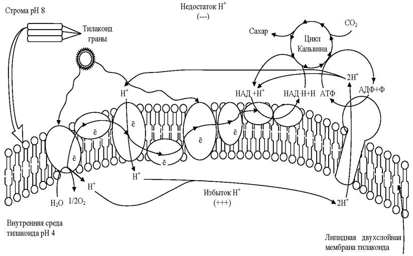
The light-dependent phase is further subdivided into photophysical and photochemical stages. In the photophysical stage, light quanta are absorbed by chlorophyll molecules P700 (Photosystem I) and P680 (Photosystem II), raising these molecules to an excited state.
In the photochemical stage, both Photosystems function in a coordinated manner.
Photosystem I. The excited P700 molecule transfers an electron to an acceptor. From there, via a chain of carriers, the electron is delivered to the outer side of the thylakoid (facing the stroma). Concurrently, the P700 molecule is oxidized, converting P700 into P+700:

Photosystem II. The excited P680 molecule transfers an electron to an acceptor. From there, via a chain of carriers, the electron is transferred to photosystem I to reduce the P700 molecule. As a result, P700 returns to its initial state and regains The ability to absorb light and become excited.
Meanwhile, by releasing an electron, the P680 molecule is converted into P+680. It is reduced back using electrons stripped from Water molecules through a process known as photolysis, or photooxidation:
2Н2O - 4е- —> 4Н+ + O2.
Due to photolysis, protons accumulate inside the thylakoid and molecular oxygen is generated, which then diffuses into the atmosphere. Oxygen is a byproduct of photosynthetic reactions.
Protons produced during photolysis cannot cross the thylakoid membrane and thus accumulate inside, forming an H+ reservoir. Consequently, protons and electrons accumulate on opposite sides of the membrane, generating an electrochemical Membrane Potential. When this potential reaches 200 mV, the electric field forces protons through the channels of the ATP synthase enzyme embedded in the thylakoid membrane, initiating the operation of the proton pump. A high energy level is generated at the exit of the proton channel, which is utilized to phosphorylate ADP molecules present in the chloroplast matrix:

The transported protons interact with electrons and a NADP molecule (the coenzyme nicotinamide adenine dinucleotide phosphate) to form the NADP•H + H+ complex:
НАДФ+ + 2е- + 2Н+ —> НАДФ•Н + Н+.
Thus, electrons activated by light energy are used to attach a hydrogen atom to its carrier, i.e., to reduce NADP+ to NADP•H + H+.
Ultimately, the light-dependent phase of photosynthesis yields ATP, reduced NADP coenzyme, and oxygen as a byproduct. The resulting ATP and reduced NADP coenzyme are then utilized in the light-independent phase of photosynthesis.
The light-independent (dark) phase of photosynthesis occurs concurrently with the light-dependent phase. This complex process, taking place in the chloroplast stroma without the direct absorption of light, involves numerous reactions that reduce CO2 to organic compounds utilizing the energy of ATP and NADP•H+H+ synthesized during the light-dependent phase. There are various reduction pathways, the primary one being the so-called C3 pathway, or The Calvin Cycle (Fig. 6).
Fig. 6. The Calvin cycle

The reductive pentose phosphate cycle, or Calvin cycle, is a series of biochemical reactions that occur during photosynthesis in photo- and chemosynthetic organisms (plants, Algae, purple Bacteria, cyanobacteria, etc.). It represents the most widespread (though not the sole) mechanism of autotrophic carbon dioxide assimilation. The American biochemist Melvin Calvin was awarded the Nobel Prize for discovering the chemical pathway of carbon dioxide assimilation by plants during photosynthesis.
The Calvin cycle consists of three stages:
1. Carboxylation. CO2 is added to ribulose-1,5-bisphosphate in a reaction catalyzed by the enzyme ribulose-1,5-bisphosphate carboxylase-oxygenase (RuBisCO). This protein constitutes the major fraction of chloroplast proteins and is presumably the most abundant enzyme in nature. The reaction yields an unstable intermediate compound that immediately breaks down into two molecules of 3-phosphoglyceric acid (PGA).
2. Reduction of PGA. First, PGA is phosphorylated by ATP in a reaction catalyzed by phosphoglycerate kinase to form 1,3-bisphosphoglyceric acid (BPGA). Subsequently, in the presence of triose phosphate dehydrogenase and NADPH, the acyl-phosphate group of BPGA is dephosphorylated and reduced to an aldehyde group, yielding glyceraldehyde 3-phosphate (G3P), a phosphorylated carbohydrate.
3. Regeneration of the CO2 acceptor. This stage involves 5 molecules of G3P which, through The formation of 4-, 5-, 6-, and 7-carbon intermediates, are reorganized to regenerate 3 molecules of ribulose-1,5-bisphosphate, consuming 3 ATP in the process.
The synthesis of a single glucose molecule requires two molecules of G3P, 6 molecules of CO2, 12 molecules of NADPH + H+, and 18 molecules of ATP across 6 turns of the Calvin cycle.
Thus, during the light-independent phase of photosynthesis, the energy of high-energy ATP bonds is converted into the chemical energy of organic compounds—effectively trapping and conserving energy within organic chemical bonds.
The rate of photosynthesis is influenced by various environmental factors, including light intensity, water availability, mineral nutrients, Temperature, and CO2 concentration.
The uniqueness and overarching Biological Significance of photosynthesis stem from the fact that all life on our planet owes its existence to this process. It serves as the primary source of primary organic matter and remains the sole source of free oxygen on Earth. Atmospheric oxygen gave rise to and maintains the ozone layer, which shields living organisms from shortwave ultraviolet radiation. Furthermore, photosynthesis plays a key role in regulating atmospheric CO2 levels.
Major contributions to the study and elucidation of the photosynthetic mechanism were made by the Russian scientist K. A. Timiryazev, the American researcher M. Calvin, the Australian scientists M. D. Hatch and C. R. Slack, as well as the Belarusian academicians T. N. Godnev and A. A. Shлык [Note: Shlyk].
2.2. Energy Metabolism in Cells
Energy metabolism in the cells of All living organisms involves the release of potential energy stored in the chemical bonds of organic or inorganic substances. Locked within these chemical bonds, this energy is not directly accessible for immediate cellular use—not even in microalgae and plants, which originally converted this energy from light into chemical form. Therefore, the potential energy of organic molecules must be released and transformed into a usable form [5, 15].
The generation and accumulation of cellularly accessible energy occurs through The process of cellular Respiration. To carry out cellular respiration, most organisms require oxygen—a process known as aerobic respiration or aerobic energy release. However, certain organisms can derive energy from nutrients without utilizing free atmospheric oxygen, a process referred to as anaerobic respiration (anaerobic energy release).
Thus, the starting Materials for respiration are energy-rich organic molecules whose formation previously required an energy input. Glucose serves as the primary substance utilized by cells for energy generation.
Aerobic (oxygen-dependent) respiration. Aerobic respiration can be conditionally divided into several sequential stages. The First stage is the preparatory stage, or digestive stage, which involves The breakdown of polymers into monomers. These processes take place within the Digestive System of animals or the Cytoplasm of cells. No Energy is stored in ATP molecules at this stage.
The next stage is the oxygen-free, or incomplete, stage. It occurs in the Cell Cytoplasm without the involvement of oxygen.
During this stage, the respiratory substrate undergoes enzymatic breakdown. A prime example is Glycolysis, a multi-step anaerobic breakdown of glucose. The intermediate steps of glycolysis yield three-carbon intermediates utilized in The Biosynthesis of various compounds.
In the reactions of glycolysis, a six-carbon glucose molecule (C6) is split into two molecules of pyruvic acid (C3). In this process, four hydrogen atoms are removed from each glucose molecule, and two ATP molecules are formed. The hydrogen atoms bind to the carrier NAD (nicotinamide adenine dinucleotide), converting it into its reduced form NADH + H+ (NAD is structurally very similar to NADP, the hydrogen carrier in photosynthesis).
The overall reaction of glycolysis can be represented as follows:
С6Н12O6 + 2АДФ + 2Н3РO4 + 2НАД+ —> 2С3Н4O3 + 2АТФ + 2ННА • Н + Н+ + Н2O.
The net energy yield of this stage is two ATP molecules, while 40–60% of the released energy is dissipated as heat.
The most critical phase is the oxygen-dependent stage of aerobic respiration, which strictly requires the presence of oxygen. The end product of glycolysis, pyruvic acid, retains a substantial portion of its energy, and its further release takes place within the Mitochondria.
Under aerobic conditions, pyruvic acid undergoes decarboxylation (losing CO2 molecules), binds to specific coenzyme A to form the acetyl-CoA complex, and enters the Krebs cycle.
The Tricarboxylic Acid Cycle, or Krebs cycle, is a core stage of energy metabolism in all aerobic organisms. It not only releases and stores significant amounts of energy within cells, but also drives the synthesis of precursor molecules involved in other biochemical transformations, producing vital compounds such as Amino Acids and Fatty acids. The tricarboxylic acid cycle was discovered by the German biochemist Hans Krebs, for which he was awarded the Nobel Prize in 1953.
Complete combustion of an energy substrate implies The oxidation of an acetyl group attached to coenzyme A (acetyl-CoA) down to CO2 and water.
In eukaryotic organisms, all Reactions of the Krebs cycle take place inside the mitochondria. This cycle consists of eight sequential reactions (Fig. 7).
The tricarboxylic acid cycle begins with the attachment of acetyl-CoA to oxaloacetate (the salt of oxaloacetic or ketosuccinic acid) to form citrate, a tricarboxylate salt. Subsequently, citrate undergoes a series of consecutive transformations accompanied by two decarboxylation steps—that is, the release of CO2—ultimately leading to the regeneration of oxaloacetate.
As can be seen, one full turn of the cycle, consisting of eight enzymatic reactions, results in the Complete oxidation of a single molecule of acetyl-CoA.
Fig. 7. The Krebs cycle

Overall equation of the tricarboxylic acid cycle:
2С3Н4О3 + 6Н2О + 8НАД- + 2ФАД+ —> 6СО2 + 8НАД • Н2 + 2ФАД • Р + 2ААТФ.
Carbon dioxide is released from the mitochondria into The Cell cytoplasm and then into the external environment.
For the cycle to run continuously, a steady supply of acetyl-CoA is required, and the Coenzymes (NAD and FAD) that have been reduced must be repeatedly reoxidized. This oxidation is carried out by The electron transport system (or Respiratory Chain) localized within the mitochondria.
Hydrogen atoms accepted by NAD and FAD (the coenzyme flavin adenine dinucleotide) enter a chain of reactions whose ultimate outcome is the synthesis of ATP. ATP synthesis is driven by a process known as Oxidative Phosphorylation, which proceeds in the following sequence (Fig. 8):
✵ hydrogen atoms are split off from NAD and FAD and captured by carriers embedded in The inner mitochondrial membrane, where they undergo oxidation:
Н —> Н+ + е-;
✵ H+ ions are transported by carriers to the outer surface of the cristae and accumulate in the intermembrane space, forming a proton reservoir;
✵ electrons (e-) from the hydrogen atoms return via the respiratory chain into the matrix and attach to oxygen atoms that are continuously supplied to the mitochondria, rendering the oxygen atoms negatively charged:
1/2 О2 + е- —> О2- ;
✵ a potential difference arises across the membrane. Once this potential difference reaches 200 mV, the proton channel within the ATP synthase enzyme molecules embedded in the inner membrane becomes active;
✵ through the proton channel, H- protons rush back into the mitochondrial matrix, generating a high level of energy—most of which is channeled into synthesizing ATP from ADP and phosphoric acid—while the protons themselves combine with the negatively charged oxygen species to form water, the secondary end product of cellular respiration:
36АДФ + 36Н3РO4 + Е —> 36АТФ;
24Н+ + 12O2- —> 12Н2O.
Fig. 8. Diagram of proton and electron Transport Across the inner mitochondrial membrane during the aerobic phase of cellular respiration (Electron Transport Chain)
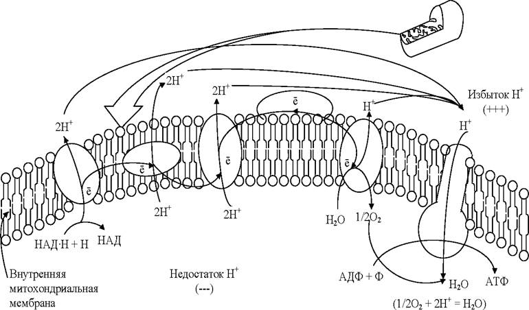
Thus, the oxygen delivered to the mitochondria is essential for accepting electrons and subsequently protons. In the absence of oxygen, the processes associated with proton and electron transport in the mitochondria halt; consequently, even the anaerobic phase cannot proceed because all hydrogen carriers remain fully loaded.
Aerobic respiration, encompassing both anaerobic and aerobic phases, can be expressed by the overall equation
С6Н12O6 + 6O2 + 6Н2O + 38АДФ + 38Н3РO4 —> 6СO2 + 12Н2O + 38АТФ.
The breakdown of a glucose molecule releases 200 kJ/mol and generates 38 ATP molecules. Of this energy, 55% is stored in ATP, while the rest is dissipated as heat.
In the absence or deficiency of oxygen—which serves as the final electron acceptor in aerobic respiration—the electron transport chain across the membrane cannot function. Consequently, no proton gradient is established to power ATP synthesis. Under these conditions, cells can still produce ATP by breaking down nutrients via Anaerobic respiration. This pathway is utilized by many bacteria, microscopic Fungi, and Protozoa. Certain cells that occasionally experience oxygen shortages (such as Muscle or plant cells) are also capable of anaerobic respiration.
Anaerobic respiration is an evolutionarily earlier and energetically less efficient method of harvesting energy from nutrients compared to aerobic respiration.
Anaerobic respiration is driven by a process in which glucose is broken down into pyruvic acid, releasing hydrogen atoms. The pyruvic acid itself acts as the hydrogen acceptor, being converted into lactic acid. The pathway of anaerobic respiration can be represented by the following chemical equations:

This process is known as Lactic acid Fermentation. It is carried out by lactic acid bacteria (such as cocci of the genus Streptococcus). Lactic acid is produced in this manner in animal cells as well, under conditions of oxygen deprivation.
Another widespread natural process is Alcoholic Fermentation, performed by Yeasts. When oxygen is absent, Yeast cells convert glucose into ethanol and CO2. The Initial Stages of alcoholic fermentation mirror those of lactic acid fermentation, but the final reactions yield ethyl alcohol. A CO2 molecule is cleaved from each pyruvic acid molecule, producing a two-carbon compound—acetaldehyde—which is then reduced to ethanol by hydrogen atoms. The overall equation is:
С6Н12O6 + 2АДФ + 2Н3РO4 —> 2С2H5OH + 2АТФ + 2СO2.
Aside from yeasts, alcoholic fermentation is carried out by certain anaerobic bacteria. This type of fermentation is also observed in plant cells when oxygen is lacking.
Glucose is the most common nutrient utilized for the anaerobic release of energy. However, it should be noted that virtually any organic compound can serve as an energy source for ATP synthesis under the appropriate conditions.
When intracellular glucose levels are low, fats and Proteins can be channeled into cellular respiration. The resulting products of fermentation include various organic acids (lactic, butyric, formic, and acetic), alcohols (ethyl, butyl, and amyl), acetone, as well as carbon dioxide and water.
2.3. Nutritional Requirements of Microalgae
For GROWTH AND REPRODUCTION, microalgae require nutrients that satisfy their demands for matter and energy. The primary chemical elements accounting for 98% of cellular structures are carbon, nitrogen, oxygen, and hydrogen, often referred to as organogenic elements. In addition, microalgal cells require phosphorus, potassium, sulfur, sodium, magnesium, and others. For normal growth and metabolic activity, all these elements must be steadily supplied to the cell in precise quantities. The chemical form in which these elements are present in the nutrient medium—organic or mineral—and whether they can be assimilated by microorganisms is also crucial. Like most plant organisms, microalgae are autolithotrophs in terms of their nutritional requirements [8, 16].
Let us briefly examine the core nutritional requirements of microalgae [8].
Carbon sources. Carbon is a vital element, making up approximately 50% of the biomass. Because microalgae are autotrophic organisms, they are capable of utilizing carbon dioxide as their sole carbon source. Furthermore, intensive microalgal growth does not require forced injection of carbon dioxide into the nutrient medium.
Nitrogen sources. Most microalgae exhibit a protein-oriented metabolism and therefore demand substantial amounts of nitrogen during cultivation. Biosynthetic processes largely depend on how well the cells are supplied with this element. Depriving the nutrient medium of nitrogen rapidly slows down microalgal growth. Once intracellular nitrogen reserves are depleted, biomass synthesis halts entirely, Protein Synthesis drops, and CARBOHYDRATES (up to 50%) and Lipids (up to 80%) accumulate intensively [8]. By regulating the duration of algae cultivation in a nitrogen-free medium, biomass with varying ratios of proteins, carbohydrates, and lipids can be obtained. Maximizing Protein Biosynthesis typically requires a higher nitrogen supply than achieving peak biomass productivity. Depending on the microalgal species, potassium nitrate, ammonium sulfate, ammonium nitrate, ammonium bicarbonate, and urea are used as nitrogen sources.
Phosphorus sources. Phosphorus is a fundamental mineral nutrient that plays a crucial role in metabolism. In photoautotrophs, phosphorus participates in the biochemical reactions of nitrogen and carbon metabolism. Phosphates form buffer systems within cells, regulating the pH of the medium. Phosphorus is also a constituent of compounds that make up the protoplasm and nucleus. Algae predominantly utilize phosphorus in the forms of PO3-4, HPO2-4, and H2PO-4, though certain algae, including Scenedesmus and Chlorella, can assimilate phosphorus from organic compounds. The uptake of phosphorus by microalgae is an energy-requiring process and thus depends on photosynthesis and respiration. In phosphorus-starved cells, absorbed phosphates are rapidly converted into reserve nutrients in the form of polyphosphates.
A shortage of phosphorus in Chlorella primarily impairs assimilation processes, whereas Cell Division usually proceeds normally. Under phosphorus deficiency, Chlorella, Anabaena, and Pediastrum exhibit an increased relative carbohydrate content, accompanied by a slight decrease in lipid quantity and shifts in their fatty acid composition. Conversely, high environmental phosphorus concentrations lead to peak intracellular levels of thiamine, biotin, riboflavin, pyridoxine, nicotinic acid, and carotene in microalgae (Chlorella, Scenedesmus, Ankistrodesmus). Typically, synthesizing 1 kg of dry chlorella biomass consumes 15–18 g of phosphorus.
Potassium sources. Potassium is an essential nutrient for microalgae. Within the cell, potassium largely dictates the colloidal and Chemical properties of the cytoplasm as well as cellular functionality by regulating internal compounds. Potassium uptake by microalgae is an active metabolic process linked to the Utilization of Light energy under autotrophic conditions. Potassium deficiency inhibits photosynthesis and accelerates respiration in microalgae, while disrupting carbohydrate utilization and thereby increasing their intracellular levels. Chlorella develops normally across a wide range of potassium concentrations; growth inhibition in continuous intensive cultures is observed only at 6200 mg/L of the element (supplied as KNO3) in the medium. Potassium deficiency manifests as cell agglutination and enlargement, signaling delayed division, alongside aberrant cell division forms and plasmolysis. Potassium is frequently added to the medium in amounts exceeding the microalgal culture's actual requirements; this is due to The Use of potassium nitrate (typically dosed according to nitrogen needs), potassium phosphates (used to boost medium buffering capacity), and potassium hydroxide (for nutrient solution neutralization).
Magnesium sources. Magnesium is a core component of chlorophyll and various other organic compounds. Bound to cellular organic matter, magnesium participates in numerous enzymatic processes, playing a particularly vital role in photosynthesis and its coupled reactions. When magnesium is deficient in the nutrient medium, Chlorella experiences reduced protein biosynthesis, leading to carbohydrate accumulation and suppressed culture growth.
Maintaining magnesium levels within 100–240 mg/L in the nutrient medium ensures normal chlorella growth. Concentrations below 10 mg/L compromise the mechanical strength of the cells. Consequently, complete element utilization in a continuous culture is unachievable, though it can reach 80–90%. Producing 1 kg of dry chlorella biomass requires 4.6 g of magnesium. Magnesium is usually applied as a sulfate, which simultaneously serves as a sulfur source, supplying both elements in the exact ratios required by Chlorella.
Sulfur sources. Sulfur is a constituent of proteins, Enzymes, Peptides, Sulfur-Containing Amino Acids, and numerous other cellular organic compounds. Certain sulfur compounds participate in redox reactions, biosynthesis, and the metabolism of various substances, with sulfur playing a particularly critical role in defining the properties and structural transformations of protein molecules. Most algae assimilate oxidized sulfur compounds, namely sulfates.
During intracellular metabolism, sulfates are reduced to the sulfhydryl level. Under specific conditions, the reverse process—the oxidation of organic sulfur compounds back into inorganic sulfate—can also occur within the cell.
The Physiological Role of sulfur in Chlorella is closely tied to cell division. In microalgal culture media, sulfur is most commonly introduced as a sulfate. Certain microalgal species can utilize alternative sulfur sources as well; Chlorella pyrenoidosa utilizes sulfur from sulfates, thiosulfates, and the amino acids Methionine and Cysteine with equal efficiency. Some algae are also capable of utilizing reduced forms of sulfur, such as hydrogen sulfide and sulfides.
During Chlorella cultivation, the sulfur content in the cells ranges from 4 to 11 g per 1 kg of dry biomass. Sulfur is typically added to the nutrient medium in the form of magnesium, potassium, or ammonium sulfates. Under production-scale cultivation conditions, potassium-magnesium fertilizer (polyhalite or similar langbeinite/kainite mixes) is commonly used.
2.4. Nutrient Media for Microalgae Cultivation
A nutrient medium is a substrate containing source substances and elements necessary to maintain metabolic processes within cells during their vital activity—specifically, energy production and exchange, as well as the synthesis of cellular structural elements. Microalgae have the ability to alter The chemical composition of their cells depending on cultivation conditions. By modifying the COMPOSITION OF THE nutrient medium, it is possible to achieve protein content variations in the cells from 8 to 60% or more, carbohydrates from 6 to 37%, and lipids from 5 to 85% [3, 8].
Industrial production of microalgae biomass is carried out in liquid nutrient media. Considering the PHYSIOLOGICAL CHARACTERISTICS OF microalgae, synthetic nutrient media are employed for their cultivation. These media incorporate inorganic mineral salts as sources of nitrogen (KNO3, ammonium sulfate, urea), phosphorus (phosphates), potassium (KNO3), sulfur (MgSO4, KSO4, NH3SO4), and others. There are also developments regarding the cultivation of microalgae biomass in wastewater [11].
Nutrient media for microalgae are formulated depending on the target product. Based on this criterion, nutrient media can be di
vided into two groups. The first group includes media rich in nitrogen, which allow for The production of a large quantity of microalgae biomass, including biomass enriched with protein. Such media include Tamiya, TAP, and A-5. The Chemical composition of these media is presented in Table 1.
The second group of nutrient media is designed for microalgae cultivation under stress conditions aimed at lipid accumulation within the cells. A characteristic feature of these media is a reduced concentration of nitrogen sources. Examples of such media include Myers' medium and low-nitrogen Tamiya medium. The chemical composition of these media is presented in Table 1.
1. Nutrient media for microalgae cultivation
Medium Name |
Macronutrient Composition, g/L |
Micronutrient Solution Composition |
Tamiya Medium |
KNO3 - 5.0; MgSO4•7H2O - 2.5; КН2РО4 - 1.25; FеSO4•7Н2О - 0.003 |
1 mL of Arnon's micronutrient solution per 1 L of medium: H3BO3 - 2860 mg/L; MnCl2•4H2O - 1810 mg/L; ZnSO4•7H2O - 222 mg/L; MoO3 - 17.64 mg/L; NH4VO3 - 22.96 mg/L |
Tamiya Medium with Urea |
Urea - 3; MgSO4•7H2O - 2.5; КН2РО4 - 1.25; FеSO4•7Н2О - 0.003 |
1 mL of Arnon's micronutrient solution per 1 L of medium: H3BO3 - 2860 mg/L; MnCl2•4H2O - 1810 mg/L; ZnSO4•7H2O - 222 mg/L; MoO3 - 17.64 mg/L; NH4VO3 - 22.96 mg/L |
Tamiya OPTIM Medium |
KNO3 - 3.2; MgSO4•7H2O - 0.125; КН2РО4 - 0.25; FeSO4•7H2О - 0.013 |
1 mL of micronutrient solution per 1 L of medium: H3BO3 - 2860 mg/L; MnCl2•4H2O - 1810 mg/L; ZnSO4•7H2O - 222 mg/L; MoO3 - 17.64 mg/L; NH4VO3 - 22.96 mg/L |
TAP Medium |
2.42 g 2-amino-2-(hydroxymethyl)propane-1,3-diol |
Hunter's micronutrient solution: |
(buffer solution); |
EDTA 50 g in 250 mL of water; |
|
25 mL of solution No. 1 (salts); |
ZnSO4•7H2O - 22 g in 100 mL of water; |
|
0.375 mL of solution No. 2 (phosphates); |
H3BO3 - 11.4 g in 200 mL of water; |
|
1.0 mL of solution No. 3 (micronutrients); |
MnCl2•4H2O - 5.06 g in 50 mL of water; |
|
1.0 mL of glacial acetic acid; |
CoCl2•6H2O - 1.61 g in 50 mL of water; |
|
water - 1 L. |
CuSO4•5H2O - 1.57 g in 50 mL of water; |
|
1. Salt solution No. 1. |
FeSO4•7H2O - 4.99 g in 50 mL of water |
|
NH4Cl - 15.0 g; МgSO4•7H2O - 4.0 g; |
||
СаСl2•2Н2O - 2.0 g; water - 1 L. 2. Phosphate solution No. 2 К2НРO4 - 28.8 g; КН2РO4 - 14.4 g; water - 100 mL |
||
M-8 Medium |
КNO3 - 3; |
1 mL of micronutrient solution per 1 L of medium: |
КН2РO4 - 0.74; |
Al2(SO4)3•18H2O - 3.58 g/L; |
|
Na2HРO4•2H2O - 0.26; |
MnCl2•4H2O - 12.98 g/L; |
|
СаСl2•2Н2O - 0.13; |
CuSO4•5H2O - 1.83 g/L; |
|
Fе•ЕDТA - 0.01; |
ZnSO4•7H2O - 3.2 g/L |
|
FеSO4•7Н2O - 0.13; МgSO4•7H2O - 0.4 |
||
N-8 Medium |
КNO3 - 1; |
1 mL of micronutrient solution per 1 L of medium: |
КН2РO4 - 0.74; |
Al2(SO4)3•18H2O - 3.58 g/L; |
|
Na2HРO4•2H2O - 0.26; |
MnCl2•4H2O - 12.98 g/L; |
|
СаСl2•2Н2O - 0.013; |
CuSO4•5H2O - 1.83 g/L; |
|
Fе-ЕDТА - 0.01 |
ZnSO4•7H2O - 3.2 g/L. |
|
MgSO4•7H2O - 0.05 |
||
Benecke's Medium |
Ca(NO3)2 - 0.5; К2НРО4 - 0.2; МgSО4•7Н2О - 0.1; iron citrate - 0.0033; citric acid - 0.0033 |
- |
Medium No. 3 |
Urea - 3; КН2РО4 - 1.5; МgSО4•7Н2О - 0.75; FеSO4•7Н2О - 0.009; EDTA - 0.037 |
1 mL of Arnon's micronutrient solution per 1 L of medium: Н3ВO3 - 2860 mg/L; МnСl2•4Н2O - 1810 mg/L; ZnSO4•7Н2O - 222 mg/L; МоO3 - 17.64 mg/L; NН4VO3 - 22.96 mg/L |
Medium No. 4 |
(NH4)2SO4 - 0.2; Ca(H2PO4)2 - 0.03; NaHCO3 - 0.1; MgSО4•7Н2О - 0.008; KCl - 0.025; FeCl, 1% solution 1 mL; soil extract 0.5 mL |
- |
BG-11 Medium |
NaNO3 - 0.3 g/L; K2HPO4•3H2O - 0.04 g/L; MgSO4•7H2O - 0.075 g/L; CaCl2•4H2O - 0.036 g/L; citric acid - 0.006 g/L; Fe2(SO4)3 - 0.006 g/L; NH4Cl - 0.3 g/L; Na2CO3 - 0.02 g/L; Na2EDTA (Trilon B) - 0.001 g/L |
Micronutrient solution 1 mL/L: H3BO3 - 2.86 g/L; МnСl2•4Н2O - 1.81 g/L; CuSO4•7Н2O - 0.022 g/L; СuSO4•5Н2O - 0.079 g/L; Na2Мо4•2Н2O - 0.39 g/L; Со(NO3)2•6Н2O - 0.05 g/L |
Prát's Medium |
КNO3 - 0.1; КН2РО4 - 0.01; MgSО4•7Н2О - 0.01; FеСl3•6H2O - 0.001; Agar-agar - 1.2% |
- |
A5 Medium |
Urea - 1.8; КН2РО4 - 0.4; MgSО4•7Н2О - 0.5; NН4Н2РО4 - 0.12; 3MgСО3•Mg(OН)2•3Н2O - 0.05 |
1 mL of concentrated micronutrient solution M-1: Iron citrate - 23,272 mg/L; МnSO4•7H2O - 1009.4 mg/L; СuSО4•5H2О - 78 mg/L; ZnSО4•7H2О - 176 mg/L; Na2МоO4•2H2O - 50 mg/L; H3ВО3 - 571 mg/L; VOSО4 - 78 mg/L; СoSО4•7H2О - 95 mg/L |
Myers' Medium |
КNO3 - 1.213; MgSО4•7Н2О - 1.204; КН2РО4 - 1.224; Fе2(SO4)3 - 0.0747 |
1 mL of Arnon's micronutrient solution per 1 L of medium: H3ВO3 - 2860 mg/L; МnСl2•4H2O - 1810 mg/L; ZnSO4•7H2O - 222 mg/L; МоO3 - 17.64 mg/L; NH4VO3 - 22.96 mg/L |
Yaguzhinsky Medium |
КNO3 - 0.5; MgSО4•7Н2О - 0.1; Nа2HРO4 - 0.2; FеSО4•7H2О - 2 |
- |
2.5. Stoichiometry of the Microalgae Cultivation Process
The primary objective of calculating stoichiometric ratios in biotechnology is to determine the yield of biomass or product relative to a given amount of substrate [7]. Reverse calculations are also sometimes applied.
The stoichiometric equation for any microbiological process can be written as follows [7]:

where k represents the stoichiometric coefficients.
In biology, just as in chemistry, the law of conservation of mass applies: atoms of organogenic elements (oxygen, nitrogen, carbon, hydrogen, phosphorus, sulfur, etc.) are rearranged, but the total number of elements incorporated into cellular structures remains equal to the amount the cell assimilated from the nutrient medium. Biomass consists of numerous diverse substances—proteins, Nucleic Acids, lipids, etc.—whose exact composition and proportions cannot be definitively specified. Therefore, when calculating stoichiometric coefficients, an empirical conventional biomass "formula" is utilized. The biomass "formula" indicates the mass of the respective element contained in the biomass, normalized to the atomic mass of that element. Coefficients in the biomass formula are typically fractional and normalized to a single carbon atom (the so-called C-mole). As a rule, calculations employ the average biomass formula proposed by Stouthamer for a C-mole—CH1.8O0.5N0.2—along with the generalized substrate formula CmHnOpNq.
Energy yield of biomass is The ratio of the energy contained in the substrate to the energy contained in the biomass:
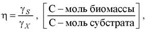
where ys and yx are the degrees of reduction of the substrate and biomass, respectively, equal to the sum of the number of electrons in the atoms of the substances (the equivalent of chemical valence).
Since energy yield is a dimensionless quantity, one can transition from it to mass yield:

where Mx and Ms are the molecular masses of the biomass C-mole and the substrate, respectively.
Example. Calculate the amounts of carbon dioxide consumed and oxygen released during the cultivation of photosynthetic organisms (microalgae) under stress conditions aimed at non-polar lipid accumulation.
1. Approximately 65% of all fatty acids in Chlorella are unsaturated acids [39, 40]. Since the proportion of unsaturated acids in the fatty acid profile of Chlorella is high, and the ratio of atoms constituting these acids varies very little, let us calculate the average formula of Chlorella lipids taking into account the glycerol residue. The atomic indices in the average fatty acid formula are found as the arithmetic mean of the atomic indices of the respective acids (palmitic C15H31COOH, myristic C13H27COOH, oleic C17H33COOH, lauric C11H23COOH):

Thus, the average formula for fatty acids is: C15H30O2.
We can derive the average lipid formula from the residues of the average acid formula and glycerol, which can be conventionally represented by the equation
СзН5(ОН)з + 3С14Н29СООН = СзН5(ОСОС14Н29)3 + 3Н2О.
Thus, the average formula for microalgal lipids takes the form С48Н92О6 or С24Н46О3.
Adjusting the indices of the resulting formula to a single C-mole, we obtain СН1,92О0,13.
2. Let us refine Stouthamer's formula for microalgal biomass with a lipid content of 30% of dry matter:

The average C-mole formula for the biomass of Chlorella, synthesizing about 30% of lipids in dry matter, becomes СН1,84O0,39N0,14.
3. Let us calculate The amount of carbon Nutrition (carbon dioxide) required to produce 1 g of biomass [14]:

Mass fraction of carbon in carbon dioxide
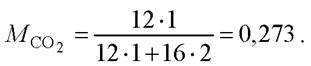
The mass of СО2 required to produce 1 g of biomass will be

If a gas-air mixture (GAM) with a carbon dioxide content of 0.8% is used, the required amount of gas mixture for the production of 1 ton of biomass will be Gгвс = 1,98 / 0,008 = 247,5 (t).
Volume of the GAM used
VСO2 = 22,4 • 247500 / (44 • 0,008) = 15 750 (L) = 15,75 (m3).
Last update: 13/08/2026
Editorial and Educational Adaptation: This material has been compiled based on the primary/original source text. The project team performed an editorial review, corrected technical inaccuracies, structured sections, and adapted the content for an educational format.
What was processed:
- elimination of formatting defects (OCR errors, structural breaks, corrupted characters);
- editorial organization of content;
- standardization of terminology in accordance with academic sources;
- verification of factual statements against the original source text.
All mentions of the author, publication year, and origin of the primary text have been preserved in accordance with the source.