Human Anatomy and Physiology - I. V. Gayvoronsky 2011
Brief Historical Outline
History of Anatomy
The history of anatomy is deeply rooted in antiquity. Primitive societies already possessed some understanding of The Human Body's Structure and the function of its parts. Apparently, The Emergence of human cognitive activity is associated with the butchering of killed animals and the comparison of the acquired knowledge regarding the shape and structure of Organs in animals and wounded humans. This is confirmed by cave and rock paintings found in Spain and China (dating back to 1400–2600 BC). Against the Background outlines of the depicted animals, the Location and shape of certain Internal Organs (The Heart, respiratory organs, Stomach, Kidneys, etc.) are shown with surprising accuracy.
In the 4th–2nd millennia BC, centers of science and culture emerged in Ancient Egypt, Ancient Babylon, and Ancient Palestine. During this time, a distinct caste of physicians—priests—arose, who widely applied Water, oils, herbal infusions, and other remedies to treat illnesses. The literal Introduction/27.html">Translation of the word "physician" meant "one who knows water" or "one who knows oils." Healing required studying the basics of Human Body Structure. In Ancient Egypt, corpse embalming became widespread: small incisions were made in the body, the internal organs and Brain were removed, and the deceased's body was treated with saline solutions and resinous compounds.
Anatomical knowledge from this period was largely highly fantastical and incorrect. In particular, the heart was considered The Organ of thought, through which Blood, mucus, air, water, and urine supposedly passed.
Records concerning the deliberate study of human body structure date from the 5th–4th centuries BC to the 2nd century AD and are tied to the history of Ancient Greece and Ancient Rome.
Anatomy in Ancient Greece and Ancient Rome. Alcmaeon of Croton is considered the founding father of ancient Greek anatomy and physiology. At the turn of the 6th to the 5th century BC, he wrote a treatise on The structure of animal bodies. He was the first to point out that the brain serves as the primary organ of perception and thought, and he described individual nerves and their importance for the function of the Senses.
Class="center">
Hippocrates (460–377 BC)
Hippocrates is regarded as the "father" of medicine. His numerous scientific works were compiled into the "Hippocratic Corpus" a century after his death. Of great interest to anatomists are his treatises "On Glands," "On the Intestines," "On The Nature of the Child," "On Teething," and others. He described certain cranial bones and the STRUCTURE OF THE heart. However, his views on Blood Circulation were erroneous; for instance, it was believed that inhaled air served to cool the heart.
In his quest to find the "soul," Herophilus (born 304 BC) dissected more than 600 corpses and was the first to synthesize data on human body structure in his work Anatomica. For this reason, Herophilus is considered the founder of Anatomy as a Science. He described the External structure of the brain, its membranes, the Dural Venous Sinuses, and the brain ventricles, distinguished Arteries from Veins, named the Pulmonary veins, and described the Prostate Gland, Seminal Vesicles, and duodenum. The works of ancient Greek scholars laid the groundwork for anatomical research. Yet, these studies remained highly fragmented, frequently contained misconceptions, and required refinement.
Claudius Galen (130–200 AD) served as a physician to wealthy Romans and gladiators. His primary achievement as an anatomist was that he synthesized and systematized all anatomical knowledge accumulated during the antiquity period. His principal works bear the general title On Anatomical Procedures, presented in 16 books. Galen's name is associated with numerous discoveries: the Classification of Bones, descriptions of the back Muscles, the identification of three arterial layers, and descriptions of the vagus and facial nerves, among others. He thoroughly studied the structure of the brain membranes and cerebral veins; consequently, one of the cerebral veins is named after him.
Because dissecting human corpses was prohibited in Ancient Rome, much of Galen's data was derived from animal anatomy and mechanically applied to the human body. This explains the errors and inaccuracies in his writings. These errors were not corrected for a long time—not until 1,400 years later—because Galen's works were deemed infallible by the medieval church and served as textbooks for training physicians in the world's leading medical schools for 14 centuries.
Medieval Anatomy. Anatomy during the Middle Ages languished under the yoke of the Inquisition. No significant discoveries were made during this era. Much attention was devoted to writing commentaries on the works of Hippocrates and Galen, yet even from Galen's writings, only what the church deemed infallible was utilized. Dissenting from Galen meant disagreeing with religion, and such dissent was severely punished. Human dissections were strictly forbidden. Consequently, Galen's teachings, transformed into dogma, retarded The Development of medical science in Europe for many centuries, right up until the Renaissance.
Meanwhile, in the Muslim East, science developed more freely. One of the greatest figures among Eastern scholars during the Middle Ages was Abu Ali Ibn Sina (Avicenna). Avicenna (980–1037) was a towering Tajik scholar, philosopher, and physician who authored over 100 works on astronomy, literature, and medicine. His primary medical masterpiece is considered The Canon of Medicine. Its first volume is dedicated to anatomy and physiology. This Canon summarizes and analyzes data on human body structure discovered by ancient Greek scientists and incorporates Avicenna's own observations.
Renaissance Anatomy. The Renaissance was marked by sweeping scientific discoveries and a surging interest in art and literature. Outstanding anatomists of this period included Leonardo da Vinci, Andreas Vesalius, and others.
Leonardo da Vinci (1452–1519) was a genius artist and scientist. As an artist, he took a deep interest in plastic anatomy and produced many precise anatomical drawings accompanied by explanatory notes. Applying the laws of mechanics, he described the curvatures of THE Vertebral Column, developed a Classification of Muscles, and provided the first Description of the Thyroid Gland. Unfortunately, his anatomical works only came to light 300 years later. Because of persecution by the Inquisition, they were hidden in a cache and therefore failed to exert a significant influence on his contemporaries or subsequent generations.
Andreas Vesalius, professor of anatomy at the University of Padua, is regarded as the reformer and "father of scientific descriptive anatomy." He performed human autopsies and dissections, sketching bones, muscles, internal organs, Blood Vessels, and nerves. In 1538,
A. Vesalius published a small anatomical atlas titled Tabulae Anatomicae (Anatomical Tables), presenting his own findings established precisely through autopsies and dissections. Years of intense labor culminated in his renowned masterpiece, De humani corporis fabrica (On the Fabric of the Human Body), published in Basel in 1543. This work delivered a crushing blow to scholastic anatomy and charted the course for anatomical development over the following century.

Andreas Vesalius (1514–1564)
Following A. Vesalius, numerous scientists pursued anatomical research, deepening the understanding of the human body's Organization. Gabriel Fallopius (1523–1562), a pupil of Vesalius, was the first to describe the uterine tubes, the Facial Nerve canal, and the Cytology/cytology/67.html">Development and Structure of bones. Bartolomeo Eustachius (1510–1574) discovered and described the auditory tube and the valve of the INFERIOR VENA CAVA, studied tooth development and Kidney structure, and described the azygos vein.
By the mid-17th century, Holland had become the epicenter of anatomical research. Under the guidance of Frederik Ruysch (1638–1731), an anatomical museum was established, which contemporaries hailed as the "eighth wonder of the world." F. Ruysch mastered unique techniques for embalming corpses and injecting organ blood vessels with various coloring agents. Peter the Great purchased Ruysch's collection of specimens. The greater part of this collection is housed in St. Petersburg's Kunstkamera, while a smaller portion is kept in the fundamental museum of the Department of Normal Anatomy at the Military Medical Academy.
The Development of Anatomy in Russia in the 17th–19th Centuries. Until the 17th century, medicine as a science did not exist in Russia. Wealthy patients were treated by foreign physicians, but hiring them proved costly for the state. Moreover, these foreigners had a poor grasp of the Russian language and lacked consistent professional training. Consequently, the need arose to train domestic medical personnel. To this end, the Apothecary Chancery was established in Moscow in 1620 to regulate medical care and pharmaceutical manufacturing for the army. In 1654, Russia's first "School for Russian Physicians" was founded. At this school, anatomy was taught using A. Vesalius's textbook, which was translated into Russian that same year by Epiphanius Slavinetsky.
Peter the Great deserves special credit for organizing the training of native medical practitioners. By his decree in 1707, a medical school was established at a Moscow hospital under the direction of Nikolaas Bidloo. Instruction in anatomy and surgery followed a manuscript guide titled Instructions for Those Studying Surgery in the Anatomical Theatre, compiled in 1710. For its time, the book contained crucial and extensive surgical information alongside detailed descriptions of various anatomical structures. Later, medical schools opened in St. Petersburg (1717) and Kronstadt (1719), known as hospital schools. By the late 18th century, 20 hospital schools were operating in Russia. Anatomy was considered the primary subject and was referred to as materia medica.
Martyn Ilyich Shein is credited with creating the first domestic anatomical atlas. He prepared an anatomical atlas titled Syllabus, or Index of All PARTS OF THE Human Body. The atlas featured 26 plates illustrated with drawings and accompanied by brief descriptions. He is also credited with translating L. Heister's anatomy textbook from Latin into Russian, titled Compendious Anatomy, Comprehending the Entire Anatomical Business Briefly. This was the first textbook translated into Russian and it served as the primary manual for teaching anatomy in the hospital schools.
Subsequently, anatomists began to study The Structure of the human body with greater focus, yielding several vital scientific discoveries. In 1782, A. M. Shumlyansky defended his dissertation On the Structure of Kidneys, in which he provided the first accurate description of their internal architecture. A. P. Protasov (1724–1796) became the first Russian academician-anatomist, dedicating his research to The Heart and blood vessels.
In 1786, hospital schools were reorganized into medico-surgical colleges and university medical faculties, where the department of "anatomy, physiology, and surgery" was established for the first time. The transformation of the St. Petersburg Main Medical School into the Medical-Surgical Academy (MSA) in 1798 was a milestone event, as by that time it had already become an independent unit with a scientifically grounded curriculum. Pyotr Andreyevich Zagorsky (1764–1846) stood at the origins of the Department of Physiological Anatomy, making a tremendous contribution to the educational process and authoring the first anatomy textbook in Russian.
An outstanding anatomist of the 19th century was Ilya Vasilyevich Buyalsky (1789–1866). He refined the techniques of dissection, embalming, vascular injection using solidifying masses (corrosion casts), and the engraving of bone anatomical specimens.
N.I. Pirogov was a corresponding member of the Russian Academy of Sciences, a brilliant Russian surgeon, anatomist, and educator, who headed the Institute of Practical Anatomy at the MSA from 1846 to 1856, and became the founder and creator of applied anatomy—topographic anatomy and operative surgery. He authored the book Surgical anatomy of Arterial Trunks and Fasciae (1838), a masterpiece that remains highly relevant today. To create his atlas Illustrated Topographic anatomy of Slices Made in Three Directions Through a Frozen Human Body (1852–1859), N.I. Pirogov pioneered the method of freezing-body slicing and sculptural anatomy, in which the respective organ was carved out of a frozen specimen.
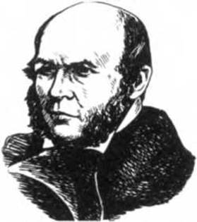
Nikolay Ivanovich Pirogov (1810–1881)
The history of anatomy in Russia during the Soviet period and up to the present day. In the Soviet era, higher medical educational institutions were established across virtually all union republics, alongside specialized morphological departments and laboratories. All of this contributed to the flourishing of anatomy. New promising scientific directions were successfully developed, and advanced morphological Research Methods were introduced. Anatomy as a science firmly established itself as Functional Anatomy. Scientists began widely incorporating microscopic, X-ray, biometric, biochemical, and functional research methods into their experimental work.
Outstanding representatives of Soviet-era anatomy include V.N. Tonkov, G.M. Iosifov, V.P. Vorobyov, G.F. Ivanov, D.A. Zhdanov, M.F. Ivanitsky, R.D. Sinelnikov, M.G. Prives, E.A. Dyskin, V.V. Kupriyanov, M.R. Sapin, and many others.
V.N. Tonkov was an Academician of the USSR Academy of Medical Sciences, HEAD of the Department of Normal Anatomy at the Military Medical Academy (1915–1950), founder and leader of the anatomical school studying collateral circulation, founder of the Department of Normal Anatomy educational museums, and Head (President) of the Military Medical Academy (1917–1925). In 1896, V.N. Tonkov became one of the first Russian anatomical researchers to apply X-rays to study skeletal GROWTH AND DEVELOPMENT; he was the first to study the Development of the Spleen, upper limb arteries, and Skull bones in birds, provided the first precise description of the angioarchitecture of Lymph Nodes AND the Pancreas, developed The Theory of collateral circulation, and experimentally investigated the potential Properties of the arterial bed in numerous body regions
and individual organs in animals. His Textbook of Human Normal Anatomy (1946–1962) and Guide to the Practical Study of Human Vessels and nerves went through six editions each. V.N. Tonkov's scientific and pedagogical school produced 30 professors, many of whom became heads of anatomy departments at leading universities of the former USSR. V.N. Tonkov was the organizer and first chairman of the All-Union Society of Anatomists, Histologists, and Embryologists.
Vladimir Petrovich Vorobyov (1876–1937) was a prominent representative of the Kharkiv school of anatomists. He proposed an original macro-microscopic research method for anatomical specimens, made a major contribution to The Study of the peripheral and, particularly, Autonomic Nervous system, developed a three-dimensional measurement method (stereotopometry), and introduced novel techniques for embalming organs and corpses. He was the first to create the five-volume Atlas of Human Anatomy (1938–1946).

Vladimir Nikolayevich Tonkov (1872–1954)
Gordey Maximovich Iosifov (1870–1933) was the head of the Soviet school of lymphologists and the founder of the comparative anatomy of The Lymphatic system. He was the first to comprehensively describe the lymphatic drainage pathways from various organs and established the regularities of lymphatic vessel architectonics.
Georgy Fyodorovich Ivanov (1893–1955) was a professor at the 1st Moscow Medical Institute named after I.M. Sechenov. He worked on The problem of collateral circulation and studied the structure of The Nervous System. A significant portion of his work is devoted to the innervation of The Cardiovascular system and the study of receptors.
Boris Alexeyevich Dolgo-Saburov (1900–1960) was a Corresponding Member of the USSR Academy of Medical Sciences and Head of the Department of Normal Anatomy at the Military Medical Academy (1950–1960). He continued and advanced V.N. Tonkov's theory of collateral circulation. His school was the first to study the development patterns of collaterals in the Venous system, apply functional research methods to collateral circulation, and elucidate the Influence of the nervous system on The formation of collateral blood flow.
Mikhail Fyodorovich Ivanitsky (1895–1969) was a professor first at the 3rd and later at the 4th Moscow Medical Institutes, and the founder of scientific sports anatomy. He pioneered the course of dynamic anatomy and published several monographs on the subject.
Rafail Davidovich Sinelnikov (1896–1981) carried on the ideas of V.P. Vorobyov. He made a significant contribution to the further development of the macro-microscopic Anatomy of The Musculoskeletal System, endocrine, vascular, and autonomic systems. He prepared and published an original three-volume Atlas of Human Anatomy, which went through six editions and remains an essential reference book for anatomists today.
Dmitry Arkadievich Zhdanov (1908–1971) was an Academician of the USSR Academy of Medical Sciences, head of the largest school of Soviet morphologists, and the founder of the ultramicroscopic Anatomy of the lymphatic system.
Mikhail Grigorievich Prives (1904–1999) led a major school of Leningrad anatomists. He made a substantial contribution to the development of X-ray anatomy, devised a technique for in vivo radiolymphography, was among the first to use electroroentgenography and ultrasonic echolocating in anatomy, and developed a method for preserving anatomical specimens while retaining their natural color.
Vasily Vasilyevich Kupriyanov (1912–2006) was an Academician of the Russian Academy of Medical Sciences (RAMS), founder of The Doctrine of structural foundations of microcirculation, and head of a major school of anatomists in Russia and neighboring countries. His primary works focus on the Study of the microcirculatory bed of organs in normal, pathological, and experimental conditions.
Efim Anatolyevich Dyskin (born 1923) is a Hero of the Soviet Union and Head of the Department of Normal Anatomy at the Military Medical Academy (1968–1988). The research conducted by E.A. Dyskin's school addresses theoretical and applied issues in military medicine.
Mikhail Romanovich Sapin (born 1925) is an Academician of the RAMS and Head of the Department of Human Anatomy at the I.M. Sechenov Moscow Medical Academy. His research interests revolve around the lymphatic and immune systems, as well as the anatomical and functional state of the Circulatory system. M.R. Sapin devotes considerable effort and attention to organizing and optimizing the educational process in human anatomy departments.
Valentin Lvovich Kolesnikov (born 1940) is an Academician of the RAMS, professor, and Head of the Department of Human Anatomy at the Moscow State University of Medicine and Dentistry. He serves as President of the All-Russian Scientific Medical Society of Anatomists, Histologists, and Embryologists, and Chairman of the Commission on Anatomical Terminology. His scientific works focus on Dental Anatomy, Morphology of the digestive tract, and medical anthropology.
Overall, the history of anatomy from its inception to the present day (according to V.V. Kupriyanov) can be divided into two periods.
Period I is the era of ancient anatomy, or the prehistory of scientific anatomy, characterized by the accumulation of empirical knowledge;
Period II is the era of scientific anatomy, beginning in the 16th century with the work of A. Vesalius and continuing to the present day.
The aforementioned periods are associated with the Introduction of new research methods into anatomy and the general level of medical development.
Last update: 08/08/2026
Editorial and Educational Adaptation: This material has been compiled based on the primary/original source text. The project team performed an editorial review, corrected technical inaccuracies, structured sections, and adapted the content for an educational format.
What was processed:
- elimination of formatting defects (OCR errors, structural breaks, corrupted characters);
- editorial organization of content;
- standardization of terminology in accordance with academic sources;
- verification of factual statements against the original source text.
All mentions of the author, publication year, and origin of the primary text have been preserved in accordance with the source.