Operative Surgery and Topographic Anatomy - 2016
Content Module № 2. Topographic Anatomy and Operative Surgery of the Regions and Organs of the Thoracic Cavity
Topic 9. Topographic Anatomy of the Thoracic Cavity. Topography of the Heart and Pericardium. Surgical Approaches to the Heart. Congenital and Acquired Heart Defects, Their Surgical Treatment. Mitral Commissurotomy. Coronary Artery Bypass Grafting. Heart Transplantation
1. Relevance of the topic: the rapid development of thoracic surgery associated with the Surgical Treatment of Diseases of the Lungs, Heart, and Esophagus requires detailed knowledge of the topography of thoracic Organs, as well as The Study of surgical approaches and techniques for operations performed on them.
2. Specific objectives:
1. Explain the Topography of the Heart and great vessels.
2. Analyze modern cardiac operations for congenital and acquired defects, as well as coronary artery disease.
3. Explain how to perform surgical approaches to The Heart.
4. Explain how to perform exposure of The Heart and suturing of cardiac wounds.
5. Explain how to perform a mitral commissurotomy.
3. Tasks for independent work during preparation for the Class.
3.1. List of key terms, parameters, and characteristics that the student must master when preparing for the class:
Term |
Definition |
1. Mitral commissurotomy 2. Pericardiocentesis |
1. Widening of the left atrioventricular orifice in case of its stenosis. 2. Puncture of the pericardial sac for diagnostic and therapeutic purposes. |
3.2. Theoretical questions for the class:
1. Surgical anatomy of the heart (skeletotopy, syntopy, Blood supply, innervation, pathways of venous and lymphatic drainage). Congenital and Acquired heart defects.
2. Anatomical and physiological rationale for surgical approaches to the heart.
3. Pericardiocentesis (Fig. 45).
4. Suturing of heart wounds.
5. Mitral commissurotomy.
6. Coronary artery bypass grafting.
7. Principles of heart transplantation.

Fig. 45. Puncture of the pericardial cavity.
a - anterior view; b — on a sagittal section.
3.3. Practical skills acquired during the class:
1. Suture a cardiac wound.
2. Perform a mitral commissurotomy.
4. Topic Outline
On the heart specimen, students dissect the right and left coronary Arteries, which originate as the first branches from the ascending aorta. The cardiac Veins merge into the coronary sinus, which empties into the right atrium. The brachiocephalic trunk branches off the aortic arch to the right, dividing into the right common carotid and right subclavian arteries. To the left of the aortic arch, the left common carotid and left subclavian arteries arise independently.
On anatomical specimens, students examine the pulmonary trunk, which originates from the right ventricle. The Pulmonary veins leave each lung as two trunks and lead into the left atrium. Students study congenital and acquired heart defects and great vessel anomalies.
Next, on the isolated heart, students place 2–3 interrupted sutures on the cardiac wound using an atraumatic needle. This is performed by excising the apex of the left atrial appendage and digitally dilating the left atrioventricular (mitral) orifice.
Students review mitral commissurotomy, which is performed using a commissurotome (valvulotome). Following an anterolateral thoracotomy in the 4th intercostal space, the Pericardium is incised posterior to the phrenic nerve, extending from the Water/144.html">Origin of the pulmonary trunk to the apex of the left ventricle. A Satinsky clamp is applied to the Base of the left atrial appendage, with a purse-string suture placed just above it. The tip of the appendage is excised with scissors. The index finger is inserted into the left atrium while removing the Satinsky clamp. After digital exploration of the left atrioventricular orifice, the valve commissures are divided with the finger (Fig. 46).
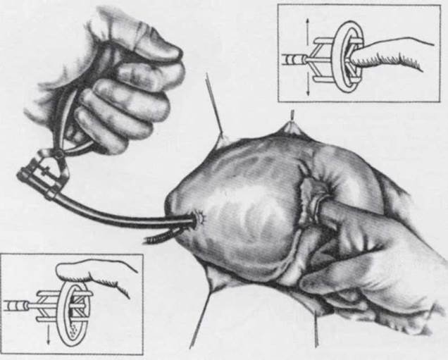
Fig. 46. Transventricular mitral commissurotomy.
Position of the dilator and its blades at the moment of dilation of the left venous orifice.
Nowadays, instrumental commissurotomy is commonly performed. For this Procedure, a U-stitch (mattress suture) is placed on the left ventricle near the apex, and a valvulotome is inserted between its strands until it contacts the finger previously introduced through the left atrial appendage, as described above. Commissurotomy is then performed. The U-stitch on the ventricle and the purse-string suture on the left appendage are tied. Various commissurotomes can be introduced exclusively through the appendage to perform the commissurotomy.
Aortocoronary bypass surgery is performed to treat coronary artery disease. Using the great saphenous vein, a bypass graft is created between the aorta and the coronary artery, bypassing the site of occlusion. Typically, two or three bypass grafts are placed (to the right and left coronary arteries, and the anterior interventricular branch of the left coronary artery) (Figs. 47, 48).

Fig. 47. Schematic representation of the reconstructed anterior interventricular artery.
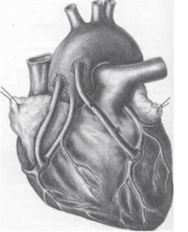
Fig. 48. Schematic representation of a double aortocoronary bypass.
The first heart transplantation was performed on December 3, 1967, by C. Barnard. The technique was developed by Lower and Shumway and involves transplanting the donor heart onto the recipient's preserved atrial remnants. Over 5,000 heart transplantations have now been performed in developed countries worldwide.
5. Self-Assessment Materials
A. Self-Assessment Tasks
Test No. 1
A surgeon is suturing a wound of the heart. What type of suture is used?
a. Donati suture
b. Simple interrupted full-thickness suture
c. Full-thickness horizontal mattress suture
d. Mattress suture
e. According to Multanovsky
Test No. 2
A physician is performing a pericardiocentesis using Larrey's approach. In which direction should the needle be inserted?
a. Perpendicular to the Skin
b. Medially
c. Laterally
d. Superiorly
e. Inferiorly
Test No. 3
A patient presents with Impaired blood supply to the papillary Muscles of the right ventricle and the posterior papillary Muscle of the left ventricle. A disruption of BLOOD FLOW IN which artery caused this condition?
а. A. coromria dextra
в. R. circumflexus
c. R. auriculares
d. A. coronaria sinistra
e. R. interventricularis anterior
Test No. 4
A 50-year-old patient was admitted to the hospital complaining of retrosternal pain and shortness of exertion. Angiography revealed pathological Changes in the posterior interventricular artery. Which areas of the heart are affected?
а. Posterior wall of the right and left ventricles
в. Left atrium
c. Anterior wall of the right and left ventricles
d. Right atrium
e. Right atrioventricular valve
Test No. 5
A 52-year-old patient was admitted to the hospital complaining of severe retrosternal pain and dyspnea. Following physical examination, the patient was diagnosed with a myocardial infarction of the anterior wall of the left ventricle. Which cardiac artery is affected?
а. Anterior interventricular branch of the left coronary artery
v. Posterior interventricular branch of the right coronary artery
c. Right coronary artery
d. Circumflex branch of the left coronary artery
e. Atriodiaphragmatic artery
B. Self-Assessment Tasks
Task 1. A patient with suspected blood in the pericardial cavity was admitted to the thoracic department. What procedure should the surgeon perform to diagnose this pathological condition?
Task 2. A patient with a penetrating chest injury was admitted to the surgical department. Physical examination reveals a 2.5x1 cm stab wound in the 4th intercostal space along the parasternal line. The patient complains of a constricting pain behind the Sternum. He is somewhat lethargic, pale, and has a pulse rate of 115 bpm. Auscultation: vesicular breath sounds on the right, absent breath sounds on the left; heart sounds are muffled and regular. Establish a provisional Diagnosis. What tissue layers did the knife penetrate during the chest injury?
Task 3. A patient with a gunshot wound to the chest was admitted to the thoracic department, complaining of dull, constricting pain behind the sternum. Objective findings: the patient is pale; There is a 1.3x1.3 cm entry wound in the 5th intercostal space along the parasternal line; pulse is 117 bpm; blood pressure is 85/60 mm Hg; Respiration rate is 20 breaths/min; auscultation reveals vesicular breath sounds on both the right and left; heart sounds are distant and regular; Percussion dullness is detected on the left between the 4th and 6th intercostal spaces, extending from the parasternal to the midclavicular line. Establish a diagnosis.
Essential
1. Operative Surgery and Topographic Anatomy; edited by M.S. Skrypnikov. — K.: Vyshcha Shkola, 2000. — P. 270-280.
2. Operative Surgery and Topographic Anatomy; edited by M.P. Kovalsky. — K.: Medytsyna, 2010. — P. 178-190.
Supplementary
1. Operativnaya khirurgiya i topograficheskaya anatomiya; edited by K.I. Kulchytsky. — K., 1994. — P. 159-168.
2. Operativnaya khirurgiya i topograficheskaya anatomiya; edited by G.E. Ostroverkhov. — Rostov-on-Don, 1998. — P. 435-488.
3. Toporov G.N. Klinicheskaya anatomiya grudi / G.N. Toporov. — Kharkov, 2007.
4. Elizarovsky S.I. Operativnaya khirurgiya i topograficheskaya anatomiya / S.I. Elizarovsky, R.N. Kalashnikov. — M., 1979.
5. Matyushin I.F. Vvedenie v kurs operativnoy khirurgii i topograficheskoy anatomii / I.F. Matyushin. — Gorky, 1976.
6. Tomashuk I.P. Rukovodstvo po operativnoy tekhnike dlya nachinayushchikh khirurgov / I.P. Tomashuk, I.I. Tomashuk. — K.: Iz-vo Evropeyskogo universiteta, 2001. — 860 p.
7. Frauchi V.Kh. Kurs topograficheskoy anatomii i operativnoy khirurgii / V.Kh. Frauchi. — M., 1976.
Last update: 10/08/2026
Editorial and Educational Adaptation: This material has been compiled based on the primary/original source text. The project team performed an editorial review, corrected technical inaccuracies, structured sections, and adapted the content for an educational format.
What was processed:
- elimination of formatting defects (OCR errors, structural breaks, corrupted characters);
- editorial organization of content;
- standardization of terminology in accordance with academic sources;
- verification of factual statements against the original source text.
All mentions of the author, publication year, and origin of the primary text have been preserved in accordance with the source.