BIOCHEMISTRY - Textbook - Ostapchenko L. I. - 2012
Chapter 12. CARBOHYDRATE METABOLISM
12.1. Catabolic pathways of carbohydrates
12.1.2. Regulation of blood glucose levels
Blood glucose concentration serves essentially as an integrative index of overall Carbohydrate METABOLISM, which lies at the core of animal metabolism. Therefore, carbohydrate metabolism is governed by a diverse set of tightly coordinated and interconnected regulatory mechanisms. Foremost among these is the regulatory (glycogenic) function of the Liver—an evolutionary ancient and relatively simple mechanism that ensures the REGULATION OF CARBOHYDRATE Metabolism in lower animals, yet proves insufficient for controlling Carbohydrate Metabolism in highly developed animals and humans. Consequently, evolutionary development gave rise to a more sophisticated neurohumoral regulation mechanism that influences both the regulatory function of The Liver and the capacity of other Tissues to utilize glucose.
The regulatory function of the liver is tied to The Mechanism of utilizing excess glucose—or more precisely, glucose-6-phosphate—by converting it into a compact, mobile reservoir of metabolic energy known as Glycogen, and subsequently mobilizing this energy as free glucose whenever the need arises.
Glucose concentration plays a decisive role in regulating hepatic glycogen metabolism. A high glucose concentration in hepatocytes stabilizes the inactive form of Glycogen phosphorylase and activates glycogen synthase, which are the primary Enzymes of glycogen metabolism. This shift inhibits glycogen phosphorolysis while stimulating its synthesis.
The conversion of glucose-6-phosphate along the glycogen synthesis pathway proceeds through several sequential enzymatic reactions, outlined as follows:
Class="center">
Initially, mediated by phosphoglucomutase, glucose-6-phosphate undergoes reversible isomerization into glucose-1-phosphate. Acting via glucose-1-phosphate uridylyltransferase, glucose-1-phosphate then reacts with UTP to form the activated glucose derivative, uridine diphosphate glucose (UDP-glucose). This reaction is irreversible because the pyrophosphate cleaved from UTP is hydrolyzed by pyrophosphatase into orthophosphate, driving the synthesis of UDP-glucose through The energy released by this Hydrolysis.
In the final step of glycogen Biosynthesis, The transfer of a glycosyl residue from UDP-glucose to the non-reducing end of a glycogen polysaccharide chain is catalyzed by glycogen synthase (UDP-glucose-glycogen glucosyltransferase), which forms a new
α-1,4-glycosidic bond between carbon atom 1 of the UDP-glucose glycosyl residue and carbon atom 4 of the terminal glycosyl residue in the glycogen molecule.
The incorporation of a single glycosyl residue into a glycogen molecule is coupled with the hydrolysis of two γ-phosphate groups from ATP and UTP molecules. Once the linear chain of a glycogen molecule reaches a length of 10 glycosyl residues, amylo-(1,4→1,6)-transglucosylase (the branching enzyme) transfers a 6-7 residue fragment to an adjacent chain, establishing an α-1,6-linkage (a branch point) that gives rise to a new branch in the molecule (Fig. 12.1). Glycogen synthase then continues to attach further glycosyl residues. The biochemical rationale behind branching lies in enhancing molecular compactness and increasing the number of non-reducing ends, which greatly facilitates the catalytic action of glycogen metabolism enzymes, namely phosphorylase and glycogen synthase.

Fig. 12.1. Scheme of glycogen synthesis
Under normal conditions, Cells almost invariably harbor a population of pre-existing glycogen polysaccharide chains that act as acceptors for newly added glycosyl residues. However, under extreme conditions (such as 24-hour fasting or exhaustive physical exertion), hepatic glycogen reserves may become entirely depleted. In such scenarios, initiation relies on polypeptide chains that serve as primary acceptors; primer Oligosaccharides are initially synthesized on these chains, providing a foundation onto which the polysaccharide chains of the glycogen molecule are subsequently built (Fig. 12.2).

Fig. 12.2. Scheme of glycogen biosynthesis on polypeptide chains
The liver of an average adult human (weighing 70 kg) can store approximately 100 g of glycogen, accounting for 3–5% of the total organ mass. An additional 250–300 g of this polyglucan can accumulate in Muscle tissue, where the aforementioned reactions of glycogen biosynthesis also take place using glucose supplied to muscle cells via the bloodstream. Nevertheless, functionally speaking, muscle glycogen—which constitutes 0.5–1.0% of tissue mass—differs from hepatic glycogen in that it serves primarily as an energy source for Muscle contraction.
Glycogen mobilization in the liver typically occurs during the postabsorptive period when peripheral tissues experience a demand for glucose. Glycogen breakdown is carried out through the concerted action of three enzymes: glycogen phosphorylase, oligo-1,6-glucosidase (oligo-transferase), and amylo-1,6-glucosidase (Fig. 12.3).
Phosphorylase catalyzes the Cleavage of glycosyl residues in the form of glucose-1-phosphate (G-1-P), starting from the peripheral ends of the outer branches. As it approaches a branch point, the enzyme halts, allowing oligo-transferase to rearrange the phosphorylase-truncated glycogen branch and expose the glycosyl residue linked via an α-1,6-bond. Following the hydrolysis of this bond by amylo-1,6-glucosidase, the phosphorolysis of α-1,4-glycosidic bonds resumes along the next polyglycosyl chain of the glycogen molecule until the next branch point is reached. The glucose-1-phosphate molecules liberated during glycogen phosphorolysis are subsequently isomerized into glucose-6-phosphate by phosphoglucomutase.

Fig. 12.3. Scheme of glycogen phosphorolysis reactions
Consequently, the vast majority (up to 90%) of hepatic glycogen undergoes phosphorolytic cleavage, whereas a significantly smaller fraction (about 10% of the α-1,6-bonds) is subject to hydrolytic cleavage. It is worth noting that glycogen hydrolysis occurs predominantly in the gastrointestinal tract under the action of specific Hydrolases.
Glucose-6-phosphate—a pivotal metabolic intermediate—can follow several alternative pathways, with the dominance of any particular route typically dictated by the immediate physiological needs of the liver itself and the Organism as a whole. First, glucose-6-phosphate can undergo dephosphorylation to free glucose, catalyzed by glucose-6-phosphate, an enzyme present in the Cells of the liver, Kidneys, and intestines; this free glucose is then released into the bloodstream whenever blood sugar levels drop. Second, glucose-6-phosphate can be catabolized via Glycolysis into Pyruvate and its oxidative decarboxylation products—acetyl-CoA and CO2—which, alongside the glycolytic intermediate dihydroxyacetone phosphate, can be channeled into the synthesis of triacylglycerols, Phospholipids, and Cholesterol. Third, glucose-6-phosphate may undergo direct oxidation via the Pentose Phosphate Pathway, yielding ribose-5-phosphate and other pentose phosphates, alongside reducing equivalents in the form of NADPH required for reductive biosynthetic reactions involving various macromolecules, most notably nucleotide and nucleic acid synthesis.
Glycogen phosphorylase plays a decisive role in glycogen mobilization, acting as a key regulatory enzyme. Its activity is controlled through two molecular mechanisms: covalent modification (phosphorylation) and Allosteric Regulation.
Covalent Modification of the enzyme molecule, which consists of two identical subunits, involves the phosphorylation of Serine-14 residues in both subunits. Two specific enzymes—cAMP-dependent phosphorylase kinase and phosphorylase phosphatase—mediate the interconversion of the two forms of glycogen phosphorylase: inactive phosphorylase b and active phosphorylase a. Phosphorylase b is converted into phosphorylase a with the participation of cAMP-dependent phosphorylase kinase, whereas phosphorylase phosphatase catalyzes the reversible transition of active phosphorylase a back to the inactive form b:

The rate of glycogen mobilization is governed by The ratio of active phosphorylase a to inactive phosphorylase b. The second mechanism regulating glycogen phosphorylase is characteristic of muscle tissue. Inactive muscle phosphorylase b can be activated via non-covalent binding to an allosteric modulator, AMP, which induces a conformational change in the enzyme molecule. ATP and glucose-6-phosphate act as allosteric inhibitors, competing with AMP for the binding site. Consequently, The activity of phosphorylase b in Muscles is determined by the ATP-to-AMP ratio. Under resting conditions, when the ATP/AMP ratio is high, the enzyme remains in its inactive form. During muscle contraction, which is accompanied by ATP hydrolysis, the intracellular concentration of AMP increases, altering the nucleotide ratio and, consequently, the activity of phosphorylase b. Unlike AMP-dependent phosphorylase b, muscle phosphorylase a is an AMP-independent form (Fig. 12.4).
The enzyme can exist in a catalytically inactive form b or an active conformation a. Both subunits are phosphorylated at serine-14 residues by phosphorylase kinase, which is stimulated by Ca2+ ions. Notably, liver phosphorylase is also regulated through allosteric modulation by glucose, which acts as an allosteric inhibitor.

Fig. 12.4. Schematic representation of glycogen phosphorylase regulation in muscles
The Neurohumoral regulation of glycogen metabolism involves neuroendocrine structures of the Hypothalamus and Pituitary gland, as well as peripheral Endocrine glands—specifically the cells of the pancreatic islets of Langerhans, The Thyroid Gland, and the Adrenal Glands—which rapidly and efficiently respond to any fluctuations in systemic blood glucose levels.
The metabolic centers of the hypothalamus and Medulla Oblongata are the first to detect changes in blood sugar levels. Through neural impulses and tropic Hormones of the adenohypophysis (corticotropin and somatotropin), they stimulate peripheral endocrine glands, prompting them to release respective hormones into the bloodstream. These primarily include Glucagon and Insulin (Peptide Hormones synthesized by α- and β-cells of the pancreatic islets of Langerhans), adrenaline and noradrenaline (amine hormones from The adrenal medulla), corticosteroids synthesized in the adrenal cortex, and thyroxine, the thyroid hormone. Hormonal Regulation of blood glucose is based on the antagonistic relationship between "diabetogenic" hormones—a group that promotes elevated blood glucose—and insulin, the sole hormone with a pronounced hypoglycemic effect. Insulin facilitates glucose uptake by hepatocytes and insulin-sensitive cells in adipose tissue and skeletal muscles. In hepatocytes, insulin stimulates the induction of glucokinase, cyclic nucleotide phosphodiesterase activity, and glycogen synthase, thereby enhancing glycogen biosynthesis.
The regulatory Action of Hormones on glycogen metabolism is exerted by modulating the activity of Key Enzymes: Glycogen synthase and glycogen phosphorylase. Their activities are regulated reciprocally; that is, when one enzyme (e.g., phosphorylase) is inactive, the other (glycogen synthase) exhibits full activity, and vice versa. In this case, simultaneous and opposing Changes in the activities of these enzymes are achieved through covalent modification—the phosphorylation-dephosphorylation of the active sites of glycogen synthase and glycogen phosphorylase.
The action of glucagon and adrenaline in the liver is mediated by specific receptors located on Cell/30.html">The Plasma Membrane of hepatocytes. The receptor type determines the Molecular Mechanism of hormonal signal Transduction. For instance, hormone binding to $\alpha_1$-receptors activates the Inositol phosphate pathway, whereas interaction with $\beta_2$-receptors stimulates the adenylate cyclase Intracellular Signaling system (Fig. 12.5).
When glucagon or adrenaline binds to specific receptors on the Plasma Membranes of hepatocytes or myocytes, forming a hormone-receptor complex, adenylate cyclase (AC) or phospholipase C (PLC) is stimulated. This stimulation results in a rapid 2- to 3-order-of-magnitude surge in intracellular second messengers—cAMP, 1,2-diacylglycerol (DAG), and inositol-1,4,5-trisphosphate ($IP_3$)—well above their basal levels. These messengers activate protein Kinases either through allosteric modulation (cAMP, cGMP) or via a $Ca^{2+$-dependent mechanism (protein kinase C).
Cyclic nucleotide-dependent protein kinase, composed of four subunits (two regulatory and two catalytic), reversibly dissociates in the presence of cAMP (cGMP), releasing the catalytic subunits. The thus-activated protein kinase, in turn, phosphorylates phosphorylase kinase and glycogen synthase, leading to the activation of phosphorylase and the simultaneous inhibition of glycogen synthase activity.
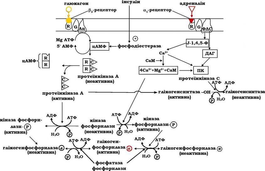
Fig. 12.5. Regulation of Glycogen phosphorolysis by glucagon and adrenaline
When adenylate cyclase stimulation by adrenaline or glucagon ceases, the enzyme returns to an inactive state, and elevated intracellular cAMP levels drop back to basal values due to the action of phosphodiesterase. The cytosolic enzyme cyclic nucleotide phosphodiesterase is activated by insulin, which thereby halts the enzymatic cascade of hormonal signal Amplification that stimulates glycogen phosphorolysis. Dephosphorylation of the enzymes is carried out by Phosphatases, which catalyze the hydrolysis of phosphoryl groups at the active sites of phosphorylase kinase, glycogen phosphorylase, and glycogen synthase. This converts active phosphorylase kinase and phosphorylase a into their inactive forms while activating glycogen synthase.
The interaction of adrenaline with $\alpha_1$-receptors and The formation of the hormone-receptor complex activate phospholipase C, which catalyzes the hydrolysis of plasma membrane phosphatidylinositol bisphosphate, releasing inositol-1,4,5-trisphosphate and 1,2-diacylglycerol. These serve as secondary messengers in the inositol phosphate signaling pathway (Fig. 12.5).
Inositol trisphosphate triggers the release of $Ca^{2+}$ ions from The Endoplasmic reticulum, which, together with 1,2-diacylglycerol, activate the $Ca^{2+}$- and phospholipid-dependent protein kinase (protein kinase C). Furthermore, the elevated concentration of ionized calcium in the Cytoplasm promotes the binding of the cation to the $Ca^{2+}$-binding protein calmodulin (CaM), imparting it with The ability to regulate $Ca^{2+}$-dependent enzymes. The activation of phosphorylase kinase and calmodulin-dependent protein kinases leads to the phosphorylation of glycogen phosphorylase b, converting it into the active form a, as well as the phosphorylation of glycogen synthase, rendering it inactive.
Consequently, the molecular mechanism ensuring the coordinated Synthesis and degradation of glycogen is based on the phosphorylation-dephosphorylation of serine residues at the active sites of glycogen metabolism enzymes. In higher animals and humans, this control mechanism is governed by the neurohumoral regulatory system.
Glucagon accelerates glycogen phosphorolysis in the liver without affecting its breakdown in muscles. The stress hormone adrenaline stimulates glycogen phosphorolysis and inhibits its synthesis in the liver, skeletal muscles, and myocardium. The regulation of Glycogen metabolism in muscles is mediated by Two Types of molecular mechanisms: covalent modification and allosteric modulation.
Insulin deficiency in the body leads to a systemic condition known as Diabetes Mellitus, characterized by impaired biosynthesis and storage of glycogen in tissues, activated glycogen phosphorolysis in the liver and skeletal muscles, increased mobilization of Fatty acids in adipocytes, and enhanced breakdown of tissue Proteins. A high degree of protein glycation, particularly of Hemoglobin, observed in the progression of this disease leads to impaired protein function. Therefore, the primary characteristic symptoms of diabetes mellitus are hyperglycemia, azotemia, and hyperlipidemia, resulting from systemic disorders in carbohydrate, protein, and Lipid Metabolism.
Hereditary disorders of glycogen metabolism—Glycogen Storage Diseases (glycogenoses)—are caused by the absence or deficiency of specific enzymes involved in glycogen synthesis or degradation. For instance, the deficiency of glucose-6-phosphatase in the liver leads to the accumulation of abnormally large amounts of glycogen (Von Gierke's disease) against the Background of pronounced hypoglycemia during the postabsorptive period. In certain glycogenoses, structurally abnormal glycogen molecules are synthesized in tissues, featuring either shortened outer branches due to the absence of amylo-1,6-glucosidase (Cori's disease) or abnormally long, sparsely branched chains resulting from a deficiency of amylo-(1,4$\rightarrow$1,6)-transglucosidase (Andersen's disease).
Glycogenoses associated with a deficiency of liver glycogen synthase (Hers' disease / liver glycogen synthase deficiency) result in a significant reduction in hepatic glycogen content, accompanied by prolonged hyperglycemia in the absorptive period and hypoglycemia in the postabsorptive state. When muscle phosphorylase activity is absent (McArdle disease), patients are unable to perform intense physical exertion due to debilitating muscle cramps. Some glycogenoses present with much more severe symptoms leading to critical outcomes (e.g., Von Gierke's disease) or even fatal consequences in early childhood (Pompe disease, caused by a deficiency of lysosomal $\alpha$-1,4-glucosidase; Andersen's disease).
Last update: 06/08/2026
Editorial and Educational Adaptation: This material has been compiled based on the primary/original source text. The project team performed an editorial review, corrected technical inaccuracies, structured sections, and adapted the content for an educational format.
What was processed:
- elimination of formatting defects (OCR errors, structural breaks, corrupted characters);
- editorial organization of content;
- standardization of terminology in accordance with academic sources;
- verification of factual statements against the original source text.
All mentions of the author, publication year, and origin of the primary text have been preserved in accordance with the source.