Practical Course in Zoology: Study Guide - T. A. Dauda 2014
Multicellular Animals
Chordates
Subphylum Vertebrates (Craniata) – Superclass Tetrapods – Class Birds
Birds are a specialized branch of higher vertebrates adapted for flight. They evolved from reptiles and are genetically close to them. At the same time, birds exhibit progressive organizational traits that fundamentally distinguish them from reptiles. Birds have a higher level of Nervous system development and, consequently, more diverse and sophisticated adaptive behavior.
Birds are warm-blooded animals. Their high and constant body Temperature is associated with a significantly higher metabolic rate. Birds are not only capable of flight but also move exceptionally well on solid substrates. Birds possess a more advanced mode of reproduction (incubating eggs and feeding nestlings).
Morphologically, birds are characterized by a body covered in feathers, forelimbs modified into wings, pneumatic bones, a four-chambered Heart with a single right aortic arch, and an absence of Teeth in modern birds, which are functionally replaced by a horny beak.
Modern birds (approximately 9,000 species) are divided into three distinct superorders: Carinate birds (Carinatae), Ratite birds (Ratitae), and Penguins (Impennes).
To study the Morphology and Biological features of birds, wild or domestic pigeons can be used.
Rock Dove (Columba livia).
Various breeds of domestic pigeons have been developed through the domestication of the wild rock dove. The pigeon—a granivorous bird distributed across almost the entire globe—is a vector of Viruses that cause ornithosis, as well as flagellated Protozoa and Toxoplasma species that cause Toxoplasmosis.
Materials and equipment. For every one to two students, the following are required: a freshly euthanized pigeon; a dissecting board; a scalpel; forceps; scissors; dissecting needles; a Glass tube with a drawn-out tip; absorbent cotton; gauze wipes; a hand lens; twine; jars with Water for collecting feathers; waste jars; and wall charts: "Internal Anatomy of the Pigeon," "Diagram of the Circulatory system," and "Structure OF THE Brain."
Assignment. Study and sketch the external anatomy of the pigeon. Dissect the bird and sketch the general arrangement of its Internal Organs. Dissect individual Organ Systems. Open the cranium, examine, and sketch the brain. Identify the ancient "reptilian" traits of birds (using the pigeon as an example) as well as the new "avian" traits that distinguish birds from reptiles. Note the Progressive features of avian Organization and their adaptation for flight. Complete the following drawings: "Internal Anatomy of the Pigeon," "Diagram of the Circulatory System," and "Structure of the Brain."
Study of external anatomy using an anesthetized pigeon. Pay attention to the pigeon's adaptations for flight: streamlined body shape, well-developed wings, and the imbricate arrangement of contour feathers, which form a dense, durable covering important for both flight and thermoregulation. Observe the division of the body into regions: HEAD, neck, trunk, limbs (the anterior ones modified into wings), and a short tail region. Examine the pigeon's head—it is relatively small, ending in a horny beak at the base of which lie slit-like nostrils opening into the Oral Cavity. The beak consists of heavily elongated jaw bones covered with a horny substance and is divided into the upper Mandible (Maxilla) and lower mandible. Examine the Base of the upper mandible—it is covered by a whitish membrane (cere) and serves as an Organ of Touch in the pigeon. The shape of the beak in birds is extremely diverse and depends on The Nature of their food and Methods of obtaining it.
Open the beak and run a finger and the handle of a scalpel along the jaws—they are devoid of teeth. A narrow, pointed Tongue is visible on the floor of the oral cavity. Pluck the pigeon's feathers slightly behind the eyes and locate the External ear openings. Examine the pigeon's eyes, upper and lower eyelids; the lower eyelids are more developed than the upper ones. In the inner corner of the eye lies the third eyelid, in the form of a translucent nictitating membrane. If this membrane is grasped with forceps and pulled out, the eye will close.
Note that the pigeon's neck is long and flexible while the trunk remains immobile. The cervical vertebrae are extremely mobile, which can be verified by turning the pigeon's head. When grasping food, the bird bends its neck rather than its body. The pigeon's trunk is compact and rigid. Its immobility is due to the fusion of many vertebrae with one another, as well as with the sacrum and pelvic bones. Examine the tail region of the body—this is where long tail feathers, known as rectrices, begin. At the base of the tail, the rectrices are covered (both above and below) by coverts. Locate the transverse slit in the posterior part of the trunk on the ventral side—the cloacal opening.
Examine the hind limbs (legs)—they have four toes equipped with claws; of these, three toes point forward and one backward. The upper part of the legs is covered with feathers, while the lower part is covered with horny scales, similar to the epidermal scales of reptiles. The forelimbs of birds have evolved into a specialized flight organ—the wings; however, the wing, like the forelimbs of terrestrial vertebrates, consists of the arm, forearm, and hand.
Locate the large contour feathers on the wings (Fig. 133). Their lateral edges overlap one another; this arrangement creates a dense, resilient wing surface that is essential for flight. Spread the pigeon's wing and observe its large surface area for flight. The large contour feathers growing along the trailing edge of the wing are called remiges; the feathers attached to the BONES OF THE hand are primary remiges (or large remiges), while the secondary remiges (or small remiges) attach to the forearm. A rudimentary first digit bears the alula (bastard wing).
Class="center">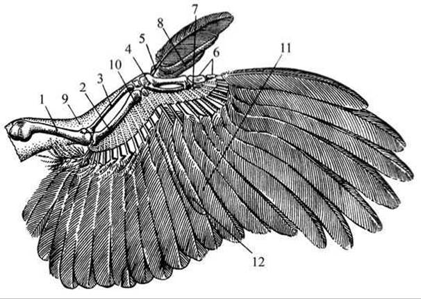
Fig. 133 Structure of a bird's wing:
1 — humerus; 2 — ulna; 3 — radius; 4 — carpometacarpus; 5 — part of the carpus; 6, 7 — Phalanges of the digits; 8 — alula; 9 — patagium (wing membrane); 10 — bases of the remiges; 11 — primary remiges; 12 — secondary remiges.
Note that the pigeon's Skin is dry and devoid of glands, much like that of reptiles. The only exception is the uropygial (preen) gland located at the base of the tail. You can feel it above the base of the rectrices by parting the feathers. The pigeon lubricates its feathers with the oily secretions of this gland. On the bodies of most birds, feathers are arranged in specific tracts: areas covered with feathers are called pterylae, while featherless areas are called apteria. This feather arrangement facilitates Muscle contraction during flight.
Pluck a small contour feather. Examine it using a hand lens and sketch the shaft (the structural basis of the feather), the vane, and the calamus (quill) (Fig. 134). Locate the opening at the tip of the calamus: it contains a dermal papilla that nourishes the feather during its growth. The flat blade of the feather, or vane, consists of barbules.

Fig. 134 Structure of a bird's contour feather:
1 — shaft (rachis); 2 — primary barbs; 3 — secondary barbs with hooklets; 4 — hooklets (barbicels); 5 — calamus (quill); 6 — vane.
Cut off a small section of the vane with scissors, place it on a glass slide, separate the barbs using dissecting needles, add a drop of water with a pipette, and examine it under low magnification using a Microscope. The primary barbs, which originate from the main shaft, bear secondary barbs equipped with tiny hooklets. The interlocking of Primary and secondary barbs via these hooklets forms a strong, elastic feather plate that is largely impermeable to air.
Examine a down feather—it shares the same basic structural type as a contour feather, but differs in that its shaft is poorly developed, and its barbs are soft, lacking hooklets and failing to interlock with one another. Down feathers play a primary role in reducing heat loss.
Dissection. Study the Internal Structure of a bird using a dissected specimen, as well as a preserved wet mount of a dissected pigeon, an injected specimen, and a diagram of the circulatory system.
Place the pigeon used for the external anatomy study ventral side up on a dissecting board, and use twine to tie its wings and legs to the hooks located at the corners of the board.
Pluck the feathers along the midline of the body on the pigeon's belly, chest, and neck using hands moistened with water. Place the plucked feathers and down into a beaker of water to keep them from blowing around.
Holding and lifting the pigeon's skin with forceps, use scissors to make an incision from the cloacal opening to the head (perform the dissection with great care, especially in the neck area where it is easy to rupture the thin-walled crop, whose walls are in contact with the skin). Grasp the edges of the incision with forceps, trim them with scissors and a scalpel, and fold them back to expose the musculature.
Musculature. Examine the pigeon's musculature. In the pectoral region, powerful breast Muscles stand out, extending from the keel of the Sternum to the upper part of the Pectoral Girdle. The keel serves for muscle attachment. The pectoralis major muscles serve to depress the wing. The pectoralis minor muscles lie beneath the pectoralis major. To observe them, make a deep cut along the pectoralis major. The pectoralis minor and deltoid muscles (extending from the coracoid to the humerus) elevate the wing.
Using large scissors, cut the Abdominal muscles along the midline from the cloacal opening to the lower edge of the sternum (exercise caution to avoid damaging internal organs). Cut the pectoral muscles along the edges of the sternum (and remove them), the ends of the Ribs starting from the lower section of the sternum, as well as the bones of the pectoral girdle. Lifting the sternum by its posterior end with your hand or forceps, carefully separate it from the underlying organs with a scalpel, cut through its base, and remove it. Blood oozing from severed vessels during dissection should be wiped away with cotton swabs.
Observe the general arrangement of the internal organs. Draw the outline of the pigeon's body and sketch in the internal organs with their corresponding labels as you examine them.
In the neck region, locate the Esophagus with the crop, the long Trachea, and the upper and lower (syringa) Larynx—the thickening of the trachea just before it branches into the Bronchi (Fig. 135). Incise the trachea, insert a glass pipette into it, and blow air: passing through the trachea and Lungs, the air enters the thin-walled air sacs located among the organs; as they fill with air, they lift the viscera. The air sacs are extensions of the inner lining of the bronchi beyond the lungs. They play a major role in the avian respiratory mechanism, particularly in relation to flight, enabling double respiration in birds. In addition, filled with air, they help reduce the bird's specific gravity and protect the body from overheating during flight. To examine the internal organs, cut open the air sacs if they have not collapsed.
Locate The Heart within the pericardial sac, with the Blood Vessels emerging from it. Flanking the heart are the pinkish-red lungs. In the Abdominal cavity, the bilobed brownish-red Liver and loops of the intestines are visible, with the muscular Stomach situated beneath the left lobe of the liver.

Fig. 135 Internal structure of a bird (pigeon):
A — dissected pigeon; B — section of the pigeon's stomach; 1 — trachea; 2 — esophagus; 3 — crop; 4 — lung; 5 — air sacs; 6 — heart; 7 — proventriculus; 8 — gizzard.
Lift The Stomach and locate the duodenum extending from it. Near it, the Pancreas and the oval Spleen are visible. Organs that dry out during dissection should be periodically moistened with cotton soaked in water. Discard used cotton, along with excised and studied viscera, into the trash bucket.
Digestive System. For ease of examining the digestive and other Organs of the pigeon, cut the end of the esophagus and the Large Intestine just anterior to the cloaca with scissors. Uncoil the dissected intestine and place it on the dissecting board next to the pigeon. While examining the digestive system, pay attention to its adaptation to the bird's lifestyle. The oral cavity leads into a short Pharynx, which transitions into a long, slender esophagus. An expansion of the esophagus forms the crop, where unground food (birds lack teeth) undergoes softening. The food then passes into the proventriculus, where digestive Enzymes act upon it; in the subsequent gizzard, the food undergoes mechanical breakdown as well. After examining the liver, remove it using forceps and a scalpel. The spleen is usually removed along with it.
Open the gizzard (muscular stomach), separate it from the intestine, and make an incision along its margin using a sharp scalpel. A cross-section reveals that the walls are thick and muscular, lined on the inside with a tough horny cuticle. The stomach may contain pebbles and sand swallowed by the bird, which assist in grinding food, thus replacing teeth.
From the gizzard, food passes into the duodenum, into which the ducts of The Liver and pancreas open. Unlike most bird species, the pigeon lacks a Gallbladder. The food then moves along the Small Intestine, where it is digested and absorbed. At the junction between the small and large intestines are small caeca. Undigested food residues collect in the very short rectum; they are not retained there and are quickly expelled. The pigeon's intestine is short, being only about 4 times the length of its trunk.
The unique structure and functioning of the avian digestive tract are due to adaptations for flight and lifestyle characteristics.
Circulatory system. Lift The Heart and locate the right aortic arch emerging from the left ventricle (the left aortic arch, present in reptiles, is reduced in birds). Grasp the heart with forceps, sever the vessels with scissors, and remove it from the pericardial sac. Collect any blood spilling from the vessels and heart using cotton. The avian heart is four-chambered, consisting of two atria and two ventricles. Examine the pigeon's heart, noting a narrow fat groove running transversely across the heart at the boundary between the ventricles and atria. Cut the heart transversely across the ventricular region with a scalpel. The cross-section shows that the ventricles are completely isolated from each other, that the lumen of the left ventricle is circular in cross-section with thick walls, while the walls of the right ventricle are significantly thinner and its lumen is crescent-shaped. Deoxygenated blood passes through the right half of the heart, while arterial (oxygenated) blood passes through the left.
Examine the SYSTEMIC AND PULMONARY circulations using the injected specimen and diagram (see Fig. 136). The systemic Circulation begins at the left ventricle with a short common aortic trunk that gives rise to two innominate Arteries (right and left). Each of these gives rise to the carotid, subclavian, and pectoral arteries. The aorta forms an arch that bends to the right (the right aortic arch) and runs along THE Vertebral Column as the dorsal (descending) aorta. Arteries branching from it supply blood to all internal organs of the body. Gas exchange occurs in the capillaries.
Venous blood from the anterior part of the body via the paired anterior venae cavae, and from the posterior part via the unpaired posterior vena cava, flows into the right atrium and, upon its contraction, into the right ventricle. The Pulmonary Circulation begins at the right ventricle with two pulmonary arteries, through which blood from the right ventricle enters the lungs. Following Gas Exchange in the lung capillaries, oxygen-rich blood returns to the left atrium via the Pulmonary Veins. From the left atrium, blood enters the left ventricle. Thus, the systemic circulation originates at the left ventricle, supplies blood to all body organs and Tissues, and terminates in the right atrium. The pulmonary circulation runs from the right ventricle to the lungs, where gas exchange occurs, and back to the left atrium.
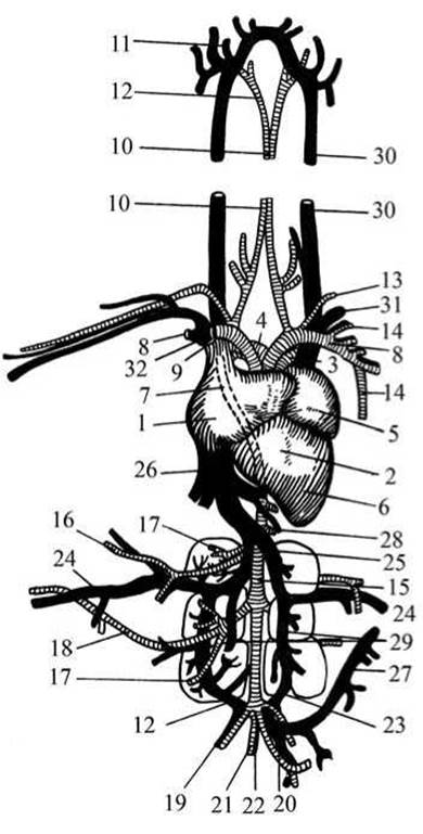
Fig. 136 CIRCULATORY SYSTEM OF a bird (pigeon):
1 — right atrium; 2 — right ventricle of the heart; 3 — left pulmonary artery; 4 — right pulmonary artery; 5 — left atrium; 6 — left ventricle of the heart; 7 — right aortic arch; 8, 9 — innominate arteries; 10-12 — carotid arteries; 13 — Subclavian Artery; 14 — left pectoral artery; 15 — aorta; 16 — right femoral artery; 17 — renal artery; 18 — sciatic artery; 19 — iliac artery; 20 — posterior mesenteric artery; 21 — caudal artery; 22 — caudal vein; 23 — renal portal vein; 24 — femoral vein; 25 — iliac vein; 26 — posterior vena cava; 27 — intestinal vein; 28 — supra-intestinal vein; 29 — renal vein; 30 — jugular vein; 31 — subclavian vein; 32 — anterior vena cava.

Fig. 137 Respiratory organs of the pigeon:
1 — trachea; 2 — lungs; 3-11 — air sacs.
Compare with the circulatory system of fish, amphibians, and reptiles.
Respiratory system. Continuing the dissection, examine the pigeon's respiratory organs — the lungs and their associated airways.
Air enters the nostrils, passes from the Nasal cavity through the choanae into the Oral Cavity and into the trachea. Examine the trachea, a long tube whose walls are supported by cartilaginous rings; the larynx located at the upper end of the trachea; and the syrinx (lower larynx), which is present only in birds (Fig. 137).
The bronchi enter the lungs and branch within them, forming microscopic tubes called bronchioles. Locate the pigeon's lungs. They are pink, spongy in texture, and permeated by a dense network of capillaries. The respiratory surface area of the avian lung is very large. Lifting the lungs reveals that they are fused to the dorsal wall of the thoracic cavity. The volume of a bird's lungs cannot change significantly during inhalation and exhalation. Air passes through them and enters the air sacs.

Fig. 138 Urogenital System of a male (A) and female (B) pigeon:
1 — Adrenal gland; 2 — Testis; 3 — vas deferens; 4 — Kidneys; 5 — Ureter; 6 — cloaca; 7 — opening of the vas deferens; 8 — opening of the ureter; 9 — Ovary; 10 — oviduct funnel; 11 — oviduct; 12 — opening of the oviduct; 13 — vestige of the right oviduct.
Reproductive System. Deep within the abdominal cavity, on either side of the vertebral column, lie the two bean-shaped Testes of the male pigeon. Vasa deferentia extend from the testes and empty into the cloaca. In the female, locate the single, cluster-like ovary situated on the left side of the body cavity (the right ovary is reduced). The oviduct begins with a funnel near the ovary and opens into the cloaca (Fig. 138). Fertilization in birds is internal.
Excretory system. Remove the reproductive organs and examine the trilobed kidneys (metanephros) — they are dark red and located on either side of the vertebral column in the depressions of the iliac bones. Extending backward along the kidneys are the Ureters (thin white cords) that open into the cloaca. Birds lack a Urinary Bladder. Locate the Adrenal Glands — Endocrine glands situated at the anterior border of the kidneys.
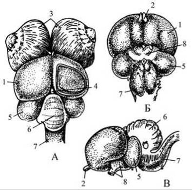
Fig. 139 Brain of a bird (pigeon):
A — dorsal view; B — ventral view; C — lateral view;
1 — cerebral hemispheres; 2 — olfactory lobes; 3 — eyeballs; 4 — Pineal Gland (epiphysis); 5 — optic lobes; 6 — Cerebellum; 7 — Medulla Oblongata; 8 — optic nerves.
Nervous system. Sever the bird's head along with a portion of the neck. Grasp the skin of the neck with forceps, pull it forward toward the head, and carefully peel it off like a glove to expose the Skull bones. Hold the pigeon's head in your left hand with the beak facing you, and use a sharp scalpel to shave away the skull bones from back to front (much like sharpening a pencil). Using forceps, carefully break away the bone fragments piece by piece until the pigeon's brain is exposed.
Examine the brain (Fig. 139). Note that the cerebral hemispheres are highly developed — they cover and even push aside the lobes of the Midbrain. The olfactory lobes of the Forebrain are very small (compare with the olfactory lobes of fish, amphibians, and reptiles). The midbrain is represented by the optic lobes, which form a prominent optic tectum and are large and well developed. Note the pigeon's cerebellum — it is very large, covers a significant portion of the medulla oblongata, and features transverse furrows. The Development of the cerebellum is associated with complex body movements.
Last update: 13/08/2026
Editorial and Educational Adaptation: This material has been compiled based on the primary/original source text. The project team performed an editorial review, corrected technical inaccuracies, structured sections, and adapted the content for an educational format.
What was processed:
- elimination of formatting defects (OCR errors, structural breaks, corrupted characters);
- editorial organization of content;
- standardization of terminology in accordance with academic sources;
- verification of factual statements against the original source text.
All mentions of the author, publication year, and origin of the primary text have been preserved in accordance with the source.