Practical Guide to Zoology: Study Guide - T. A. Dauda 2014
Multicellular Animals
Chordates
Subphylum Vertebrates (Craniates) - Superclass Tetrapods - Class Reptiles
Reptiles, alongside birds and mammals, form the group of higher terrestrial vertebrates.
All higher vertebrates (amniotes) exhibit internal Fertilization. Their reproduction occurs on land, and only a few viviparous species (such as cetaceans) reproduce in Water. The embryonic development of amniotes is characterized by The Emergence of specialized extraembryonic membranes. These membranes ensure that the embryo can develop in a gaseous environment in egg-laying species.
Reptiles clearly display all the defining traits of higher vertebrate animals. They possess a comparatively more advanced Brain, marked by the appearance of a secondary cerebral roof, the neopallium, which serves as the precursor to the Cerebral Cortex.
A well-developed cervical region has evolved in the Skeleton, granting the HEAD greater mobility. The Skin features a cornified epidermal layer and horny scales that protect the body from desiccation. Respiration is exclusively pulmonary. A rib cage is present, providing a more efficient breathing mechanism than that of amphibians.
The Heart contains a septum (incomplete in most species) dividing the left and right ventricles, from which three independent arterial trunks emerge, rather than a single one as seen in amphibians.
At the same time, the body Temperature of reptiles is variable because the presence of mixed Blood in the trunk Arteries limits their capacity for thermoregulation.
Modern reptiles represent a tiny fraction of the Class that flourished during the Mesozoic era and are classified into four orders: Rhynchocephalia, Squamata, Crocodilia, and Chelonia.
To study the external features and internal Organization of reptiles, any local lizard species is best suited as a model.
Sand Lizard (Lacerta agilis).
The sand lizard, widely distributed across the middle latitudes and southern regions of Russia, can be used as a study object. It inhabits open, dry forest patches and meadows, living in burrows. Its body temperature is variable. It exhibits diurnal activity, and its development proceeds without metamorphosis. It feeds on insects, their larvae, and other invertebrates. It hibernates during the winter.
Materials and Equipment. For every one to two students, the following are required: a lizard anesthetized with ether or preserved in 75% alcohol; a dissecting tray; forceps; a scalpel; scissors; dissecting needles; pins (10–15 pcs.); a hand lens; waste dishes; charts: "Internal Structure of a Lizard," "Diagram of the REPTILIAN Circulatory system," and "Brain Structure.">
Assignment. Study the external Anatomy of the lizard. Dissect the lizard, expose, and sketch its Internal Organs. Open the cranium, examine, and draw the brain. Study The structure of the circulatory system and the heart. Throughout your study, identify similarities with amphibians alongside the novel, progressive traits that emerged in reptiles in connection with their transition to a fully terrestrial lifestyle. Complete the following drawings: "Internal STRUCTURE OF THE Lizard," "Diagram of the Circulatory System," and "Brain Structure.">
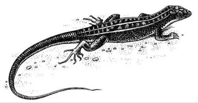
Fig. 129 Sand lizard (Lacerta agilis)
Study of external anatomy using living and anesthetized (or preserved) lizards. Observe the lizard's body—it consists of a head, an elongated trunk separated from the head by a true neck (a novel feature compared to amphibians), a long tail, and paired (fore and hind) pentadactyl limbs (Fig. 129). Note that the lizard's head is mobile, and the difference in length between the forelimbs and hindlimbs is relatively small (compare with amphibians). Observe that both the front and rear legs possess five digits equipped with small claws. This grants the lizard The ability to move swiftly across the ground and climb inclined surfaces.
In the course of evolution, the decrease in environmental moisture prompted the disappearance of Skin glands in reptiles. Notice that reptile skin is dry and covered in horny scales. Examine the ventral scales as well as those covering the back and sides: they differ in shape and size.
Observe the Mouth, nostrils, and the eyes located on the sides of the head, which are equipped with eyelids. The upper eyelid is less mobile than the lower one, and a nictitating membrane (third eyelid) is situated at the anterior corner of the eye. In a preserved lizard, the eyelids are difficult to examine. Using a hand lens, locate the auditory slits behind the eyes, where the tympanic membrane is positioned. Take the anesthetized lizard in your hand with its head facing toward you, open its mouth slightly, and insert scissors to make a horizontal cut on both the right and left sides extending back to the auditory slits. Fold the lower jaw downward, and locate the Tongue, which is forked at the tip (an Organ of Touch). While gently pulling the tongue out with forceps, examine the laryngeal (respiratory) cleft and the entrance to the Esophagus; the openings of the internal nares (choanae) are visible in the anterior part of the palate. Examine the jaws and palate using a lens; small conical Teeth are situated on the jaws. In preserved specimens, the Oral Cavity is not examined.
Locate the cloacal opening at the Base of the tail between the hind limbs.
Dissection. Internal structures should be studied on a freshly dissected lizard, while also utilizing a wet-mounted dissected specimen, an injected specimen, and a circulatory system diagram.
Secure the lizard in the dissecting tray ventral-side up by pinning down its limbs. Using scissors, make two skin incisions: a transverse one just in front of the cloaca, and a longitudinal one extending from the cloaca to the lower jaw. Carefully separate the skin flaps from the Muscles, using a scalpel where necessary, and pin them back to the sides. Cut through the musculature in the same manner, severing the BONES OF THE Pectoral Girdle and the Sternum. Fold the muscular flaps, along with the skin, outward and pin them to the bottom of the tray. Cover the preparation with water to facilitate clear observation of the internal organs. Preserved lizards are not submerged in water.
Sketch the outline of the lizard's body and, as you proceed with your study of its internal anatomy, fill in the arrangement of the organs (Fig. 130).
Circulatory System. The heart is visible in the upper region of the body cavity. Free it from the pericardial sac by making a small incision; locate the two thin-walled atria and the more massive, thick-walled ventricle.
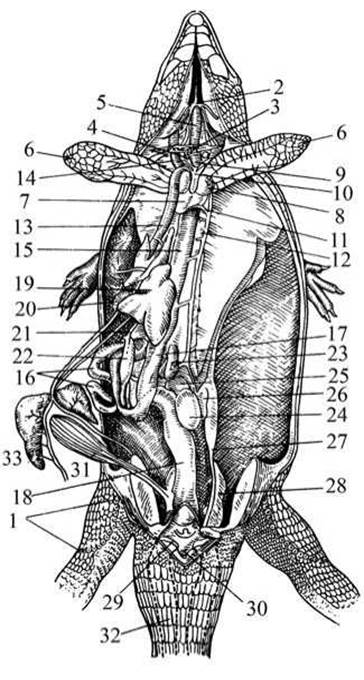
Fig. 130 Internal structure of a lizard:
1 — femoral pores; 2 — Hyoid bone; 3 — Thyroid Gland; 4 — Thymus gland; 5 — Trachea; 6 — lung; 7, 8 — heart; 9 — carotid artery; 10 — left aortic arch; 11, 12 — dorsal aorta; 13 — hepatic vein; 14 — jugular vein; 15 — esophagus; 16 — Small Intestine; 17 — Stomach; 18 — rectum; 19 — Liver; 20 — Gallbladder; 21 — Bile ducts; 22 — Pancreas; 23 — Spleen; 24, 25 — Testes; 26 — Adrenal Glands; 27 — vasa deferentia; 28 — Kidneys; 29 — urogenital aperture; 30 — cloaca; 31 — Urinary Bladder; 32 — tail; 33 — fat body.
The heart is three-chambered, but due to the presence of an incomplete septum in the ventricle, blood mixing is only partial and occurs to a lesser extent than in amphibians. The lizard's body is supplied with mixed blood that has a higher oxygen content than the mixed blood of a frog. The sinus venosus is largely incorporated into the right atrium. The conus arteriosus is absent (unlike in amphibians). The systemic Circulation vessels (two aortic arches — right and left) and Pulmonary Circulation vessels (paired pulmonary artery) emerge independently from the ventricle.
Examine the SYSTEMIC AND PULMONARY circulations using the diagram and the injected specimen. The pulmonary artery emerges from the right half of the ventricle. Shortly after leaving the heart, it divides into the right and left arteries, which carry deoxygenated blood to the Lungs. Blood enriched with oxygen in the lung capillaries returns via the Pulmonary Veins to the left atrium. Thus, the pulmonary circulation begins in the right half of the ventricle and terminates in the left atrium.
The right aortic arch originates from the left side of the ventricle and carries oxygenated blood. It gives rise to two arteries (right and left) that supply oxygenated blood to the head. Below the carotid arteries, the subclavian arteries branch off from the right aortic arch, directing blood to the forelimbs. The left aortic arch originates at the boundary between the right and left halves of the ventricle and carries mixed blood. It crosses the right aortic arch, loops around the esophagus on the left side, and extends downwards. Both aortic arches unite below the heart to form the dorsal aorta. Not far from the junction of the arches, the celiacomesenteric artery branches off from the left arch. Numerous arteries branch from the latter and from the dorsal aorta, supplying blood to the internal organs and hindlimbs. Deoxygenated blood from the anterior part of the body collects into two anterior venae cavae, and from the posterior part — into the unpaired posterior vena cava, emptying into the right atrium. Thus, the systemic circulation originates from the left side of the ventricle and terminates in the right atrium.
Respiratory organs. Remove The Heart and examine the respiratory organs: the glottis, Larynx, long trachea formed by incomplete cartilaginous rings, short Bronchi, and elongated sac-like lungs. The lungs are saccular in shape, but their walls are more compartmentalized (cellular) than those of amphibians, since cutaneous respiration is absent in reptiles.
Digestive System. Locate the esophagus situated behind the trachea. Insert a thin tube or probe into the Pharynx and push it down into the esophagus. In the dissected lizard, it is clearly visible that the esophagus transitions into The Stomach. To examine the stomach, move aside the massive liver, which is partially divided into two lobes. Locate the oval gallbladder on the inner side of the liver. The stomach leads into the duodenum. The pancreas is visible within the loop of the duodenum. Trace how the short small intestine transitions into the Large Intestine. Note that the posterior part of the large intestine receives the ducts of the Urogenital System and forms the cloaca.
Locate the elongated, dark-red spleen near the posterior end of the stomach. It is clearly visible that the intestine is attached to the DORSAL SIDE OF the body cavity by peritoneal folds.
Reproductive System. Cut through the large intestine and mesentery, and reflect the entire intestine and liver to the side. In the male, locate a pair of bean-shaped testes positioned on either side of THE Vertebral Column in the lumbar region, suspended asymmetrically by a specialized mesentery—one Testis slightly higher than the other (see Fig. 131). Alongside the testes on the outer side, coiled epididymides (rudiments of the pronephros/mesonephros) are visible, giving rise to coiled vasa deferentia that empty into the Ureters (which are not visible). The ureters open into the cloaca.
In the female, locate the paired, botryoidal (grape-like) Ovaries situated in the lumbar region on either side of the Vertebral Column and suspended by a mesentery. Examine the oviducts—highly convoluted tubes opening via funnels into the body cavity; their other ends open into the cloaca. Fertilization in lizards is always internal. Locate the adrenal glands, which appear as elongated orange-yellow structures adjacent to the Gonads of both male and female specimens.
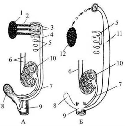
Fig. 131. Diagram of the urogenital system of reptiles:
A — male; B — female; 1 — testis; 2 — efferent ductules; 3 — Epididymis; 4 — Wolffian duct (vas deferens); 5 — remnant of the pronephros (primary Kidney); 6 — Blood Vessels; 7 — Ureter; 8 — urinary bladder; 9 — cloaca; 10 — secondary kidney (metanephros); 11 — Müllerian duct (oviduct); 12 — Ovary.
Cut through the pelvic bones along the midline and locate the secondary kidneys (metanephros) in the pelvic region, from which ureters emerge and empty into the cloaca. Starting with reptiles, pelvic kidneys replace trunk kidneys. Examine the thin-walled urinary bladder opening into the cloaca, provided it has not collapsed.
Remove the lizard from the dissecting pan, make a skin incision at the border of the Head and Neck region, and skin it. Cut the vertebral column at the base of the Skull. Insert scissors into the foramen magnum and make cuts toward the eyes. Break away the skull bones with forceps, rinse the brain with water, and examine it dorsally. It is evident that the cerebral hemispheres and Cerebellum are more developed compared to amphibians. The cerebral hemispheres partially cover the Diencephalon, and the cerebellum covers a significant portion of the Medulla Oblongata (Fig. 132). The olfactory lobes are smaller than those of a frog. With careful dissection, the parietal organ is noticeable, structurally similar to an eye and capable of perceiving light stimuli. The optic tectum (Midbrain) appears as two rounded colliculi.
Draw a DORSAL VIEW OF the brain.
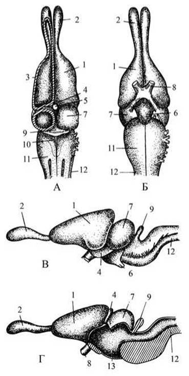
Fig. 132. Brain of a lizard:
A — dorsal view; B — ventral view; C, D — lateral view; 1 — cerebral hemispheres; 2 — olfactory lobes; 3 — corpus striatum; 4 — diencephalon; 5 — parietal organ; 6 — Pituitary Gland (hypophysis); 7 — optic tectum (midbrain); 8 — optic nerves; 9 — cerebellum; 10 — Fourth ventricle; 11 — medulla oblongata; 12 — Spinal Cord; 13 — Third ventricle.
Last update: 13/08/2026
Editorial and Educational Adaptation: This material has been compiled based on the primary/original source text. The project team performed an editorial review, corrected technical inaccuracies, structured sections, and adapted the content for an educational format.
What was processed:
- elimination of formatting defects (OCR errors, structural breaks, corrupted characters);
- editorial organization of content;
- standardization of terminology in accordance with academic sources;
- verification of factual statements against the original source text.
All mentions of the author, publication year, and origin of the primary text have been preserved in accordance with the source.