Molecular Biology of the Cell - Volume 1 - Alberts B., Bray D., Lewis J., Raff M., Roberts K., Watson J. 1994
Introduction to Cell Biology
Small Molecules, Energy, and Biosynthesis
Coordination of Catabolism and Biosynthesis
2.5.1. METABOLISM is an organized and regulated process
Some idea of how exquisitely designed The Cell is, when viewed as a chemical machine, can be gained from Figure 2-35, which shows a map containing only a fraction of all metabolic pathways. All these numerous reactions occur within a cell whose diameter does not exceed 0.1 mm, with each reaction requiring different Enzymes, which themselves are products of a whole series of information-transfer and protein-synthesis reactions. Take any small molecule, such as The amino acid Serine; there are half a dozen or more enzymes capable of chemically modifying it in various ways. Serine can bind to AMP (adenylation at the stage preceding its incorporation into Protein Synthesis), or be cleaved to Glycine, or converted to Pyruvate before being oxidized; it can be acetylated by acetyl-CoA or transferred to a fatty acid to form phosphatidylserine. All these different pathways compete for the same serine molecule, while at the same time a similar competition is underway for thousands of other small molecules. One might think that the system as a whole requires such delicate balancing that any minor disruption, such as a temporary change in diet, would lead to its demise.
Class="center">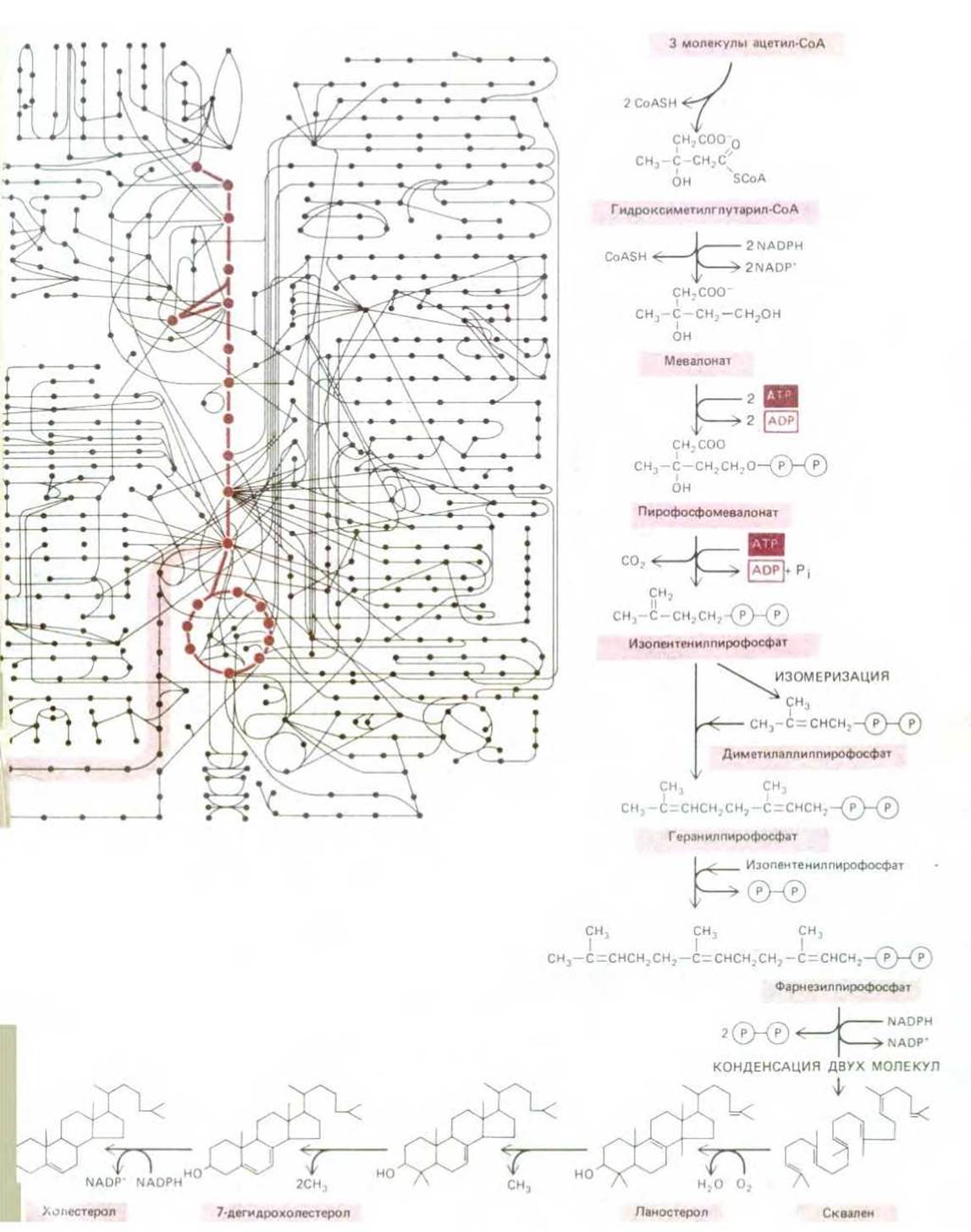
Figure 2-35. Some of the Chemical Reactions occurring in a cell. A. About 500 common metabolic reactions diverge in different directions from The Glycolytic Pathway and The Citric Acid Cycle (shown in darker color). Each chemical reaction is represented as a solid circle. A typical mammalian cell synthesizes more than 10,000 Proteins, most of which are enzymes. In an arbitrarily chosen area (highlighted in a lighter color) of this complex web of metabolic pathways, Cholesterol is synthesized from acetyl-CoA. To the right and below the "maze", this area is shown in more detail on an enlarged scale (B). →
In reality, however, the cell possesses striking stability. In response to any external influence, the cell's reaction is directed toward restoring its original state. It can adapt and continue to function in a coordinated manner during starvation or disease. Many Selection/21.html">Types of Mutations can lead to the elimination of individual reaction sequences, and yet the cell survives, provided that certain minimum requirements are met. This is possible due to a complex system of mechanisms that regulate and coordinate chemical reactions in Cells. Some of the higher levels of control are discussed in subsequent chapters. Here, we address only the simplest mechanisms that regulate the flow of small molecules in Various metabolic pathways of the cell.
2.5.2. Metabolic pathways are regulated by changes in enzymatic activity [20]
The concentrations of various small molecules in the cell are remarkably stable, which is achieved through feedback regulation. Regulatory molecules of this type adjust the flow of metabolites along a specific metabolic pathway by temporarily increasing or decreasing The activity of Key Enzymes. For example, the first enzyme in a given reaction sequence is usually inhibited by the end product of that metabolic pathway via negative feedback; thus, if too much end product accumulates, the further entry of precursors into this metabolic pathway is automatically inhibited (Figure 2-36). In the case of branching or intersecting metabolic pathways, which occurs quite frequently, there are typically several points where control is exerted by different end products. Structure/19.html">The Importance of such feedback regulation processes is illustrated in Figure 2-37, which shows the REGULATION OF ENZYMATIC Activity in the reaction sequences leading to Amino acid synthesis.
Feedback regulation can operate almost instantaneously and is reversible; furthermore, a specific end product can inhibit the enzymes that catalyze its synthesis while simultaneously acting as an activator for enzymes of another metabolic pathway. The Molecular Basis of this type of control in cells is well understood; however, we will not address this issue here, as it requires an understanding of Protein Structure. Therefore, we will defer this topic to Chapter 3.
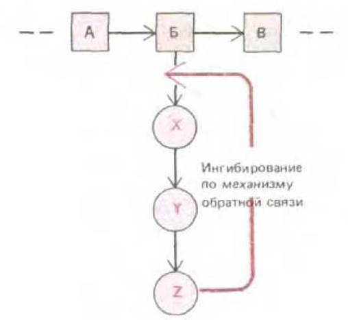
Figure 2-36. Feedback inhibition in a single biosynthetic pathway. The end product Z inhibits the first enzyme required for its synthesis, thereby regulating its own level in the cell.
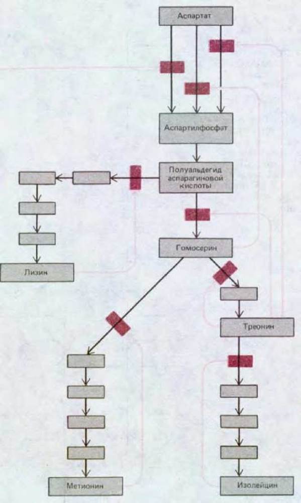
Figure 2-37. Feedback inhibition in the Synthesis of the Amino Acids Lysine, Methionine, Threonine, and isoleucine in Bacteria. Colored arrows indicate the sites where enzymes are inhibited by reaction products. Note that the initial reaction is catalyzed by three different enzymes (called Isoenzymes), each of which is inhibited by its own end product.
2.5.3. Catabolic reactions can be reversed by the input of energy [21]
Large-scale changes affecting the metabolism of the entire cell can be achieved by regulating key enzymes. For example, a specific feedback regulation scheme allows the cell to switch from glucose breakdown to its Biosynthesis, or Gluconeogenesis. The Need for such a pathway reversal is particularly acute both during periods of intense exercise, when glucose required for Muscle contraction is synthesized in Liver cells, and during starvation, when glucose must be formed from Fatty acids and amino acids for the body's survival. The normal breakdown of glucose to pyruvate during Glycolysis is catalyzed by several different sequentially acting enzymes. Most of the Reactions Catalyzed by these enzymes are easily reversed; however, three of them (steps 1, 3, and 9 in Figure 2-20) are virtually irreversible. In fact, The process of glucose breakdown is usually driven by a large negative free-energy change in these reactions. For this process to run in the opposite direction and produce glucose from pyruvate, a bypass (shunt) must exist around each of these reactions, and the bypass reactions must run "uphill"—an energy-consuming process (Figure 2-38). Thus, while The breakdown of one glucose molecule into two pyruvate molecules yields two ATP molecules, reversing the reaction during gluconeogenesis requires the Hydrolysis of four ATP molecules and two GTP molecules. This is equivalent to the hydrolysis of six ATP molecules for each newly synthesized glucose molecule.
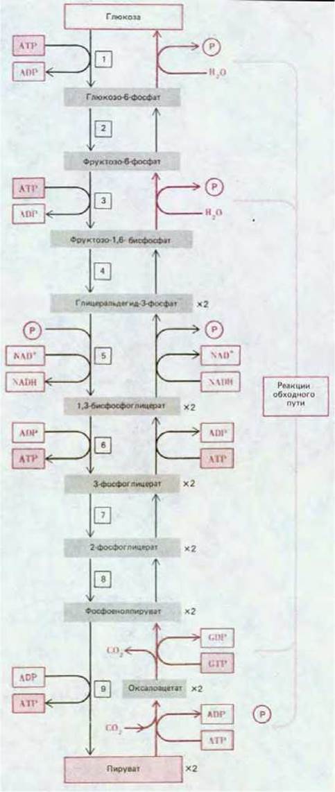
Figure 2-38. Comparison of the reactions leading to glucose synthesis during gluconeogenesis with the reactions of glucose breakdown during glycolysis. Glucose breakdown reactions (glycolytic reactions) are energetically favorable (the free-energy change is less than zero), whereas synthesis reactions require energy input. Glucose synthesis requires various bypass enzymes that shunt reactions 1, 3, and 9 of glycolysis. The overall direction of the reactions between glucose and pyruvate is determined by feedback control mechanisms operating at these three key steps.

Figure 2-39. The sudden addition of glucose to an extract containing glycolytic Enzymes and Coenzymes can cause significant fluctuations in the concentration of certain intermediates, such as NADH. Such metabolic oscillations can be due, in particular, to the positive feedback Regulation of the glycolytic enzyme Phosphofructokinase.
The bypass reactions (Figure 2-38) must be tightly controlled so that glucose is rapidly broken down in the event of "energy starvation" and synthesized when the cell is rich in nutrients. If the forward and reverse reactions could proceed without restriction, they would wastefully cycle many metabolites back and forth through futile cycles, consuming vast amounts of ATP and generating useless heat.
The elegance of such control mechanisms can be illustrated by a single example. Step 3 of glycolysis is one of the reactions that must be bypassed during glucose formation. Normally, this step involves The addition of a second phosphate group from ATP to fructose 6-phosphate; the reaction is catalyzed by the enzyme phosphofructokinase. This enzyme is activated by AMP, ADP, and inorganic phosphate, and inhibited by ATP, citrate, and fatty acids. In other words, the enzyme is activated when energy stores are low and ATP breakdown products accumulate; it is inactivated when there are abundant energy stores (in the form of ATP) or nutrients in the form of fatty acids or citrate (derived from amino acids). The enzyme that catalyzes the reverse (bypass) reaction (The formation of glucose by hydrolysis of fructose 1,6-bisphosphate to fructose 6-phosphate) is fructose 1,6-bisphosphatase. The activity of this enzyme is regulated by feedback in the same manner as phosphofructokinase, but with the opposite effect, so that fructose 1,6-bisphosphatase is active when phosphofructokinase is inactive.
Note that phosphofructokinase is activated by ADP, which is a product of the reaction catalyzed by this enzyme (ATP + fructose 6-phosphate → ADP + fructose 1,6-bisphosphate), and inhibited by ATP, one of the substrates of this reaction. As a result, this enzyme can activate itself, being subject to complex positive feedback control. Under certain conditions, such feedback control causes unusual oscillations in the activity of this enzyme, leading to corresponding fluctuations in the concentrations of various glycolytic intermediates (Figure 2-39). The physiological significance of such specific oscillations is unclear. However, they demonstrate how a biological oscillator can be created using just a few enzymes. Such oscillations could, in principle, serve as a kind of "internal clock," allowing the cell to "keep time" and, for example, perform specific Functions at particular intervals.
2.5.4. Enzymes can be switched to an active or inactive state by covalent modifications [22]
The types of feedback control discussed above allow for the continuous Regulation of Metabolic pathway rates in response to second-by-second fluctuations in metabolism. In addition, cells possess special regulatory mechanisms for situations requiring long-term (from several minutes to several hours) changes in enzyme activity. Such mechanisms involve reversible covalent modifications of enzymes, which are often (though not always) achieved by attaching a phosphate group to a specific (serine, threonine, or Tyrosine) amino acid residue of the enzyme. The phosphate is derived from ATP, and its transfer is catalyzed by enzymes called protein Kinases.
In the next chapter, we will examine how The change in enzyme shape upon phosphorylation enhances or suppresses its activity. The subsequent removal of the phosphate group, which reverses The Effect of phosphorylation, is achieved by another enzyme called phosphoprotein phosphatase. Covalent Modification of enzymes represents regulation in a new dimension, as it enables the Regulation of Specific reaction pathways by signals (such as Hormones) that are not metabolic intermediates.
2.5.5. Reactions are compartmentalized both at THE CELLULAR LEVEL and at the level of the whole Organism [23]
Not all metabolic reactions in a cell occur within the same subcellular compartments (distinct subcellular structures). Since different enzymes are localized in different cellular compartments, the flow of chemical intermediates is directed physically as well as chemically.
The simplest form of such spatial segregation is observed when two enzymes catalyzing two sequential reactions form a single enzyme complex; consequently, the product of the first enzymatic reaction does not need to diffuse through the Cytoplasm to encounter the second enzyme. As soon as the first reaction ends, the second begins immediately. Some large enzyme aggregates carry out an entire sequence of reactions while remaining in contact with the substrate. For example, The conversion of pyruvate to acetyl-CoA occurs in three steps, each taking place on the same enzyme complex (Fig. 2-40), and in fatty acid synthesis, an even longer sequence of reactions is catalyzed by a single multienzyme assembly. It is not surprising that some of the largest enzyme complexes are responsible for the synthesis of macromolecules such as proteins and DNA.
At the next level of spatial segregation within the cell, functionally related enzymes are concentrated in the same membrane or within the membrane-bound aqueous compartments of Organelles. This can be illustrated by the metabolism of glucose (Fig. 2-41). Pyruvate produced by glycolysis is actively transported from the Cytosol into the mitochondrial matrix, which contains all the enzymes and metabolites of The Citric Acid cycle. Furthermore, The inner mitochondrial membrane itself contains all the enzymes that catalyze the sequential reactions of Oxidative Phosphorylation, including Electron transfer from NADH to O2 and ATP synthesis. Consequently, the entire mitochondrion can be viewed as a tiny factory producing ATP. Similarly, other cellular organelles, such as The Nucleus, the Golgi apparatus, and Lysosomes, can be considered specialized compartments housing functionally related enzymes to perform specific tasks. In a sense, a living cell is like a modern city with many specialized services concentrated in different districts and linked to one another by an extensive communication network.

Fig. 2-40. Structure of pyruvate dehydrogenase, a large multienzyme complex in which reaction intermediates pass directly from one enzyme to another. This enzyme complex catalyzes the conversion of pyruvate to acetyl-CoA.

Fig. 2-41. Spatial segregation of the three stages of glucose breakdown in a Introduction/5.html">Eukaryotic Cell. Glycolysis occurs in the cytosol, whereas the Reactions of the citric acid cycle and oxidative phosphorylation take place exclusively in Mitochondria.
In Multicellular Organisms, spatial Organization extends far beyond the individual cell. Different Tissues of the body possess diverse sets of enzymes and contribute in various ways to the survival of the organism. In addition to differences in specialized products, such as hormones or Antibodies, There are also significant differences in common metabolic pathways among different cell types of the same organism. Although virtually all cells contain enzymes for glycolysis, the citric acid cycle, lipid Synthesis and degradation, and Amino acid metabolism, the rates of all these processes are regulated differently in various tissues. Nerve Cells, perhaps the most "fastidious" cells in the body, contain extremely small reserves of Glycogen or fatty acids, relying entirely on glucose supplied by the Blood. Liver cells supply glucose to actively working muscle cells. In addition, they use lactic acid produced in the Muscles to synthesize glucose (Fig. 2-42). Each cell type has its own specific metabolic features, and they cooperate extensively under normal conditions, as well as during exercise, stress, or starvation.
Summary
Thousands upon thousands of different biochemical reactions occurring simultaneously within a cell are closely coordinated. A variety of control mechanisms regulate the activity of cellular enzymes as conditions within the cell change. The most common form of regulation is readily reversible feedback inhibition, where the first enzyme of a metabolic pathway is inhibited by the end product of that pathway. A longer-term form of regulation involves the chemical modification of one enzyme by another, which often occurs through phosphorylation. Combinations of regulatory mechanisms can produce profound and lasting changes in cellular metabolism. Not all cellular reactions occur within the same intracellular compartments, and the Spatial Compartmentalization of the cell by internal membranes allows organelles to specialize in their biochemical functions.

Fig. 2-42. Schematic representation of the metabolic interaction between liver and muscle cells. Glucose serves as the primary "fuel" for actively working muscle cells, with a significant portion supplied by liver cells. Lactic acid—the end product of anaerobic glucose breakdown in muscles during glycolysis—is converted back into glucose via gluconeogenesis in the liver.
General
Herriott J.; Jacobson G; Marmur J.; Parsom W. Papers in Biochemistry. Reading, MA: Addison-Wesley, 1984.
*Lehninger A.L. Principles of Biochemistry. New York: Worth, 1982.
Stryer L. Biochemistry, 3rd ed. New York: W.H. Freeman, 1988.
Wood W.B.; Wilson J.H.; Benbow R.M.; Hood I.E. Biochemistry: A Problems Approach, 2nd ed. Menlo Park. CA: Benjamin-Cummings, 1981.
Cited References
1. Henderson L.J. The Fitness of the Environment. Boston: Beacon, 1927; reprinted 1958. (A classic analysis in an accessible presentation.)
2. Ingroham J. L.; Maaloe О.; Neidhardt F. C. Growth of the Bacterial Cell. Sunderland, MA: Sinauer, 1983.
3. Roehrig K. L. Carbohydrate Biochemistry and Metabolism. Westport, CT: AUI Publishing, 1984.
Sharon N. CARBOHYDRATES. Sci. Am., 243(5): 90-116, 1980.
4. Robertson R. N. The Lively Membranes. Cambridge, U. K.; Cambridge University Press, 1983.
5. Saenger W. Principles of Nucleic Acid Structure. New York: Springer, 1984.
6. Hess B.; Markus M. Order and chaos in biochemistry. Trends Biochem. Sci. 12: 45-48, 1987.
Lehninger A. L. Bioenergetics: The Molecular Basis of Biological Energy Transformations. 2nd ed. Menlo Park, CA: Benjamin-Cummings, 1971.
**Schrodinger E. What is Life? Mind and Matter. Cambridge, U. K.: Cambridge University Press, 1969.
7. Dickerson R. E. Molecular Thermodynamics. Menlo Park, CA: Benjamin, 1969.
Klotz I. M. Energy Changes in Biochemical Reactions. New York: Academic Press, 1967.
8. Raven P. H.; Evert R. H.; Eichhorn S. E. Biology of Plants, 4th ed. New York: Worth, 1981.
9. Racker E. From Pasteur to Mitchell: a hundred years of bioenergetics. Fed. Proc. 39: 210-215, 1980.
10. Fothergill-Gilmore L. A. The Evolution of the glycolytic pathway. Trends Biochem. Sci. 11: 47-51, 1986.
Shulman R. G. NMR Spectroscopy of living cells. Sci. Am. 248(1): 86-93, 1983.
11. Lipmann F. Wanderings of a Biochemist. New York: Wiley, 1971.
12. Schlenk F. The ancestry, birth and adolescence of ATP. Trends Biochem. Sci. 12: 367-368, 1987.
13. McGilvery R. W. Biochemistry: A Functional Approach, 3rd ed. Philadelphia: Saunders, 1983.
Racker E. A. New Look at Mechanisms in Bioenergetics. New York: Academic Press, 1976.
14. Kornberg H. L. Tricarboxylic acid cycles. Bioessays 7: 236-238, 1987.
15. Krebs H. A. The history of The Tricarboxylic Acid Cycle. Perspect. Biol. Med. 14: 154-170, 1970.
Krebs H. A.; Martin A. Reminiscences and Reflections. Oxford, U. K.: Clarendon Press; New York: Oxford University Press, 1981.
16. Pullman M. E.; et al. Partial resolution of the enzymes catalyzing oxidative phosphorylation. J. Biol. Chem. 235: 3322-3329, 1960.
17. Prigogine I.; Stengers I. Order Out of Chaos: Man's New Dialogue with Nature. New York: Bantam Books, 1984.
18. Eisenberg D.; Crothers D. Physical Chemistry with Applications to the Life Sciences. Menlo Park, CA: Benjamin-Cummings, 1979.
Reich J. G.; Selkov E. E. Energy Metabolism of the Cell - A Theoretical Treatise. New York: Academic Press, 1981.
19. Martin B. R. Metabolic Regulation: A Molecular Approach. Oxford, U.K.: Blackwell Scientific, 1987.
***Newsholme E. A.; Start C. Regulation in Metabolism. New York: Wiley, 1973.
20. Pardee A. B. Molecular basis of biological regulation: origins of Feedback inhibition and allostery. Bioessays 2: 37-40, 1985.
21. Hess B. Oscillating reactions. Trends Biochem. Sci. 2: 193-195, 1977.
22. Cohen P. Control of Enzyme Activity. 2nd ed. London: Chapman and Hall, 1983.
The following Russian translations are available:
*Lehninger A. Principles of Biochemistry. In 3 volumes. Moscow: Mir, 1985.
**Schrödinger E. What is Life? From a Physicist's Point of View. 2nd ed. Moscow, Atomizdat, 1972.
***Newsholme E., Start C. Regulation in Metabolism. Moscow: Mir, 1977.
Last update: 12/08/2026
Editorial and Educational Adaptation: This material has been compiled based on the primary/original source text. The project team performed an editorial review, corrected technical inaccuracies, structured sections, and adapted the content for an educational format.
What was processed:
- elimination of formatting defects (OCR errors, structural breaks, corrupted characters);
- editorial organization of content;
- standardization of terminology in accordance with academic sources;
- verification of factual statements against the original source text.
All mentions of the author, publication year, and origin of the primary text have been preserved in accordance with the source.