Molecular Biology of the Cell - Volume 1 - Alberts B., Bray D., Lewis J., Raff M., Roberts K., Watson J. 1994
Introduction to Cell Biology
Macromolecules: Structure, Shape, and Informational Functions
Molecular Recognition Processes
Cellular macromolecules differ from small molecules not only in their larger size. Proteins, Nucleic Acids, and Polysaccharides possess unique and truly remarkable properties, which bear little resemblance to those of their small constituent molecules. Biological macromolecules are built from thousands, sometimes millions, of atoms assembled into a precisely determined three-dimensional Structure. Each of these macromolecules carries specific information encoded within its structure. This information can be viewed as a series of biological messages that can be "read" through interactions with other molecules, thereby enabling a specific function essential to The Cell.
In this chapter, we will analyze The structure of macromolecules, primarily proteins and nucleic acids, and attempt to explain how they have adapted through evolution to perform their Functions. We will examine the principles by which these molecules catalyze chemical transformations, build complex macromolecular structures, generate movement, and, most importantly, store and transmit hereditary information.
Macromolecules typically have molecular weights ranging from 10,000 to 1 million; thus, in terms of size, they occupy an intermediate position between the organic molecules described in Chapter 2 and the supramolecular structures and Organelles that will be discussed in subsequent chapters (Fig. 3-1). A single small molecule, Water, accounts for 75% of the total cell mass; macromolecules make up almost all of the remaining cell mass (Table 3-1).
As described in Chapter 2, macromolecules are assembled from low-molecular-weight subunits that join one after another to form a long polymer chain (see Fig. 2-33). Typically, only subunits of a single family participate in building each chain. Thus, Amino Acids link with Other Amino Acids to form proteins; NUCLEOTIDES link with other nucleotides to form nucleic acids; and sugars combine with other sugars to form polysaccharides. Because the precise sequence of subunits (monomers) is crucial for the normal functioning of a macromolecule, macromolecular Biosynthesis must involve mechanisms that precisely determine THE POSITION OF each monomer in the polymer chain.
Class="center">
Fig. 3-1. Comparison of protein sizes with those of other cellular components. A ribosome is a macromolecular complex consisting of approximately 60 proteins and RNA molecules.
Table 3-1. Approximate Chemical composition of a typical bacterium and a typical mammalian cell
Percent of total cell mass, %
|
Component 1) |
E. coli bacterium |
mammalian cell |
|
Н2О |
70 |
70 |
|
Inorganic ions (Na+, K+, Mg2+, Са2+, Cl-, etc.) |
1 |
1 |
|
Various small metabolites |
3 |
3 |
|
Proteins |
15 |
18 |
|
RNA |
6 |
1,1 |
|
DNA |
1 |
0,25 |
|
2 |
3 |
|
|
Other Lipids |
— |
2 |
|
Polysaccharides |
2 |
2 |
|
Total cell volume |
2- 10-12 cm3 |
4-10-9 cm3 |
|
Relative cell volume |
1 |
2000 |
1) Proteins, polysaccharides, DNA, and RNA are macromolecules. Lipids are not generally considered macromolecules, although they share some of their properties; for example, most lipids are synthesized as linear polymers from smaller molecules (the acetyl group of acetyl coenzyme A) and form larger structures (membranes) through self-assembly.
3.1.1. Specific macromolecular interactions depend on weak noncovalent bonds [2]
Macromolecular chains are formed by covalent bonds, which are strong enough to maintain The sequence of macromolecular subunits over long periods. However, the information encoded within this sequence is expressed through much weaker noncovalent bonds. These weak bonds occur both between different PARTS OF THE same macromolecule and between different macromolecules. Collectively, these bonds determine both the three-dimensional structure of macromolecular chains and how these structures interact with one another.
Noncovalent bonds in biological molecules are typically classified into three types: ionic interactions, Hydrogen Bonds, and Van der Waals interactions. Another important weak interaction is created by the structure of water, which tends to force hydrophobic groups together, thereby minimizing their disruptive effect on the hydrogen-bonded network of water molecules (Panel 2-1). This exclusion from aqueous solution is sometimes considered a fourth type of weak noncovalent bond. All four types of weak bonds are shown in Panel 3-1.
In aqueous solution, each noncovalent bond is 30 to 300 times weaker than the typical covalent bonds holding biological molecules together (Table 3-2) and is only slightly stronger than the average energy of molecular collisions caused by thermal motion at 37 °C. Unlike a single covalent bond, a single noncovalent bond is too weak to withstand thermal motion, which tends to pull molecules apart; therefore, many noncovalent bonds are required to hold the surfaces of two molecules together. A large number of noncovalent bonds can form between two surfaces only when many atoms on these surfaces fit together precisely (Fig. 3-2). This explains the Specificity of biological recognition, such as that occurring between an enzyme and its substrates.
Weak noncovalent bonds determine how different regions of a single molecule are positioned relative to one another; they also determine how such a macromolecule interacts with other molecules. However, as can be seen at the top of Panel 3-1, atoms behave as hard spheres of a defined radius (the 'van der Waals radius'). The inability of two atoms to overlap limits the number of spatial arrangements of atoms (or Conformations) possible for any polypeptide chain. In principle, a long, flexible chain, such as a protein molecule, could fold in an enormous number of ways, with each conformation having a different set of weak intramolecular interactions. In practice, however, most cellular proteins fold stably in only one way; evolution has selected Amino acid sequences in which one conformation can form much more favorable interactions than any other.
3.1.2. The helix is a common structural element in biological molecules built from repeating subunits [3]
Biological structures are often formed by linking similar subunits, such as amino acids or nucleotides, into a long repeating chain (Section 2.4.5). If all subunits are identical, neighboring subunits in the chain will be linked to each other in only one way: their relative orientation will be such that the contact energy between them is minimized. Each subunit is positioned in exactly the same way relative to its neighbor, so that subunit 3 fits into subunit 2, subunit 2 into subunit 1, and so on. Because assembling subunits in a straight line is very rare, they typically form a helix—a regular structure resembling a spiral staircase, as shown in Fig. 3-3. Depending on the direction of twisting, helices can be right-handed or left-handed (Fig. 3-4). The handedness of a helix does not change if the helix is turned upside down, but it is reversed by mirror reflection.
Helices are highly common among biological structures. Helical structures are formed both by molecules consisting of covalently linked subunits (DNA) and by large Protein Assemblies held together by noncovalent bonds (Actin filaments). This is not surprising: a helix arises simply by stacking many subunits, each repeating the exact orientation of the previous one.
3.1.3. Diffusion is The First stage of Molecular recognition [4]
Before two molecules can bind to each other, they must come into contact. This is achieved through thermal motion, which causes random movements, or molecular diffusion. Because molecules in a liquid rapidly collide and bounce off one another, an individual molecule moves first in one direction and then in another, tracing a 'random walk' (Fig. 3-5). The average distance traveled by such a molecule is proportional to the square ROOT of time. In other words, if it takes a molecule an average of 1 s to diffuse 1 µm, it will take an average of 4 s to diffuse 2 µm, 100 s to diffuse 10 µm, and so on. Thus, diffusion is an efficient way for molecules to travel short distances, but an inefficient way to travel long distances.
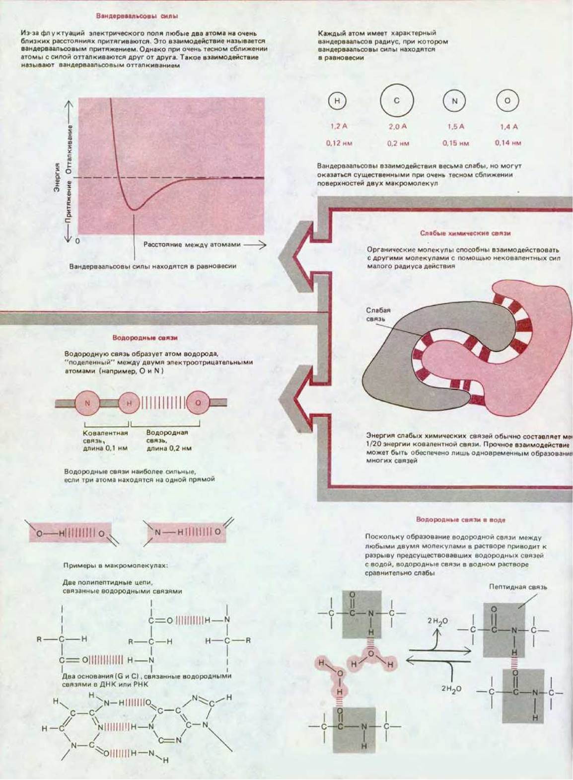
Panel 3-1. MAIN TYPES OF weak noncovalent bonds involved in macromolecular interactions.
Table 3-2. Covalent and noncovalent chemical bonds
Bond energy, kcal/mol 1)
|
Bond type |
Length, nm |
in vacuum |
in water |
|
Covalent |
0,15 |
90 |
90 |
|
Ionic |
0,25 |
80 |
1 |
|
Hydrogen |
0,30 |
4 |
1 |
|
Van der Waals |
0,20 |
0,1 |
0,1 |
1) Bond energy can be thought of as the energy required to break a bond. Here it is given in kilocalories per mole (kcal/mol). One kilocalorie is The amount of energy required to raise the Temperature of 1000 g of water by 1°C. Another widely used unit of measurement is the kilojoule (kJ), which is equal to 0,24 kcal. Individual bonds vary widely in strength depending on the specific atoms in their microenvironment, so the values shown can only serve as a rough guide. Note that the aqueous environment of the cell significantly weakens ionic and hydrogen bonds between non-aqueous molecules.
Experiments involving the Introduction of fluorescent Dyes and other labeled molecules into Cells have shown that small molecules diffuse in the Cytoplasm nearly as rapidly as they do in water. A molecule the size of ATP requires only 0,2 s to diffuse an average distance of 10 µm, which is the diameter of a small animal cell. Macromolecules, however, move much more slowly. This is due not only to their lower rate of diffusion but also to the fact that their movement is impeded by frequent collisions with many other macromolecules whose positions in the cytoplasm are fixed (Fig. 3-6).
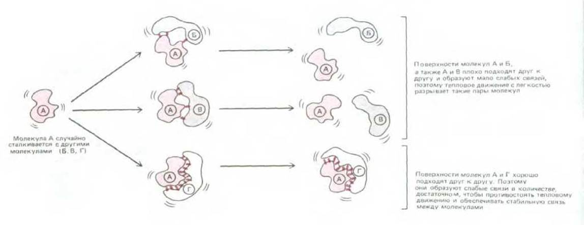
Fig. 3-2. Schematic diagram illustrating how macromolecules recognize each other through weak interactions.
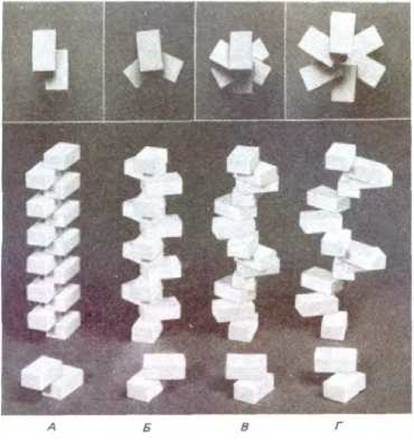
Fig. 3-3. A helix is formed when a series of subunits associate with each other in a regular manner. The interaction of two subunits is shown in the foreground, and the helices resulting from this interaction are shown in the Background. These helices have two (A), three (B), and six (C and D) subunits per turn. At the top of the figure is a top-down view of the helix. Note that helix D has a wider pitch than C.
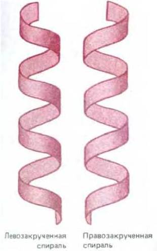
Fig. 3-4. Comparison of left- and right-handed helices. It is useful to remember that a standard screw, which tightens when turned clockwise, is right-handed. Note that a helix retains its handedness even if it is turned upside down.
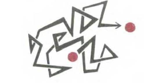
Fig. 3-5. Random movement. Molecules in solution move randomly due to constant collisions with other molecules. As a result, small molecules diffuse from one part of the cell to another in an astonishingly short time—less than a second.
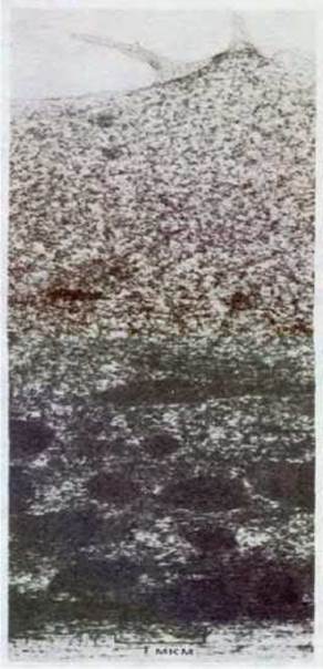
Fig. 3-6. Electron micrograph of a region of animal Cell Cytoplasm, illustrating the high concentration of proteins it contains. Macromolecules in the cytoplasm diffuse relatively slowly because they interact with other macromolecules; small molecules diffuse almost as rapidly as in an aqueous solution. The cell shown in this micrograph was prepared using a special rapid-freezing technique that preserves cytoplasmic structures. (From P. C. Bridgman and T. S. Reese, J. Cell Biol. 99: 1655-1668, 1984. Reproduced with permission of the Rockefeller University Press.)
3.1.4. Thermal motion not only brings molecules together but also pushes them apart [5]
Two macromolecules, or a macromolecule and a small molecule, collide by simple diffusion to form a complex. Complex formation can occur either immediately (in which case The rate of complex formation is said to be diffusion-limited) or with some delay if the interacting surfaces fit together only after some "adjustment" of the structure of one or both molecules. In any case, if the two interacting molecules come close enough, they form multiple weak bonds that persist until random thermal motion causes the molecules to dissociate again.
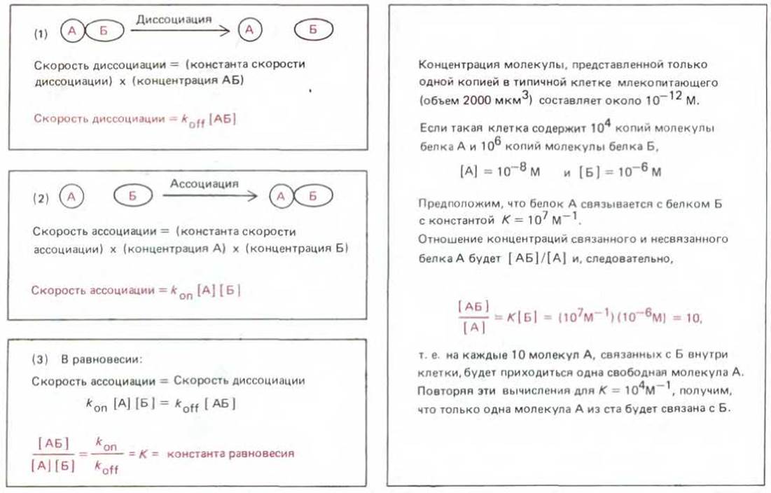
Fig. 3-7. THE PRINCIPLE OF equilibrium. The equilibrium between molecules A and B and the complex AB is maintained by two opposing reactions shown in diagrams 1 and 2. The ratio of the association and dissociation rate constants (3) is equal to the Equilibrium Constant of the reaction, K. Since molecules A and B must collide to react in reaction 2, the rate of this reaction is proportional to the product of the concentrations of A and B. As a result, the product [A] ∙ [B] appears in the final expression for K (square brackets denote concentration). By convention, the concentrations of the products are placed in the numerator, and the concentrations of the reactants in the denominator of the equilibrium constant equation. Therefore, the equilibrium constant in diagram 3 refers to the association reaction A + B → AB, and its reciprocal will be the equilibrium constant for the dissociation reaction AB → A + B. However, when dealing with simple binding interactions, it is more appropriate to speak of the affinity constant, or association constant, expressed in liters per mole; the larger the association constant (Ka), the stronger the binding between A and B. The reciprocal of Ka is the dissociation constant, expressed in moles per liter; the smaller the dissociation constant Kd, the stronger the binding between A and B.
Table 3-3. Relationship between free-energy change and the equilibrium constant (K)
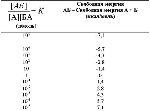
If the reaction 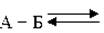 AB has reached equilibrium, the relative amounts of components A, B, and AB will depend on the difference in their Free energy, ∆G°. The values shown above are calculated for 37°C using the equation
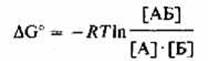
or

where ∆C0 is expressed in kilocalories per mole and represents the free-energy change of this reaction under standard conditions (the concentration of all components is 1,0 mol/L).
In general, the stronger the binding of molecules in a complex, the lower the rate of dissociation. In the limiting case where the energy of the bonds formed is negligible compared to the energy of thermal motion, the two molecules dissociate immediately after collision. In the other extreme, the bond energy is so high that virtually no dissociation occurs. Thus, the magnitude of the interaction energy is a useful indicator of the Specificity of the recognition process.
To understand how the energy of interaction is measured, let us consider the binding reaction of molecule A with molecule B. This reaction will proceed until it reaches an equilibrium state where the rates of complex formation and dissociation are equal. Using the equilibrium concentrations of molecules A, B, and the AB complex, we can determine the equilibrium constant K of the reaction (Fig. 3-7). This constant is sometimes called the affinity constant and is commonly used as a measure of the binding strength between two molecules: the stronger the binding, the higher the value of the affinity constant.
The equilibrium constant for the association of two molecules is directly related to the standard free-energy change, ∆G°, of the reaction. Using the appropriate equation (Table 3-3), ∆G° can be calculated for a range of K values. The affinity constants for simple binding reactions in biological systems typically range from 103 to 1012 liters/mole, which corresponds to binding energies of 4 to 17 kcal/mole, or The formation of an average of 4 to 17 hydrogen bonds.
The strongest interactions occur when biological function requires two macromolecules to remain tightly bound for a long time, such as when a Gene regulatory protein binds to DNA, turning the gene off (see Section 10.2.1). The weakest interactions occur when function requires rapid Changes in the STRUCTURE OF THE complex, for example, when two interacting proteins swap partners during the movements of a protein machine (see Section 1.3.1).
3.1.5. Atoms and molecules are in constant motion [6]
Chemical Reactions in the cell occur remarkably fast. For example, a typical enzyme molecule catalyzes ~1000 reactions per second, and for some Enzymes, this rate can exceed 106 reactions per second. Because each reaction requires an individual collision between an enzyme and a substrate molecule, such rates are possible only because molecules move rapidly. There are Three types of molecular motion: 1) the movement of a molecule from one place to another (translational motion), 2) the rapid back-and-forth vibrations of covalently linked atoms relative to one another, and 3) rotations. All of these motions are important for bringing interacting molecules into contact.
The rates of molecular motion can be measured by many spectroscopic Methods, which show, for example, that a large globular protein is in constant motion, rotating about its axis approximately a million times per second. The rates of diffusional collisions resulting from translational motions are proportional to the concentration of the diffusing molecules. For example, if a typical intracellular concentration is ~1 mM, then every patch on a protein molecule will experience about 106 random collisions per second with ATP molecules; if the concentration is an order of magnitude lower, the number of collisions will drop to 105 per second, and so on.
If two molecules collide and are in the correct relative orientation, a chemical reaction between them can occur very rapidly. Knowing how fast molecules move and react, we should not be surprised by the observed rate of Enzymatic Catalysis.
3.1.6. The process of molecular recognition cannot be completely error-free [7]
All molecules possess energy: kinetic energy from translational motions, vibrations, and rotations, and potential energy stored in their electron shells. Through molecular collisions, this energy is randomly distributed among different atoms, so that while the energy level of most atoms is close to the average, a small fraction of atoms will possess significant energy. Favorable conformations, or molecular states, correspond to a minimum of free energy (see Section 2.4.1), but high-energy states arise during violent collisions. Knowing the temperature, one can calculate the probability of an atom or molecule being in a given energy state (see Table 3-3). The probability of a high-energy state becomes smaller than that of a low-energy state as the difference in their free energies increases. It only becomes zero, however, when this energy difference becomes infinite.
Because of the element of chance in molecular interactions, a small number of "Side Reactions" occur from time to time. Therefore, the cell frequently makes mistakes. Sometimes even energetically unfavorable reactions occur. For example, two covalently bound atoms can be forced apart during an exceptionally violent collision. Similarly, the substrate specificity of an enzyme cannot be absolute, as The ability to distinguish one molecule from another cannot be perfect. Errors could be completely eliminated only if cells had evolved mechanisms with an infinitely large energy difference between alternative states. Since this is impossible, cells must tolerate a certain level of errors and employ various repair reactions to correct those that are most dangerous.
On the other hand, as we already know, errors play an important role in the living world. Were it not for random errors during DNA Synthesis, evolution would hardly be possible (see Section 3.2.4).
The sequence of subunits in macromolecules contains the information that determines the spatial configuration of their surfaces. It is this configuration that is used for recognition between different molecules and between different parts of the same molecule through weak noncovalent bonds. Molecules are in constant rapid motion; if recognition occurs upon collision as a result of random diffusion, they bind to each other with a strength that can be expressed by an equilibrium constant. Because recognition can be error-free only if the interaction energy increases to an infinitely large value, living cells constantly make mistakes. When necessary, these errors are corrected by special repair mechanisms.
Last update: 12/08/2026
Editorial and Educational Adaptation: This material has been compiled based on the primary/original source text. The project team performed an editorial review, corrected technical inaccuracies, structured sections, and adapted the content for an educational format.
What was processed:
- elimination of formatting defects (OCR errors, structural breaks, corrupted characters);
- editorial organization of content;
- standardization of terminology in accordance with academic sources;
- verification of factual statements against the original source text.
All mentions of the author, publication year, and origin of the primary text have been preserved in accordance with the source.