Intensive Care of Emergency Conditions - V. M. Zaporozhan 2006
Cardiopulmonary-cerebral Resuscitation
As already noted, resuscitation is The process of reviving patients from a terminal state.
According to the widely accepted three-stage Classification developed by V. A. Negovsky, terminal states include the preagonal state, agonal state, and clinical death.
These are links in a single dying process accompanied by a series of pathological alterations whose nature depends little on the causes of the terminal state, but largely reflects general biological patterns of dying.
Without understanding them, the rescuer's actions will lack conscious purpose and may prove incomplete. After all, restoring Blood Circulation and gas exchange is merely the first and far from the most complex stage of the fight for life—a task accessible even to a rescuer trained in basic resuscitation Methods without a medical Background. Following this, new and far more complex challenges arise: restoring the entire Organism to a state capable of full vital activity. For a human being, this means returning to full physical and mental health and normal social life, which entails restoring Brain Functions along with other Organs and systems. The characteristic changes associated with bodily recovery led V. A. Negovsky to coin the term "post-resuscitation disease." To achieve the fullest possible restoration of all body functions, one must have a clear understanding of the Pathophysiology of terminal states.
The primary driver of pathophysiological alterations in the body is tissue Hypoxia, which develops alongside declining Respiration and circulation. It is particularly devastating to Tissues with high metabolic demands, such as the brain. Although this organ accounts for only 2% of body weight, it consumes about 15-20% of all incoming oxygen, requiring a cerebral blood flow maintained at 15% of the Cardiac Output. This is especially true because, at the onset of a terminal state, Central Nervous system (CNS) activity increases to compensatorily stimulate vital functions. However, prolonged hypoxemia soon leads to decompensation.
Under conditions of oxygen and energy deprivation, Cellular METABOLISM is severely disrupted. The highly efficient AEROBIC GLUCOSE OXIDATION process within the Krebs Tricarboxylic Acid Cycle—which yields enough energy to convert 32 ADP molecules into ATP per glucose molecule—is replaced by anaerobic Glycolysis, capable of generating energy for the synthesis of only 2 ATP molecules per glucose molecule. Furthermore, once circulation ceases, stores of glucose and Glycogen are rapidly depleted, and lactic acid accumulates. This leads to metabolic acidosis—compounding the respiratory acidosis caused by carbon dioxide retention—as well as the dilation of cerebral arterioles and subsequently other tissues, resulting in the decentralization of blood circulation (if it was initially centralized).
Anergy leads to the failure of the sodium-potassium pump and transmineralization across The Cell membrane and extracellular space. A decrease in pO2 in capillaries to 19-17 mmHg sharply impairs mitochondrial function, and at 12-10 mmHg, it ceases entirely.
Due to a significant increase in the permeability of all Introduction/36.html">Biological Membranes, including lysosomal membranes, Enzymes are released that trigger cell lysis, thereby determining irreversibility and cell death. As for the dead Cells of The Nervous System—Neurons—they are irreplaceable.
The clinical picture of the preagonal state includes general lethargy, confusion with progressive depression of consciousness, unmeasurable blood pressure, absent peripheral pulses (though palpable in the carotid and femoral Arteries), tachypnea or bradypnea with abnormal breathing patterns, and cyanosis or pallor of the Skin and mucous membranes.
The agonal state is characterized by unresponsiveness and absent ocular Reflexes, unmeasurable blood pressure, absent peripheral pulses, markedly diminished pulses in major arteries, barely audible heart sounds, and ECG findings showing pronounced myocardial hypoxia and Cardiac Arrhythmias. Vital functions are under bulbar regulation, and a brief surge in functional activity, including CNS activity (an "agonal flare"), is frequently observed.
Clinical death occurs at the exact moment of cardiac arrest and the complete cessation of circulation, resulting in apnea and the shutdown of CNS functional activity.
Immediately following cardiac and respiratory arrest, metabolic processes drop sharply, yet they do not cease entirely because energy from anaerobic glycolysis continues to reach the tissues for some time. Consequently, clinical death is a reversible state, and its duration is determined by the survival time of the Cerebral Cortex under conditions of completely arrested circulation and respiration. At normal Temperature and barometric pressure, this does not exceed 3-4 minutes, although in cardiac arrest accompanied by body hypothermia of 10-8 °C, reversible CNS changes persist for up to 2 hours, and this window is longer in children than in the elderly.
The causes of respiratory and circulatory arrest can vary:
— hypoxia and hypercapnia resulting from overdoses of sleeping pills, narcotics, and anesthetics; traumatic brain injury; impaired breathing biomechanics, including multiple rib fractures, hemothorax and pneumothorax, tracheobronchial obstruction, suffocation, drowning, etc.;
— Hemorrhage;
— electrolyte imbalances, most commonly disruptions in the potassium-to-sodium ratio (hyperkalemia sharply reduces cardiac electrical and contractile capacity while leaving electrical activity intact);
— neurohumoral and neuroreflex factors;
— vagal reflexes — rough tracheal intubation, surgical manipulations in reflexogenic zones (heart, mesentery, etc.);
— psychological factors (fright, fear, grief, etc.);
— factors associated with overdoses of medications affecting respiratory and Cardiac Activity (Glycosides, sympathomimetics, etc.);
— myocardial damage — myocardial infarction (with small and medium-sized lesion foci, fibrillation is more common because ischemic and surrounding tissues have differing potentials, generating a continuous current and "trigger zones" that suppress cardiac automaticity; with large lesion zones, asystole is more frequently observed).
Etiological factors of terminal states, the preferential damage to specific organs and systems under their influence, and the duration of the dying process all affect pathophysiological alterations. For instance, the window of clinical death with the possibility of resuscitation following death from blood loss is 1.5-2 times shorter than that following cardiac arrest caused by Electrical Injury.
At the same time, adaptive reactions to life-threatening factors follow a uniform pattern. Most notably, this involves the reflex stimulation of the sympathetic nervous system, accompanied by a sharp surge (30-100 times or more) in blood levels of catecholamines and catabolic Hormones (ACTH, ADH, corticosteroids, Glucagon, Steroid Hormones), alongside a simultaneous decrease in anabolic hormone levels (Insulin, androgens, and estrogens). These form protective reactions, the most general of which is the centralization of circulation accompanied by reduced cardiac output. However, if the damaging factor is excessively intense or prolonged, these protective reactions exceed physiologically permissible limits and become damaging—a feature highly characteristic of the Pathogenesis of terminal states.
In rapid dying, compensatory mechanisms either fail to engage in time or prove insufficient, meaning primary alterations affect circulation and respiration alongside the aforementioned pathophysiological and biochemical disruptions.
During prolonged dying, such as in protracted hemorrhage, the protective reaction of circulatory centralization becomes prominently manifested, making it possible to sustain Shock-induced circulatory failure at the compensation stage for a certain period. In this process, the viability of the brain and heart is preserved at the expense of ischemia in other organs and systems, where the effects of hypoxia culminate in necrotic damage to the cellular elements of tissues most sensitive to oxygen deprivation. This is how "shock Liver" and "shock Kidneys" develop.
Among the pathogenetic factors in The Development of terminal states, endogenous intoxication plays a significant role. Toxins are considered to be products of distorted metabolism—ammonia, carbonic acid, lactic acid, and other organic acids, as well as BIOLOGICALLY ACTIVE SUBSTANCES (histamine, serotonin, "myocardial depressant factor", ferritin). Our observations have demonstrated that endotoxemia is accompanied by the accumulation of its recognized markers in the blood, namely middle-molecular-weight molecules, which are largely oligopeptides resulting from protein degradation due to increased proteolytic activity of blood serum. Research continues to identify and study The Nature of the so-called "stress protein", which appears during shock and terminal states, although its biological role remains unknown for now.
Great attention is also paid to The entry of Bacterial toxins into the bloodstream As a result of accelerated microorganism proliferation under conditions of reduced blood circulation and increased permeability of biological membranes, as well as indole, skatole, and Other toxins from the intestinal lumen.
Enhanced lipolysis leads to the Formation of Ketone bodies and acetone.
The Role of endotoxemia increases with the decline in the efficiency of natural detoxification mechanisms in the liver, kidneys, and reticuloendothelial system during terminal states. Endotoxemia is evidently one of the causes of irreversible changes that invariably develop before energy reserves are completely depleted and tissue metabolism ceases.
It is crucial to keep in mind that all these pathophysiological changes often do not have time to manifest during the terminal state itself, but they severely burden the post-resuscitation period and thereby reduce the effectiveness of resuscitation—the restoration of full vital activity. Therefore, cardiopulmonary resuscitation in its simplest form is far from always successful.
The application of resuscitation methods must be complemented by a rational combination with intensive care techniques aimed at eliminating post-resuscitation disease and treating the underlying condition that triggered the development of terminal states.
Resuscitation and intensive care methods have evolved over a long period. As a rule, they were developed on The basis of scientific and technical achievements in related, sometimes quite distant fields. To understand the origins of modern resuscitation methods, one can look at The most significant historical milestones: Artificial ventilation (Sylvester, 1858; Elam, 1954); direct cardiac massage in a clinical Setting (Kristan, Ingelsrud, 1901); chest compression / closed-chest cardiac massage (Kouwenhoven, Jude, Knickerbocker, 1959); endotracheal intubation using a laryngoscope (Lilienthal, 1910); direct electrical defibrillation of The Heart (Prévost, Batelli, 1899); closed-chest electrical defibrillation (N. Ya. Gurvich, G. S. Yunyev, 1946); the triple airway maneuver (HEAD tilt, jaw thrust, and Mouth opening) (P. Safar, 1953), among others. Thus, old resuscitation methods have transformed into new ones over the past 40 years, and this process is ongoing.
P. Safar, one of the founders of modern resuscitation methodology, attaching enormous importance to the organizational aspects of rescuing patients from terminal states, outlines the stages and phases of cardiopulmonary-cerebral resuscitation as follows:
I. Basic Life Support.
A — (from aer — "air") — ensuring an open airway / airway clearance.
B — (from breathing) — artificial ventilation.
C — (from circulation) — chest compressions / cardiac massage.
II. Advanced Life Support. Restoration of spontaneous circulation and stabilization of hemodynamics and respiration.
D — (from drugs) — administration of medications.
E — (from electrocardiography — "ECG") — diagnosing cardiac activity using ECG.
F — (from fibrillation) — performing electrical defibrillation in case of cardiac Muscle fibrillation.
III. Prolonged Life Support — post-resuscitation intensive care.
G — (from grade) — Assessment of the patient's central nervous system depression using the Glasgow or Glasgow-Pittsburgh scale.
H — (from human) — restoration of normal human mental function through modern brain-function recovery measures.
I — (from intensive care) — correction of organ system failures other than the brain, to facilitate brain function recovery.
The measures and stages of cardiopulmonary-cerebral resuscitation (steps A, B, C), which P. Safar refers to as the basic life support stage, are performed in terminal states. These should be recognized within 7–10 seconds based on the presence of at least any two of the three signs: cessation of breathing, pupil dilation, and absence of a pulse in the carotid artery. Since the time factor is crucial for successful resuscitation alongside technical precision, to expedite the Diagnosis of clinical death, the presence of a pulse and the state of the pupils are checked simultaneously: one hand determines the pulse while the other lifts the eyelid to examine the pupil.
The first person to discover the victim must perform the following measures as quickly and in as strict a sequence as possible to ensure The First stage of resuscitation—basic life support:
1. Place the patient on their back, obligatorily on a firm surface. Tilt the head back as far as possible by placing one hand under the neck and the other on the forehead, thereby ensuring airway patency, and check for the presence or absence of respiratory sounds (Fig. 1). In case of suspected cervical spine injury, head tilting is not recommended.
2. In the absence of respiratory sounds or with shallow, infrequent breaths, initiate mouth-to-mouth artificial ventilation (Fig. 2). To do this, the rescuer takes a deep breath, tightly seals the victim's Lips with their own (using a handkerchief, gauze, etc., as a barrier), pinches the victim's nostrils with their fingers or cheek, and delivers an energetic exhalation. The depth of the breath is monitored by the movement of the patient's chest. One can also attempt mouth-to-Nose ventilation while covering the patient's mouth.
Class="center">
Fig. 1. Initial position of the casualty for resuscitation and Auscultation of respiratory sounds
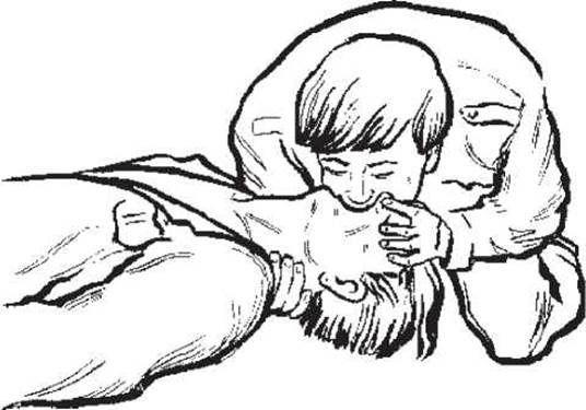
Fig. 2. Mouth-to-mouth artificial ventilation

Fig. 3. Forward Displacement of the lower jaw

Fig. 4. Cleaning the mouth and Pharynx with a finger
3. If the chest does not expand upon attempting a breath:
a) displace the lower jaw forward (Fig. 3);
b) clear the mouth and pharynx of foreign matter using a handkerchief or gauze pad wrapped around a finger (Fig. 4), or a suction device if available;
c) insert an airway (Fig. 5), and perform endotracheal intubation if possible (indicated when other means fail to maintain airway patency, or during prolonged resuscitation).

Fig. 5. Insertion of an airway: a — oral; b — nasal

Fig. 6. Palpation of the carotid pulse
4. After delivering 3-5 breaths, check for a pulse at the carotid artery (Fig. 6).

Fig. 7. External chest compressions

Fig. 8. Chest compressions and artificial ventilation performed by a single rescuer

Fig. 9. Chest compressions and artificial ventilation performed by two rescuers
5. If no pulse is detected, immediately initiate external (closed-chest) cardiac massage (Fig. 7). To do this, the rescuer, positioned at the casualty's side, places the palm of one hand—with fingers elevated—on the lower third of the Sternum directly along the midline, and places the palm of the other hand over the wrist of the first. Pressing rhythmically, apply force to the sternum using not only arm strength but the full weight of the body, depressing the sternum by 4-5 cm toward the spine. Repeat compressions at least once per second. External chest compressions are considered effective if pupillary constriction occurs, the fatal pallor of the lips disappears, and spontaneous respiratory movements eventually appear. As for internal (open-chest) massage, its use is restricted to cases of cardiac arrest occurring during certain surgical Procedures.
6. External cardiac massage must be combined with artificial ventilation. If resuscitation is performed by a single rescuer (Fig. 8), 2 rapid rescue breaths should be delivered after every 15 chest compressions (a 2:15 ratio), completing 4-5 such cycles per minute. If resuscitation is performed by two rescuers (Fig. 9), a ventilation-to-compression ratio of 1:5 is maintained, with at least 12 such cycles performed per minute. It should be noted that ensuring adequate cerebral blood flow requires a minimum of 60-70 chest compressions per minute. According to the European Resuscitation Council guidelines (2000), after isolating the airway from the Esophagus (via endotracheal intubation, etc.), simultaneous lung ventilation and chest compressions are performed in adults at a rate of 12 breaths to 100 compressions per minute.
7. Monitor for the return of spontaneous circulation by checking the carotid pulse, interrupting chest compressions for a few seconds every 2-3 minutes to do so. Cardiac massage should be continued until spontaneous heart activity is restored.
In a hospital setting or inside a specialized ambulance, the aforementioned measures can be performed with the additional use of special equipment.
Specifically, to maintain airway patency, clinicians use a suction device, standard and specialized Airways, tracheal intubation and tracheobronchial suctioning, cricothyroidotomy, tracheotomy, and bronchoscopy.
Pulmonary ventilation is supported using basic breathing devices (such as an Ambu bag or manual resuscitator), anesthesia machines equipped with a bellows or an oxygen-filled bag, as well as specialized mechanical ventilators that regulate breathing based on pressure, rate, or volume.
For prolonged mechanical ventilation (MV), modern servo-ventilators are the most appropriate choice. By monitoring gases in both inspired and expired air as well as in the blood, they allow for the individualized adjustment of respiratory parameters, thereby preventing severe complications during mechanical ventilation such as acute respiratory distress syndrome and Pneumonia.
In certain situations, high-frequency mechanical ventilation may be required. In cases of tension pneumothorax, mechanical ventilation is initiated immediately; if the patient's condition permits, however, pleural puncture is performed beforehand (in the 2nd intercostal space along the midclavicular line to evacuate air, or in the 6th intercostal space along the midaxillary line to remove fluid).
To restore Blood Circulation in the operating room when external chest compressions are ineffective or impossible, internal cardiac massage can be performed (Fig. 10), alongside The Use of specialized mechanical massage devices.

Fig. 10. Open-chest cardiac massage
The absence of signs indicating effective resuscitation using all specified methods for 30 minutes serves as an indication to terminate resuscitation efforts. If signs of effectiveness are observed, resuscitation is continued until spontaneous circulation is restored; however, first-stage measures alone are insufficient for full physiological recovery. Therefore, immediately after initiating resuscitation, a specialized resuscitation team—possessing the expertise and equipment required for the Second Stage of resuscitation—must be called.
The goal of the second stage of resuscitation is continued life support. Typically performed in a hospital setting, this stage involves restoring spontaneous circulation by supplementing first-stage measures with pharmacological therapy (stage D) and ECG Diagnostics.
In critical and, even more so, terminal states, the choice of drug administration routes is crucial. Oral, subcutaneous, and intramuscular routes are ineffective. Intravenous administration is recommended; if venous access is difficult, urgent administration should be performed endotracheally via an intubation tube or through puncture of the cricothyroid ligament, doubling the drug dose and diluting it in 10 mL of sterile Water or saline. Intracardiac injection, which was widely used in the past, is no longer recommended.
Treatment begins with "first-line" medications. These include intravenous administration of 0.1% epinephrine solution at 1 mL (1 mg) every 3 minutes, and 0.1% atropine solution administered 2–3 times throughout the resuscitation period in doses of 1–0.5–0.5 mL (corresponding to 1–0.5–0.5 mg), without exceeding a total dose of 2–3 mg.
Sodium bicarbonate (50 mL of an 8.4% solution) or an alternative buffer (such as trisamine buffer) is administered via titration when pH < 7.1, or 20–25 minutes after circulatory arrest if blood acid-base balance data are unavailable. This precaution is necessary due to the potential Adverse effects of sodium bicarbonate during cardiopulmonary and cerebral resuscitation: exacerbation of intracellular acidosis caused by increased CO2 levels, shifting of the HbO2 dissociation curve, inactivation of catecholamines, and reduced defibrillation efficacy. It must be administered exclusively via the intravenous route; tracheal administration suppresses ciliated epithelial function.
ECG diagnostics should be initiated as early as possible after starting external chest compressions. Regardless of the diverse underlying causes of clinical death, circulatory arrest manifests in only three main ECG patterns, which frequently transition into one another.
The most unfavorable type of cardiac arrest is asystole. On the monitor, it appears as a flat isoelectric line, indicating a complete absence of myocardial electrical activity (Fig. 11, a) and accounting for 41% of in-hospital cardiac arrests (according to the National Registry of Cardiopulmonary Resuscitation, 2002).

Fig. 11. ECG patterns of asystole (a), pulseless electrical activity (b), and ventricular fibrillation (c)
Pulseless electrical activity (or electrical activity without a pulse, which in modern classifications includes both electromechanical dissociation and severe bradiarrhythmia—previously referred to as an ineffective heart or agonal ventricular complexes) is a form of cardiac arrest characterized by preserved synchronous myocardial activity that fails to produce cardiac output due to various underlying causes (Fig. 11, b). It accounts for 29% of cases.
Ventricular fibrillation (pulseless ventricular tachycardia) is a prognostically more favorable type of circulatory arrest, as myocardial electrical activity and the contractility of individual, asynchronously contracting fibers are preserved (Fig. 11, c). It accounts for 30% of cases.
Clinically, these pathological states manifest identically (through signs of circulatory arrest) and require standard resuscitation protocols. However, in cases of asystole, the primary treatment objective is to stimulate cardiomyocyte electrical activity using epinephrine and atropine. If these drugs were not administered prior to ECG monitoring, they must be given immediately using the doses and schedule outlined above. If electrical activity fails to recover after three doses of epinephrine, sodium bicarbonate administration is initiated.
In cases of ventricular fibrillation, electrical defibrillation using a defibrillator must be performed In addition to standard resuscitation measures (Fig. 12). Because foreign-made defibrillators generate monopolar electrical impulses, whereas domestic devices produce bipolar asymmetric quasisinusoidal Gurvich impulses, the recommended discharge energies for these Two Types of defibrillators differ (Table 1).
If three successive defibrillation attempts fail, the European Resuscitation Council Guidelines (2000) recommend the intravenous administration of cordarone (amiodarone)—the first-choice antiarrhythmic drug—at a dose of 300 mg diluted in 20 mL of a 5% glucose solution, followed by a repeat dose of 150 mg up to a maximum total dose of 2 g. If cordarone is unavailable, lidocaine is administered at 1.5 mg/kg (maximum 3 mg/kg) or novocainamide at 30 mg/kg up to a cumulative dose of 17 mg/kg.
For ventricular fibrillation that is refractory to electrical defibrillation when hypomagnesemia is suspected, magnesium sulfate is recommended at a dose of 8 mmol (approximately 10 mL of a 25% MgSO4 solution).
Calcium chloride, which was previously used extensively during resuscitation, is now recommended only for hypocalcemia, calcium channel blocker overdose, hyperkalemia, or hypomagnesemia at a dose of 0.2 mL/kg of a 10% solution administered intravenously.
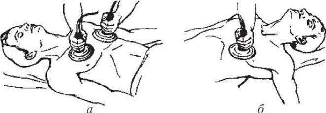
Fig. 12. Electrofibrillation with electrode placement on the chest (a) or via the anteroposterior chest-to-back method (b)
If ineffective, all subsequent shocks are delivered at the maximum energy of 360 J according to the following protocol:
drug massage shock drug massage shock... Restoring circulation during pulseless electrical activity aims to eliminate its underlying cause, as this is the only way to restore cardiac output. All such causes are divided into three groups: "empty heart", characterized by decreased venous return; "pulmonary circuit blockade", where blood from the right side of the heart fails to reach the left; and "cardiac failure", where cardiomyocytes are unable to perform mechanical pumping work.
In the first case, it is imperative to promptly expand the intravascular volume using crystalloid or hydroxyethyl starch solutions. Meanwhile, restoring formed blood elements takes a back seat.
The second group of causes may include: cardiac tamponade (requiring pericardiocentesis), tension pneumothorax (converted to an open pneumothorax or drained via the pleural cavity with continuous suction), vascular collapse (vasopressors), and Pulmonary Embolism (thrombolysis).
Table 1 Energy levels for repeated defibrillations (according to L. V. Usenko et al., 2001)
|
Defibrillator |
Waveform |
1st shock |
2nd shock |
3rd shock |
|
Lifepak-7, Edmark |
Monophasic |
200 J |
250-300 J |
360 J |
|
DKI-01, DIS-04 |
Biphasic |
65-90 J 4.0 kV |
140 J 5.7 kV |
190 J 7.0 kV |
Table 2 Glasgow Coma Scale (1977)
|
Category |
Response |
Score |
|
Eye opening |
Spontaneous |
4 |
|
To speech |
3 |
|
|
To pain |
2 |
|
|
None |
1 |
|
|
Verbal response |
Oriented |
5 |
|
Confused conversation |
4 |
|
|
Inappropriate words |
3 |
|
|
Incomprehensible sounds |
2 |
|
|
None |
1 |
|
|
Motor response |
Obeys commands |
6 |
|
Localized to pain |
5 |
|
|
Withdrawal from pain |
4 |
|
|
Flexion to pain |
3 |
|
|
Extension to pain |
2 |
|
|
None |
1 |
Note. Coma depth and prognosis categories: 1st — 14-15 points (alert/clear consciousness); 2nd — 11-13 points (obtundation); 3rd — 8-10 points (stupor); 4th — 5-7 points (coma); 5th — 3-4 points (brain death). Coma depth increases from category 1 to 5, while the prognosis becomes progressively more unfavorable.
Acute heart failure requires cardiotonics (adrenaline may be administered as an IV bolus of 1 mg every 5 min, dopamine at a dose of 2-10 µg/(kg·min), dobutamine at 2-20 µg/(kg·min), or dobutrex).
The Third Stage of cardiopulmonary cerebral resuscitation — prolonged life support — aims at cerebral resuscitation, which includes assessing the patient's condition (G), restoring normal human cognition (H), and intensive care for complications and residual deficits (I). The optimal setting for treating patients at this stage is specialized intensive care units or wards, to which such patients should be transferred whenever possible.
The primary task following the restoration of spontaneous circulation is to evaluate the patient's condition to diagnose and eliminate the causes of death, thereby preventing a recurrence of the terminal state, as well as to assess the degree and nature of bodily disorders using the standard Glasgow Coma Scale (Table 2) and Trauma Score (Table 3).
Table 3 Trauma Score (1981)
|
Parameter |
Value |
Score |
|
|
A. Respiratory rate |
10-24 |
4 |
|
|
Number of breaths per 15 s x 4 |
25-35 |
3 |
|
|
>35 |
2 |
||
|
<10 |
1 0 |
||
|
B. Respiratory effort Shallow — significantly reduced chest wall excursion |
Normal Shallow, labored |
1 0 |
|
|
Labored — use of accessory Muscles, intercostal retractions observed |
|||
|
C. Systolic blood pressure |
>90 |
4 |
|
|
By auscultation or radial palpation |
70-90 50-69 |
3 2 |
|
|
<50 |
1 |
||
|
No carotid pulse |
0 |
0 |
|
|
D. Capillary refill ("spot sign") Normal — forehead, lips, or nail bed within 2 s Delayed — more than 2 s |
2 1 |
||
|
None — capillaries do not refill |
0 |
||
|
E. Glasgow Coma Scale (GCS) |
Total GCS score |
||
|
1. Eye opening Spontaneous |
4 |
14-15 11-13 |
5 4 |
|
To speech |
3 |
8-10 |
3 |
|
To pain |
2 |
5-7 |
2 |
|
None |
1 |
3-4 |
1 |
|
2. Verbal response Oriented |
5 |
||
|
Confused |
4 |
||
|
Inappropriate words |
3 |
||
|
Incomprehensible sounds |
2 |
||
|
None |
1 |
||
|
3. Motor response Obeys commands |
6 |
||
|
Localizes pain |
5 |
||
|
Withdrawal |
4 |
||
|
Flexion |
3 |
||
|
Extension |
2 |
||
|
None |
1 |
||
|
Total GCS score (1+2+3) |
|||
Note. Trauma Score = (A+B+C+D+E).
The next two stages of cardiopulmonary cerebral resuscitation (H and I) involve the treatment of post-resuscitation disease (PRD).
Post-resuscitation disease (PRD) is a complex, distinct pathological process associated with the period of clinical resuscitation and subsequent survival. E. S. Zolotokrilina (1999) identifies the following stages of PRD:
Stage I (6-8 hours from THE START OF treatment) — the stage of unstable functions, characterized chiefly by a pronounced decrease (by 4-5 times) in tissue perfusion and the presence of circulatory hypoxia.
Stage II (10-12 hours from the start of treatment) — relative stabilization of major bodily functions. It is marked by the stabilization of vital signs and a patient improvement, often temporary. Severe impairments in tissue perfusion persist (tissue blood volume is reduced by 2-2.5 times), along with a deficit in circulating blood volume (CBV). There is an increase in K+ loss and Na+ retention, and the previously developed lactic acidosis persists. Pronounced hyperfermentemia is observed, and plasma fibrinolytic activity is likely slowed.
Stage III (late 1st-2nd day of treatment) — secondary deterioration of the patient's condition. Some patients exhibit normal body temperature alongside tachycardia, tachypnea, and elevated blood pressure, particularly in young and middle-aged individuals, accompanied by agitation. Mixed-origin hypoxia develops due to a maximal reduction in Oxygen transport resulting from altered Hemoglobin properties, impaired oxyhemoglobin dissociation, persistently low tissue perfusion, pulmonary blood shunting, and a hypodynamic circulatory state. Hemostasis and Fibrinolysis disorders peak during this stage, creating conditions for microthrombi formation in organs and tissues. Parenchymal organ dysfunction develops: Renal Dysfunction manifested as functional oliguria; worsening Acute Respiratory Failure (ARF) presenting as non-specific lung injury ("shock lung"), and less frequently, hepatic dysfunction. All these disorders are functional and reversible under a favorable clinical course.
Stage IV (3rd-4th day) follows a dual course: favorable — a period of stabilization and subsequent recovery of impaired functions, allowing for de-escalation of intensive care and uncomplicated recovery; unfavorable — a period of further clinical deterioration driven by the progression of a generalized inflammatory response and multi-system organ dysfunction.
This phase is marked by intensified Catabolism; interstitial edema of the Lungs, brain, and subcutaneous tissue; deepening mixed-type hypoxia and hypercoagulation; and the onset of purulent-inflammatory complications. Against this background, organ and system failure manifests: secondary upper gastrointestinal bleeding (erosions), psychoses with hallucinatory syndrome, secondary heart failure, worsening ARF, pancreatitis, hepatic dysfunction, and acalculous cholecystitis.
Stage V (5th-6th day) is observed exclusively in unfavorable courses of PRD. Inflammatory and purulent processes progress, including massive and frequently nosocomial, abscess-forming pneumonias; wound suppuration; soft tissue abscesses; and purulent Pleurisy. Despite the early administration of Antibiotics and antiseptics, Sepsis frequently develops against the backdrop of severe cellular and humoral immune impairment caused by prolonged, severe mixed-origin hypoxia. Sepsis typically triggers a new, secondary wave of damage to the lungs, myocardium, liver, kidneys, commonly referred to as multiple organ dysfunction syndrome (MODS).
Post-resuscitation disease is inevitably accompanied by post-hypoxic encephalopathy — a temporary or persistent central nervous system dysfunction. This key pathogenetic component unfolds in two phases:
I — acute phase (catabolic), lasting 2-3 days:
— activation of metabolic processes or their inhibition in case of adaptation failure;
— development of energy deficit in neurons;
— endogenous intoxication of intracerebral and extracerebral origin.
II — stabilization phase:
— unstable stabilization of Homeostasis (strain and exhaustion of adaptive processes), ending in death or lasting for months or years;
— stable stabilization of homeostasis, lasting from several weeks to months.
Intensive care for acute cerebral disorders involves a set of intracerebral and extracerebral measures taking into account the stage of the clinical course (according to V. I. Cherniy, 1997):
Intracerebral measures
I. Induction of protective inhibition and reduction of the brain's energy demand, protecting it against recurrent hypoxia. Subnarcotic doses of a 5% sodium thiopental solution (single dose up to 5 mg/kg) and sodium oxybutyrate (single dose 20 mg/kg) are administered alternately. Thiopental is given every 3 hours, and oxybutyrate every 2 hours. As the neurological deficit decreases, the intervals between administrations are increased and the doses are reduced.
II. Restoration of cell and vascular membrane function:
1. Reduction of their permeability (prednisolone 1-4 mg/kg per day intramuscularly), suppression of the kallikrein-kinin system activity and trypsinemia, and reduction of fermentemia (contrical 150-800 IU/kg).
2. Reduction of Lipid Peroxidation (LPO) intensity (tocopherol 2-8 ml of a 30% solution per day, unitiol 5 ml intravenously, and aevit 2 ml intramuscularly twice a day).
III. Restoration of cerebral circulation.
1. Restoration of microcirculation in cerebral vessels (euphylline 10 ml of 2.5% solution intramuscularly three times a day, xanthinol nicotinate 5-30 mg/kg per day or nicotinic acid 1-3 mg/kg per day, persantine 25-75 mg per day). Moderate hypervolemic hemodilution using crystalloids and colloids (hematocrit not below 0.30).
2. Improvement of venous outflow from the cranial cavity (sulfocamphocaine 6-25 mg/kg per day and euphylline; in case of elevated central venous pressure, furosemide intravenously or intramuscularly 0.2-0.4 mg/kg, controlled nitroglycerin infusion, and stimulation of myocardial contractility).
3. Management of cerebral edema (mannitol 0.25-0.5 g/(kg·day); glycerol 1 g/(kg·day) via a nasoenteric tube is more hazardous due to the "rebound syndrome").
4. Restoration of volumetric cerebral blood flow rate (reduction of intravascular pressure using sermion 48 mg intravenously or intramuscularly, cavinton 15-30 mg, and stugeron 75-150 mg per day).
IV. Management of ammoniemia (drip infusion of a 1% glutamic acid solution 15-30 mg/(kg·day)).
V. Restoration of nerve cell metabolism (piracetam or nootropil 8-12 g/day until consciousness is restored, followed by 4-8 g/day for 2-3 days, and another 4 g/day for 3 weeks).
VI. Hyperbaric Oxygenation (HBO) (10 sessions at 1.2-1.5 ATA, no earlier than 3-4 days after clinical death).
Extracerebral measures
I. Correction of hypovolemia and central hemodynamics disorders is performed according to standard principles, adjusted for the need to maintain moderate hypervolemic hemodilution.
II. Normalization of respiratory function. Treatment of acute respiratory distress syndrome. Ensuring adequate airway patency both during the coma and after the recovery of consciousness. In the absence or inadequacy of spontaneous breathing, mechanical ventilation is applied. Treatment of acute respiratory distress syndrome is carried out according to the principles outlined in the relevant section.
III. Correction of water-electrolyte balance and acid-base disorders is performed according to standard principles. To prevent cerebral edema and maintain hypervolemic hemodilution, the water balance must be kept at a neutral (zero) level.
IV. Correction of hemocoagulation disorders involves measures to balance the procoagulant and anticoagulant pathways, eliminate microcirculatory blockade, and restore the functional activity of the reticuloendothelial system (RES).
V. Prevention and treatment of hepatic and renal failure (measures to restore circulating blood volume (CBV), accelerate endotoxin elimination, or neutralize them). In hepatic failure, hemoperfusion and plasmapheresis are performed, while renal failure is treated with hemodialysis and ultrafiltration.
VI. Correction of immune reactivity disorders is achieved partly through medications used in the treatment regimen for other purposes (ascorbic acid, tocopherol, native plasma), as well as the administration of Decaris 150 mg/day for 3-5 days, and Metrogyl 100 ml 2-3 times a day intravenously for 5 days.
VII. Prevention and treatment of purulent-septic complications are achieved by restoring microcirculation and prescribing broad-spectrum antibiotics.
VIII. Enteral and parenteral Nutrition. Until bowel function is restored, peristalsis stimulation and parenteral nutrition are administered. Once peristalsis recovers, enteral nutrition is initiated according to the principles outlined in the relevant section.
IX. Detoxification therapy (Forced diuresis, ultraviolet irradiation of autologous blood, and other extracorporeal detoxification methods as needed).
L. V. Usenko et al. (2001) Supplement the above treatment regimen for post-resuscitation disease with perftoran infusion—a multifunctional blood substitute that improves gas exchange and tissue metabolism, enhances the oxygen-transport function of blood, restores central and peripheral hemodynamics, blood rheology, and microcirculation. It exhibits myocardial membrane-protective, sorption, and diuretic properties, acts against edema, and functions as a slow calcium channel blocker. To enhance The Effect of perftoran infusion, it is recommended to combine it with oxygen inhalation (40-60%) for 24 hours from the start of the infusion.
Prognosis of PRD:
1. Early prognosis:
— if circulatory arrest lasts up to 4 minutes — prognosis is relatively favorable;
— arrest for 5-6 minutes — prognosis is doubtful;
— more than 7 minutes — highly doubtful;
— more than 20 minutes — hopeless.
If the duration of arrest is unknown, the prognosis is based on the timing of reflex and respiration recovery. If breathing recovers no later than 20 minutes, the prognosis may be favorable.
Prognosis based on the timing of Electroencephalogram (EEG) normalization:
— within 10 minutes — favorable;
— within 30 minutes — potentially favorable;
— more than 30 minutes — unfavorable.
2. Delayed prognosis (established within the first 24 hours):
— decreasing depth of coma, absence of seizures and decerebrate rigidity — favorable;
— absence of spontaneous breathing and corneal reflexes for more than 4 hours, dilated and deformed pupils, persistent anisocoria, lack of light reaction, extensor hypertonia, seizures — severe;
— rhythmic myoclonus against the background of coma — hopeless.
As a result of treatment administered during resuscitation and the post-resuscitation period, consciousness may be restored, brain death may occur, or a vegetative (apallic) state may be established, rendering further resuscitative measures pointless.
Resuscitation can be considered unsuccessful if a flat line is registered on the ECG for at least 30 minutes, despite correctly performed resuscitation and appropriate medical therapy. Without an ECG, it is impossible to prove the irreversibility of cardiac arrest. Cardiac electrical activity may still persist in the form of agonal ventricular complexes or ventricular fibrillation, which are always considered potentially reversible.
Signs of irreversible cardiac arrest are accompanied by brain death and serve as grounds for terminating resuscitation. Determining brain death after the restoration of spontaneous circulation is significantly more complex. Dilated pupils unresponsive to light and failing to constrict for at least 12 hours after the restoration of cardiac activity—unless resulting from traumatic brain injury—indicate brain death or severe cerebral impairment following recovery. Brain death is also evidenced by persistent hypotension that does not respond to vasopressors for at least 24 hours. Neurological signs of brain death may not appear immediately, but even 2 days later, against the background of initial improvement in neurological symptoms.
In addition to clinical signs, the EEG plays a crucial role in diagnosing brain death. However, even with the necessary equipment available, obtaining a high-quality EEG recording in an intensive care unit is often impossible, making it acceptable to forego this important method. In such cases, direct evidence of brain death can be obtained through carotid angiography, which demonstrates the absence of intracranial blood flow.
The determination of brain death and the vegetative (apallic) state should definitively be made no earlier than 1-2 weeks after resuscitation, provided that comprehensive treatment proves ineffective.
Following the confirmation of biological death or brain death, organs may be harvested for transplantation with the written consent of the patient's relatives.
Medico-legal guidelines change over time and must be kept in mind, but excessive fear of the law is unjustified. Legal practice regarding medical malpractice indicates that charges of incompetent resuscitation are extremely rare, whereas cases of withholding resuscitation are frequently classified as failure to render assistance.
1. Belousova, E. Yu., Obolentsev, N. N., & Belousov, A. N. (2005). Nanotechnology as a pathway to prolonging life processes in the body. Pain, Anaesthesia and Intensive Care, (3), 5-7.
2. Bunyatyan, A. A., Ryabov, G. A., & Manevich, A. Z. (1984). Anesthesiology and Resuscitation. Moscow: Meditsina, pp. 336-366.
3. Sychov, O. S., Parkhomenko, A. N., Amosova, E. N., et al. (2004). Sudden Cardiac Death: Risk Factors and Prevention: Guidelines. Kyiv, 32 p.
4. Groer, C., & Cavallaro, D. (1996). Cardiopulmonary Resuscitation (E. B. Kleimenova, Trans.; G. A. Ryabov, Ed.). Moscow: Praktika, 127 p.
5. Cherny, V. I., Novikova, R. I., Shramenko, E. K., et al. (2003). Diagnosis and treatment of Cardiac Arrhythmias in Anesthesiology and Intensive Care: Methodological Guidelines. Donetsk, 56 p.
6. Zavgorodny, V. L., Nalapko, Yu. I., & Mamchur, S. Yu. (2002). Intensive Care Algorithms for Brain Diseases and Injuries. Luhansk: Yantar, 144 p.
7. Koryachkin, V. A., & Strashnov, V. I. (Eds.). (2002). Intensive Care of Life-Threatening Conditions. St. Petersburg: St. Petersburg Medical Publishing House, 286 p.
8. Chuev, P. N., Molchanov, V. I., Vladyka, A. S., et al. (2003). Intensive Medicine: A Textbook for Teaching Intensive Medicine to Students, Interns, and Physicians. Simferopol: Tavriya, pp. 82-120.
9. Nikonov, V. V. (1997). Emergency Medical Care. Kharkiv: KONSUM, 592 p.
10. Usenko, L. V. (Ed.). (2002). Fundamentals of Intensive Care. Ternopil: Ukrmedknyha, 332 p.
11. Lobenko, A. A., Vladyka, A. S., Novikov, A. A., et al. (1994). Practical Skills of First Medical Aid on Maritime Vessels (A. A. Lobenko, Ed.). Odessa, 71 p.
12. Safar, P., & Bircher, N. G. (1997). Cardiopulmonary and Cerebral Resuscitation. Moscow: Meditsina, 552 p.
13. Cherny, V. I., Tyumentseva, S. G., Akhlamova, Yu. I., et al. (2004). Cardiopulmonary-cerebral resuscitation. In V. I. Cherny & R. I. Novikova (Eds.), Intensive Care in Emergency Conditions. Kyiv: Zdorovya, pp. 3-33.
14. Starchenko, A. A. (2002). Reference Guide to Clinical Neuroresuscitation. St. Petersburg: St. Petersburg Medical Publishing House, 665 p.
15. Usenko, L. V., & Aryayev, L. N. (1976). First Aid on Water and Resuscitation in Drowning. Kyiv: Zdorovya, 112 p.
16. Usenko, L. V., Krishtafor, A. A., & Estrin, A. A. (2001). Cardiopulmonary and Cerebral Resuscitation: Post-Resuscitation Disease. Dnipropetrovsk, 78 p.
17. Usenko, L. V. (2004). Strategy for Restoring the Personality of Patients after Critical States. Dnipropetrovsk, 26 p.
18. Usenko, L. V., & Tsarev, A. V. (2004). Cardiopulmonary cerebral resuscitation: modern international guidelines from the perspective of evidence-based medicine. Pain, Anaesthesia and Intensive Care, (1), 50-68.
19. Bobrov, V. O., Zharinov, O. Y., Yagensky, A. V., et al. (2001). Atrial Fibrillation: Modern Classification and Management Principles: Methodological Guidelines. Kyiv: Chetverta Khvylia, 31 p.
20. Khyzhnyak, A. A., Kursov, S. V., & Mikhnevich, K. N. (2002). Cordarone in the Treatment of Cardiac Arrhythmias in Anesthesiology and Intensive Care. Kharkiv, 30 p.
21. Shifrin G. A. Standardization of Cardiopulmonary Resuscitation: A Guide to Integral Medicine. — Zaporizhzhia: Prosvita, 2003. — P. 55-57.
22. Greene T. O., Mittleman R. S. Cardioversion and Defibrillation // Intensive Care Medicine / Ed. by J. M. Rippe, R. S. Irwin, M. P. Fink, F. B. Cerra. — Vol. I. — Boston; New York; Toronto; London: Little, Brown & Co., 1996. — P. 81-92.
23. Paraskos J. A. Cardiopulmonary Resuscitation // Ibid. — P. 307-336.
24. Tchuyev P. M., Vladyka A. S. Intensive Care in Emergency. — Odessa: The Odessa State Medical University, 2005. — P. 74-90.
25. Intensive Care Medicine: Textbook / P. N. Chuev, V. I. Molchanov, A. S. Vladyka et al. — Simferopol: Tavria, 2006. — P. 80-118.
Last update: 11/08/2026
Editorial and Educational Adaptation: This material has been compiled based on the primary/original source text. The project team performed an editorial review, corrected technical inaccuracies, structured sections, and adapted the content for an educational format.
What was processed:
- elimination of formatting defects (OCR errors, structural breaks, corrupted characters);
- editorial organization of content;
- standardization of terminology in accordance with academic sources;
- verification of factual statements against the original source text.
All mentions of the author, publication year, and origin of the primary text have been preserved in accordance with the source.