Human Histology - O.D. Lutsyk 2003
General Histology
Nervous Tissue
Nervous Tissue (textus nervosus) is classified as a specialized tissue. Its elements are capable of perceiving stimuli, transforming these stimuli into nerve impulses, rapidly transmitting them, storing information, and producing BIOLOGICALLY ACTIVE SUBSTANCES. Through these Functions, nervous tissue ensures the coordinated activity of the body's Organs and systems, as well as its ADAPTATION TO ENVIRONMENTAL conditions. Nervous tissue is composed of Nerve Cells (Neurons, neurocytes) and supporting elements collectively known as neuroglia (Fig. 3.49).
Neurons (Figs. 3.49, 3.50, 3.51) are the morphological and functional units of nervous tissue. They consist of a Cell body (perikaryon) and processes. The presence of the latter is the most characteristic feature of nerve cells. It is these processes that conduct nerve impulses, often over considerable distances, with their length ranging from a few micrometers to 1–1.5 m. Neurons are typically incapable of mitotic division and have a long lifespan. Their lifespan generally coincides with that of the individual. However, this does not apply to all cells. It has recently been discovered that new nerve cells are formed in adult mammals, including humans. Nevertheless, neurogenesis (the generation of neurons from stem cells) occurs in only two regions: the subventricular zone of the Brain and the subgranular zone of the hippocampus. At certain Stages of Ontogeny (during The Development of the Central Nervous system organs), massive programmed (physiological) cell death of neurons also occurs. After birth, a human loses an average of about 10 million cells per year, and over a lifetime, the brain loses approximately 0.1% of all neurons (the total number of neurons in humans is at least 1012 (one trillion)). The size of the neuronal perikaryon varies widely—from 5–8 μm (granule Cells of the Cerebellum) to 120 μm (giant pyramidal neurons of the Cerebral Cortex). The processes of nerve cells are classified into axons and dendrites.
The axon (neurite) is a long process that can reach up to 1.5 m in length. Its name is derived from the Greek "axis". There is always only one axon per cell. The diameter of the axon remains constant throughout its length; it does not branch, but it can give off collaterals that run in other directions. The axon originates from the perikaryon at the so-called axon hillock—a cone-shaped region of Cytoplasm that lacks chromatophilic substance (see below). The axon ends in a terminal arborization containing synaptic vesicles. The axon conducts nerve impulses away from The Cell body. The expression of neuromodulin (GAP-43)—an axon-specific phosphoprotein—is a hallmark of the onset of neuronal differentiation. Initially, short processes are formed, which can potentially become either an axon or dendrites. The process that accumulates the GAP-43 protein subsequently develops into the axon. The volume of the axon can account for up to 99% of the total volume of the neuron.
Class="center">
Fig. 3.49. Cellular elements of nervous tissue: A - neurons (neurocytes); B - neuroglial cells. Cell sizes are not to scale

Fig. 3.50. A - schematic diagram of the Structural Organization of a multipolar motor neuron; B - semi-schematic representation of a histological section of nervous tissue: multipolar motor neurons of the anterior horn of the Spinal Cord, x 400

Fig. 3.51. Light Microscopy of neurocytes: A - a chain of neurons in the Connective Tissue stroma of a small intestinal villus, silver impregnation, x 100; B - neurons of the parasympathetic ganglion of the Cytology/practical/98.html">Submandibular salivary gland, histochemical reaction with concanavalin A, x 400; C - motor neuron of the anterior horn of the spinal cord, x 1200
Dendrites are mostly short processes (although long dendrites also occur) that branch in a tree-like fashion. Their name comes from the Greek word "dendron", meaning tree. The bases of dendrites have a conical expansion. These processes transmit nerve impulses toward the cell body.
Nerve cells contain a single large, round, pale Nucleus in the center of the perikaryon, with a small amount of heterochromatin and one or more nucleoli. At the same time, neurons in some ganglia of the Autonomic nervous system can contain up to 15 nuclei. The cytoplasm of the nerve cell (neuroplasm) contains Three types of organized structures: general Organelles, inclusions, and specialized organelles. Neuroplasmic inclusions may include CARBOHYDRATES (Glycogen), pigments (lipofuscin, melanin), and various secretory products (in neurosecretory cells). The specialized organelles of neurons are the chromatophilic substance and neurofibrils.
Under a Light Microscope, chromatophilic substance appears as basophilic clumps and granules of various Sizes and Shapes, localized in the perikaryon and dendrites of neurons, and is never found in axons or their initial segments. Chromatophilic substance was first described by F. Nissl in 1889, and was subsequently named after him (Nissl substance). J. Lenhossék (1895) termed it "tigroid". Chromatophilic substance is also referred to as basophilic substance. Under an Electron microscope, this Structure is revealed to be rough Endoplasmic reticulum with a parallel arrangement of its flattened cisternae (so-called ergastoplasm), where protein required for nerve cell function is intensively synthesized. Chromatophilic substance is an indicator of the functional state of the neuron. It can disappear when the nerve cell is depleted (a process known as chromatolysis or tigrolysis) and subsequently recover.
In axons, which lack organelles for Protein Synthesis, cytoplasm constantly moves from the perikaryon to the terminals at a rate of 1–3 mm per day. This is known as slow axonal transport, which carries Proteins, such as Enzymes required for neurotransmitter synthesis in synaptic terminals. In addition, there is fast axonal transport (5–10 mm/h), which mainly transports substances necessary for synaptic function, dendritic transport (at a rate of 3 mm/h), and retrograde transport, by which various cytoplasmic components are returned from the terminals to the cell body. The Transport of substances along neuronal processes is primarily mediated by microtubules.
In the perikaryon and dendrites, microtubules lack a uniform orientation, whereas in the axon, their (+)-ends face the terminals and their (-)-ends face the perikaryon. The orientation of microtubules is crucial for the distribution of various organelles within neuronal processes. Mitochondria and secretory vesicles move toward the (+)-end, while Ribosomes, multivesicular bodies, and elements of the Golgi complex move toward the (-)-end. The stabilization and parallel arrangement of microtubules are maintained by the Tau protein. Its hyperphosphorylation leads to the disorientation of microtubules and is one of the underlying causes of Alzheimer's disease.
Neurofibrils can be visualized in the cytoplasm using silver impregnation techniques. They appear as thin threads with a diameter of 0.3–0.5 μm, forming a dense network in the perikaryon and running parallel within dendrites and neurites, including their finest terminal branches. Electron microscopy has revealed that neurofibrils correspond to bundles of neurofilaments (Intermediate filaments) with a diameter of 6–10 nm and neurotubules (microtubules) with a diameter of 24 nm. Microfilaments and microtubules are part of the neuronal Cytoskeleton. The cytoskeleton also contains the protein spectrin, which is an analog of erythrocyte spectrin.
The morphological Classification of neurons is based on the number of their processes. According to this criterion, nerve cells are classified into the following types:
1) unipolar (have a single process, which is an axon);
2) bipolar (have two processes - an axon and a dendrite);
3) pseudounipolar (have a single process that divides at a certain distance from the cell body into an axon and a dendrite, so that the cell effectively has two processes, similar to a bipolar neuron);
4) multipolar (have multiple processes, one of which is an axon, while all others are dendrites).
In The Human Body, the vast majority of neurons are multipolar; bipolar cells are found only in the retina of THE EYE AND in the ganglia of the Vestibulocochlear nerve, while pseudounipolar cells are located in the spinal (dorsal ROOT) ganglia. True unipolar cells have not been found in the human body. Only neuroblasts—undifferentiated nerve cells—possess a single process.
The Functional Classification of neurons is based on The Role of the nerve cell within the reflex arc. According to this classification, the following types of neurons are distinguished:
1) afferent (sensory, receptor) perceive stimuli and transform them into nerve impulses;
2) associative (interneurons) transmit nerve impulses between neurons;
3) efferent (motor, secretory) ensure the transmission of nerve impulses to an effector structure.
A reflex arc is a chain of nerve cells that transmits a Nerve Impulse from a sensory nerve ending (receptor) to a Motor nerve ending (effector) located in an effector organ. The simplest reflex arc consists of two neurons: an afferent neuron, whose dendrite ends in a receptor and whose axon transmits the impulse to the dendrite of an efferent neuron, and an efferent neuron, which transmits the impulse via its axon to the effector in the target organ. Complex reflex arcs contain one or more association nerve cells (interneurons) between the afferent and efferent neurons. Nerve excitation is transmitted along the reflex arc in one direction only, a phenomenon known as physiological (or dynamic) polarization of neurons. As shown by A.I. Babukhin, an isolated neuron is capable of conducting impulses in any direction. The unidirectional transmission of impulses within the reflex arc is determined by The structure of the interneuronal contact called a synapse.
Synapses. A synapse (Figs. 3.52, 3.53) consists of two parts—presynaptic and postsynaptic—separated by a synaptic cleft. The presynaptic part (or terminal) is formed by the terminal branch of the axon of the transmitting neuron. It is usually expanded into a button-like or bulbous shape (synaptic bouton) and is covered by the presynaptic membrane. This terminal contains mitochondria and synaptic vesicles filled with chemical messengers called Neurotransmitters. The latter facilitate the transmission of the nerve impulse to the postsynaptic part. Synaptic vesicles vary in size, ultrastructure, and chemical composition: small clear vesicles (30–60 nm), large dense-core vesicles (80–150 nm), and clear vesicles with a dense granule (50–90 nm). The most common neurotransmitters are listed in Table 22. Dopamine, Glycine, and gamma-aminobutyric acid (GABA) act as inhibitory neurotransmitters. Recent studies have shown that insufficient dopamine secretion leads to the development of Parkinson's disease. The presynaptic membrane contains electron-dense particles 60 nm in diameter, which are interconnected by microfilaments to form a presynaptic grid for the vesicles. Apparently, this grid determines the docking sites of synaptic vesicles with the presynaptic membrane. The cytoplasmic side of the presynaptic membrane also contains small accumulations of material of moderate electron density.
The postsynaptic part of the synapse may contain significant accumulations of electron-dense material. In this case, it differs in appearance from the presynaptic part (so-called asymmetric synapses). The electron-dense material in the postsynaptic part can also be arranged in discrete patches that mirror the Topography of the patches in the presynaptic part (symmetric synapses). The postsynaptic membrane contains a specific protein—a neurotransmitter receptor—which mediates The Effect of the neurotransmitter on the postsynaptic component.
The synaptic cleft is 20–30 nm wide and filled with tissue fluid. It may contain electron-dense particles or filamentous structures extending from the surfaces of both synaptic membranes like the bristles of a brush. This structure likely helps hold the pre- and postsynaptic membranes together.
Table 22. The most common neurotransmitters
|
Small molecules |
Catecholamines |
Neuroactive Peptides |
|
Glutamate |
Dopamine |
Substance P |
|
GABA (γ-aminobutyric acid) |
Norepinephrine |
Enkephalin |
|
Glycine |
Serotonin |
Endorphin |
|
Acetylcholine |
Histamine |
Vasopressin |
|
Vasoactive intestinal peptide |

Fig. 3.52. Synaptic contacts: A—general principle of synaptic structural organization; B—diagram of a Neuromuscular Junction illustrating the Mechanism of Muscle contraction: a nerve impulse triggers the release of acetylcholine from synaptic vesicles into the synaptic cleft, leading to increased permeability of the T-system membranes to Ca2+ ions, which in turn initiate the sliding filament mechanism of Actin and Myosin filaments and the contraction of sarcomeres

Fig. 3.53. Transmission Electron Microscopy of synaptic contacts: A—synapse between a sympathetic nerve fiber and a smooth muscle cell, x 45,000; B—motor endplate (neuromuscular junction) in Skeletal Muscle, x 33,000
Upon the arrival of a nerve impulse at the presynaptic terminal, synaptic vesicles fuse with the presynaptic membrane, releasing their contents into the synaptic cleft, where the neurotransmitter acts on the postsynaptic membrane. The vesicle membrane itself is subsequently recycled.
Functionally, synapses are classified into two types: excitatory and inhibitory. Morphological types of synapses are distinguished based on the contacting neuronal parts: axodendritic (the axon of the first neuron contacts the dendrite of the second); axosomatic (the axon of the first neuron contacts the cell body of the second); and axo-axonic (the axon terminals of the first neuron end on the axon of the second). Axo-axonic synapses are believed to serve an inhibitory function. In addition, dendrodendritic and dendrosomatic synapses have been identified between certain neurons. Thus, virtually any part of a neuron can form a synapse with any part of another neuron.
In addition to the described chemical (or open) synapses, there are electrical (or closed, vesicle-free) synapses. The latter lack a synaptic cleft and contain no synaptic vesicles. In humans, they have been found between cerebellar neurons.
Neuroglia. Neurons exist in close genetic, structural, and functional association with neuroglia. This term, coined by R. Virchow in 1846, literally translates to "nerve glue," but in reality, it is the environment surrounding neurons. Neuroglia is composed of cells. Its functions include supportive, barrier (demarcating), trophic, secretory, and protective roles.
All types of neuroglial cells are divided into two groups: macroglia and microglia. In turn, macroglial cells include ependymocytes, astrocytes, and oligodendrocytes, as well as neurolemmocytes (Schwann cells) in the Peripheral Nervous System, which are a variety of the latter (Fig. 3.49). Like neurons, macroglia originates from the neural tube, whereas microglia derives from monocytes and belongs to the macrophage system (Table 23).
Ependymocytes form a dense, epithelium-like layer of cells lining the central canal of the spinal cord and all brain ventricles. Ependymocytes are the first to arise during the Histogenesis of nervous tissue from the glioblasts of the neural tube. At this stage of development, they perform barrier and supportive functions. The surfaces of the cells facing the lumen of the neural tube canal bear cilia, with up to 40 cilia per cell. The cilia are believed to facilitate the movement of fluid within the brain cavities. Long processes extend from the basal end of the ependymocyte, branching and traversing the entire neural tube to form its supportive framework. On the outer surface of the tube, these processes form the superficial glial limiting membrane, which demarcates the neural tube from other tissues.
After birth, ependymocytes function primarily to line the brain cavities. Cilia on ependymocytes are gradually lost and persist only in certain areas, such as the cerebral aqueduct. Some ependymocytes perform a secretory function. For example, ependymocytes of the subcommissural organ produce a secretion that may be involved in the Regulation of Water balance. Ependymocytes covering the choroid plexuses of the brain ventricles have a specialized structure. The cytoplasm of the basal pole of these cells forms numerous deep folds and contains large mitochondria and various inclusions. The ependymocytes of the choroid plexuses play an active role in The formation of CEREBROSPINAL FLUID and The regulation of its composition.
Table 23. Origin and Main Functions of neuroglial cells
|
Glial cell types |
Origin |
Main functions |
|
|
Oligodendrocyte |
Neural tube |
Central nervous system |
Myelin formation, electrical insulation |
|
Neurolemmocyte (Schwann cell) |
Neural tube |
Peripheral nervous system |
Myelin formation, electrical insulation |
|
Astrocyte |
Neural tube |
Central nervous system |
Support, METABOLISM, Blood-brain barrier, participation in repair processes |
|
Ependymocyte |
Neural tube |
Central nervous system |
Lining of the cavities of the central nervous system |
|
Microglia |
Hematopoietic stem cells of the Red Bone Marrow |
Central nervous system |
Macrophage activity |
Astrocytes form the supportive framework of the central nervous system. These are small, star-shaped cells with numerous processes radiating in different directions. They are classified into protoplasmic and fibrous (fibrillar) astrocytes; transitional forms (fibrous-protoplasmic) also exist. Protoplasmic astrocytes are localized primarily in the Gray matter of the brain. Their size ranges from 15 to 25 µm. Their processes are short, thick, and branched. In preparations impregnated with metal salts, these cells resemble a dense shrub. Fibrous astrocytes are located mainly in the White matter OF the brain. Their processes are long, straight, and sparsely branched or unbranched, appearing round or oval in cross-section.
The processes of astrocytes terminate on Blood Vessels, neurons, and the basement membrane that separates the brain tissue from the pia mater. In all cases, the processes expand at their ends and flatten against The surface of a capillary or neuron, covering a significant portion of it and forming a so-called astrocytic FOOT (end-foot). These astrocytic end-feet contact one another to form an almost continuous sheath around the capillary or neuron (leaving only synaptic contacts free), thereby contributing to the blood-brain barrier.
Recent studies have established that astrocytic glial cells of the hippocampus and periventricular Regions of the brain stimulate the generation of neurons from neural stem cells across all brain regions in adult mammals.
The cytoplasm of astrocytes contains fibrils composed of microfilaments. Each bundle of microfilaments begins in the end-foot, passes through the process to the perinuclear space, and then extends into another process, reaching its tip. Thus, the cytoplasm of astrocytes is packed with straight or slightly wavy bundles of intermediate filaments with a diameter of 8–9 nm. These structures clearly provide structural strength to the astrocytic processes. The astrocyte nucleus is large and pale. The cytoplasm is also quite pale, as it contains few ribosomes and elements of the rough endoplasmic reticulum. Degenerating astrocytes are occasionally observed in nervous tissue. It is likely that the processes of cell death and astrocyte renewal are balanced, allowing this cell population to slowly regenerate.
Oligodendrocytes are the most numerous group of glial cells. They are characterized by their small size and short, very thin processes. Their cell bodies are polygonal or oval. Oligodendrocytes surround neuronal cell bodies and their processes along their entire length, and are located in both the central and peripheral nervous systems. The cytoplasmic density of oligodendroglial cells is close to that of nerve cells. The cytoplasm of oligodendrocytes does not contain neurofilaments. The functions of these cells are highly diverse: trophic, insulating, involvement in Water-Salt Metabolism, and Participation in the degeneration and regeneration of nerve fibers. Oligodendrocytes that form sheaths around the processes of nerve cells are called neurolemmocytes (Schwann cells).
Microglia is a population of small cells with two or three processes that bear short secondary and tertiary branches on their surface. The nuclei of microglial cells are elongated or triangular and rich in heterochromatin. In response to nervous tissue irritation (inflammation, injury), microglial cells undergo changes: the volume of The Nucleus and cytoplasm increases, the cells become rounded and motile, and they retract their processes. Like other macrophages, microglial cells become filled with phagocytosed material. In this state, they are referred to as compound granular corpuscles (or gitter cells). Recently, the ability of microglia to participate in the synthesis of IMMUNOGLOBULINS (Antibodies) has been demonstrated.
Nerve fibers are sheathed processes of nerve cells. Depending on the sheath structure, they are divided into two main groups: myelinated and unmyelinated (Figs. 3.54, 3.55, 3.56, 3.57). Both types consist of an axial cylinder, which is a process of a nerve cell, and a sheath formed by oligodendroglial cells (neurolemmocytes).
Myelinated nerve fibers have a rather complex structure. They are present in both the central and peripheral nervous systems, i.e., within the BRAIN AND SPINAL cord, as well as peripheral nerves. These are thick fibers, with their cross-sectional diameter ranging from 1 to 20 µm. They consist of an axial cylinder, a myelin sheath, a neurolemma, and a basement membrane. The axial cylinder is a process of a nerve cell, which is most commonly an axon, but can also be a dendrite. It consists of neuroplasm containing longitudinally oriented neurofilaments and neurotubules, as well as mitochondria. The axial cylinder is covered by the axolemma (an extension of The cell membrane), which ensures the conduction of the nerve impulse.
The myelin sheath is a sleeve ranging in thickness from 0.3 to 15-20 µm that covers the axial cylinder longitudinally. It is absent where the process emerges from the perikaryon, in the region of terminal axonal branchings, and in areas known as the nodes (constrictions) of Ranvier (Fig. 3.54). The section of the fiber between two adjacent nodes is called the internodal segment. The length of the latter ranges from a few micrometers to several millimeters. The nodal constriction is 0.25-1 µm in size.
The myelin sheath contains Lipids and therefore stains black when treated with osmic acid. At certain intervals, narrow light lines running obliquely are located within the dark myelin sheath. These are the so-called Schmidt-Lanterman myelin incisures. Electron microscopy revealed that the myelin sheath has a lamellar structure. Studies on the development of myelinated nerve fibers helped to fully understand the STRUCTURE OF THE myelin sheath.
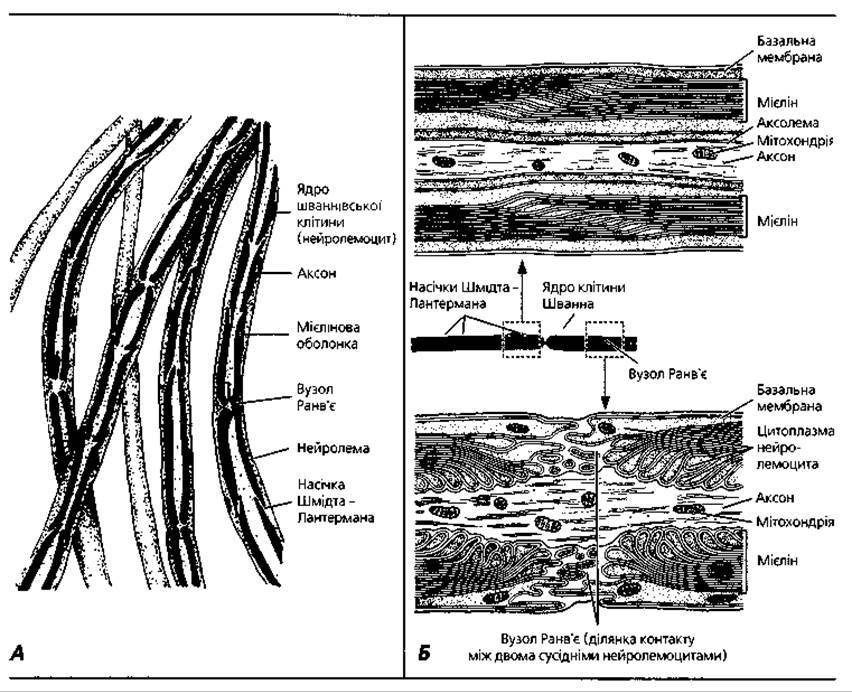
Fig. 3.54. Myelinated nerve fibers: A - semi-schematic representation of a teased histological preparation, osmium impregnation, x 600; B - diagram illustrating the structure of the nodes of Ranvier and Schmidt-Lanterman incisures

Fig. 3.55. Three-dimensional reconstruction of nerve fibers: A - successive phases of myelin sheath formation; B - region of the node of Ranvier of a myelinated nerve fiber; C - unmyelinated nerve fiber with isolated axons; D - cable-type unmyelinated fiber

Fig. 3.56. Diagrams of the ultrastructural organization of Different types of nerve fibers: A - myelinated fibers of the central nervous system: a single oligodendrocyte forms myelin sheaths around 3-50 nerve fibers. Myelinated (B) and unmyelinated (C) fibers of the peripheral nervous system: the sheath around the axons is formed by neurolemmocytes (Schwann cells)

Fig. 3.57. Electron microscopy of nerve fibers: A - myelinated nerve fiber, cross-section, x 35 000; B - myelinated and Unmyelinated nerve fibers within a peripheral nerve fragment, x 20 000
During the development of a myelinated fiber, the axon invaginates into the neurolemmocyte, depressing its membrane and forming a deep fold (Fig. 3.55, A). This double fold (duplicature) of the neurolemmocyte Plasmalemma is called the mesaxon. During further development, the neurolemmocyte slowly rotates around the axon, wrapping the mesaxon around it multiple times. The cytoplasm and Nucleus of the neurolemmocyte remain at the periphery, forming the neurolemma. Thus, the myelin sheath is formed by tightly concentric layers of mesaxon wraps around the axon, which constitute the lamellae of the myelin layer.
Each wrap of the mesaxon is about 8-12 nm wide and corresponds to the lipid layers of the two leaflets of the neurolemmocyte plasmalemma. Under an electron microscope, thin dark lines formed by protein molecules can be observed in its center and on its surface. Myelin incisures correspond to the areas where the mesaxon wraps are separated by the neurolemmocyte cytoplasm. The sheath of a single nerve fiber is formed by many neurolemmocytes. They contact each other at the nodes of Ranvier. The internodal segment corresponds to the area covered by a single neurolemmocyte.
In a Longitudinal section of a myelinated nerve fiber near the node of Ranvier, There is a region where the mesaxon wraps sequentially contact the axon. The attachment sites of the deepest wraps are furthest from the node, while each subsequent wrap gradually approaches it. This is because the mesaxon becomes layered during the growth of both the axon and the neurolemmocytes. Thus, the inner layers of the mesaxon are shorter than the outer ones. The adjacent margins of two neighboring neurolemmocytes, contacting at the node, form finger-like processes about 50 nm in diameter. The lengths of these processes vary. Together, they have the characteristic appearance of a "fluffy collar".
The neurolemma is a thin, light (when treated with osmic acid) Sheath of the nerve fiber located external to the myelin layer. The neurolemma is formed by the cytoplasmic portions of the neurolemmocytes and their nuclei. The basement membrane, covering the outer surface of the nerve fiber, merges with the Collagen fibers of the endoneurium (the connective tissue surrounding nerve fibers).
The structure described above is characteristic of myelinated nerve fibers of the peripheral nervous system. Myelinated fibers of the central nervous system have several distinctive features: their sheath is formed by typical oligodendrocytes instead of neurolemmocytes (the former have less cytoplasm and are smaller); myelin incisures and a basement membrane are absent; the nodes of Ranvier are larger, and the internodal segments are shorter; furthermore, a single oligodendrocyte can simultaneously form sheaths around several nerve processes (Fig. 3.56, A).
Unmyelinated nerve fibers are typical of the autonomic nervous system. The fiber diameter is 1-4 µm, meaning they are thinner than myelinated fibers. Unmyelinated fibers consist of an axon, a neurolemma, and a basement membrane. The neurolemma is formed by a strand of closely apposed neurolemmocytes. By invaginating the neurolemmocyte membrane, the axon becomes deeply embedded in this strand, and the glial cell envelops the process like a sheath. The neurolemmocyte membrane forms a deep fold, the mesaxon, similar to the one described above for the myelinated fiber. If the strand of lemmocytes encloses several axons (10-20) rather than just one, such unmyelinated fibers are called polyaxonal, or cable-type fibers. On the outside, the unmyelinated nerve fiber, like the myelinated one, is covered by a basement membrane (Figs. 3.55, 3.56, 3.57).
The conduction velocity of nerve impulses along myelinated nerve fibers is significantly higher (5-120 m/s) than along unmyelinated ones (1-2 m/s). This is because in an unmyelinated fiber, the depolarization wave propagates continuously along the entire plasmalemma, whereas in a myelinated fiber, it propagates saltatorily (by leaps), occurring only at the nodes of Ranvier.
Nervous tissue develops from the neural plate, which is a thickening of the ectoderm on the DORSAL SIDE OF the embryo. The neural plate sequentially transforms into the neural groove and the neural tube, which, upon closure, detaches from the cutaneous ectoderm (see Chapter 2). A portion of the neural plate cells remains between the neural tube and the cutaneous ectoderm as a loose cluster of cells known as the neural crest (or ganglionic plate). The crest cells migrate in lateral and ventral directions, giving rise to the following derivatives: cranial nerve nuclei, neurons of spinal and autonomic ganglia, neurolemmocytes (neuroglia), and Skin pigment cells.
The cells of the neural tube, which is composed of pseudostratified neuroepithelium, are called ventricular or neuroepithelial cells. They are columnar in shape; their apical portions border the lumen of the neural tube and are joined by Gap Junctions, while their basal portions contact the subpial limiting membrane, and they exhibit cyclic nuclear migration. The proliferative capacity of these cells decreases during embryonic development and after birth. Morphologically similar ventricular cells differentiate into various types of nervous tissue cells. Some of them give rise to neurons, while others give rise to glial cells (ependymocytes, astrocytes, oligodendrocytes). In certain brain regions, ventricular cells give rise to so-called subventricular and extraventricular neurogerminal cells, which retain proliferative activity longer and persist for some time after birth. They give rise to certain types of neurons and neuroglia.
Key terms to remember
1. Nervous tissue. 2. Neuron (neurocyte). 3. Perikaryon. 4. Axon. 5. Dendrite. 6. Chromatophilic substance (Nissl substance, tigroid). 7. Neurofibril. 8. Axonal transport. 9. Dendritic transport. 10. Unipolar neuron. 11. Bipolar neuron. 12. Pseudounipolar neuron. 13. Multipolar neuron. 14. Afferent (sensory) neuron. 15. Interneuron (associative neuron). 16. Efferent (motor) neuron. 17. Reflex arc. 18. Synapse. 19. Presynaptic part. 20. Postsynaptic part. 21. Synaptic cleft. 22. Axosomatic synapse. 23. Axodendritic synapse. 24. Axo-axonal synapse. 25. Dendro-dendritic synapse. 26. Dendrosomatic synapse. 27. Neuroglia. 28. Macroglia. 29. Ependymocyte. 30. Astrocyte. 31. Protoplasmic astrocyte. 32. Fibrous astrocyte. 33. Oligodendrocyte. 34. Neurolemmocyte (Schwann cell). 35. Microglia. 36. Nerve fiber. 37. Myelinated nerve fiber. 38. Myelin sheath. 39. Node of Ranvier. 40. Internodal segment. 41. Myelin incisure. 42. Mesaxon. 43. Neurolemma. 44. Unmyelinated nerve fiber. 45. Neural plate. 46. Neural groove. 47. Neural tube. 48. Neural crest (ganglionic plate). 49. Ventricular (neuroepithelial) cell. 50. Extraventricular (neurogerminal) cell.
Last update: 09/08/2026
Editorial and Educational Adaptation: This material has been compiled based on the primary/original source text. The project team performed an editorial review, corrected technical inaccuracies, structured sections, and adapted the content for an educational format.
What was processed:
- elimination of formatting defects (OCR errors, structural breaks, corrupted characters);
- editorial organization of content;
- standardization of terminology in accordance with academic sources;
- verification of factual statements against the original source text.
All mentions of the author, publication year, and origin of the primary text have been preserved in accordance with the source.