Human Anatomy and Physiology - N. I. Fedyukovich 2003
Nervous System
Peripheral Nervous System
Cranial Nerves
Nerves emerging from the Brainstem are called Cranial Nerves. In humans, there are 12 pairs of cranial nerves, designated by Roman numerals in order of their Location. Cranial nerves have various Functions because they consist of motor fibers, sensory fibers, or a combination of both. Therefore, some are classified as motor nerves (pairs III, IV, VI, XI, and XII), others as sensory nerves (pairs I, II, and VIII), and the rest as mixed nerves (pairs V, VII, IX, and X).
The olfactory nerves (nn. olfactorii) are the I pair of cranial nerves (Fig. 118).
Class="center">
Fig. 118. Olfactory nerve:
1 — olfactory bulbs; 2 — olfactory nerves
Functionally, they are sensory nerves formed by the central processes of olfactory Cells located in the nasal mucosa. These processes form nerve fibers that, as part of 15–20 olfactory nerves, pass through the foramina of the cribriform plate into the cranial cavity to reach the olfactory bulb (see "Olfactory Organ").
The Optic nerve (n. opticus) is the II pair of sensory nerves (Fig. 119).

Fig. 119. Optic nerve (diagram):
1 — Eyeball; 2 — optic nerve; 3 — orbital part; 4 — intracanalicular part; 5 — intracranial part; 6 — optic chiasm
It is composed of the axons of retinal ganglion cells. After passing through the choroid and sclera, the optic nerve fibers enter the cranial cavity through the optic canals, where they form an incomplete optic chiasm. Post-chiasmatically, the nerve fibers continue as the optic tracts (see "Organ of Vision").
The Oculomotor nerve (n. oculomotorius) is the III pair (Fig. 120). Part of the nerve originates from the motor Nucleus, and another part from the autonomic (parasympathetic) nucleus, both located in the Midbrain. It emerges onto the Base of the Skull from the sulcus of the same name on the Medial surface of the cerebral peduncle and enters the Orbit through the superior orbital fissure, where it divides into superior and inferior branches, innervating the extraocular Muscles. Autonomic fibers branch off from the inferior division of the oculomotor nerve to form the oculomotor (parasympathetic) ROOT, which travels to the ciliary ganglion.
The Trochlear nerve (n. trochlearis), pair IV, is a motor nerve (see Fig. 120). It originates from the trochlear nucleus in the midbrain, emerges from the dorsal surface of the brainstem, and courses along the base of the skull toward the orbit. Within the orbit, the nerve enters through the superior orbital fissure, reaches the superior oblique Muscle, and innervates it.

Fig. 120. Oculomotor and trochlear nerves:
1 — decussation of trochlear nerves; 2 — trochlear nerve; 3 — oculomotor nerve; 4 — sympathetic root; 5 — optic nerve (part); 6 — short ciliary nerves; 7 — ciliary ganglion; 8 — inferior branch of oculomotor nerve; 9 — nasociliary root; 10 — Trigeminal nerve; 11 — superior branch of oculomotor nerve
The trigeminal nerve (n. trigeminus), pair V, is a mixed nerve. The motor fibers of the trigeminal nerve originate from its motor nucleus, which is located in the Pons.
The sensory fibers of this nerve project to the mesencephalic and spinal nuclei of the trigeminal nerve.
The nerve emerges at the base of the Brain from the lateral surface of the pons via two roots: sensory and motor. On the anterior surface of the petrous part of the Temporal bone, the sensory root expands to form the trigeminal ganglion. This ganglion contains The Cell bodies of sensory Neurons, whose central processes form the sensory root, while their peripheral processes contribute to the three Branches of the trigeminal nerve arising from the ganglion: 1) the ophthalmic nerve;
2) the maxillary nerve; and 3) the mandibular nerve. The first two branches are purely sensory, whereas the third is mixed because it is joined by the motor fibers.
The first branch, the ophthalmic nerve (Fig. 121), enters the orbit through the superior orbital fissure, where it divides into three main branches: the lacrimal nerve, frontal nerve, and nasociliary nerve. These branches innervate the orbital contents, the eyeball, the Skin and conjunctiva of the upper eyelid, the skin of the forehead and Nose, and the mucosa of PARTS OF THE Nasal cavity, frontal, and sphenoid sinuses.
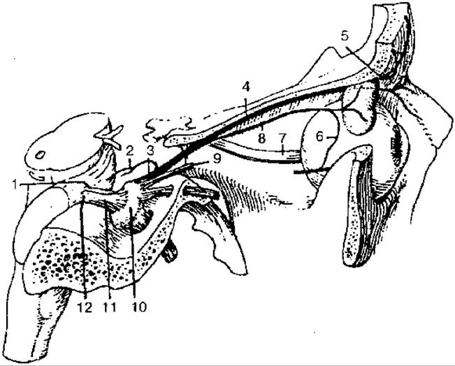
Fig. 121. Ophthalmic nerve (the first branch of the trigeminal nerve):
1 — motor root; 2 — tentorial (meningeal) branch; 3 — ophthalmic nerve; 4 — frontal nerve; 5 — supraorbital nerve; 6 — communicating branch (with zygomatic nerve); 7 — optic nerve; 8 — lacrimal nerve; 9 — nasociliary nerve; 10 — trigeminal ganglion; 11 — trigeminal nerve; 12 — sensory root
The second branch, the maxillary nerve (Fig. 122), passes through the foramen rotundum into the pterygopalatine fossa, where it gives off the infraorbital and zygomatic nerves, as well as ganglionic branches to the pterygopalatine ganglion.
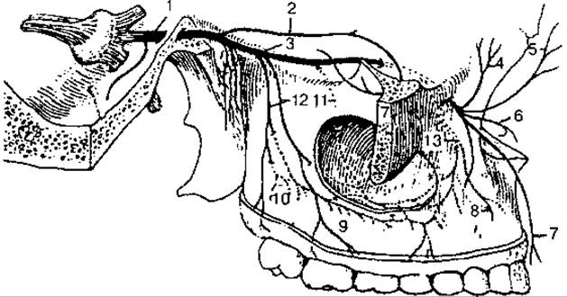
Fig. 122. Maxillary nerve (Second Division of the trigeminal nerve):
1 — maxillary nerve; 2 — zygomatic nerve; 3 — infraorbital nerve; 4 — inferior palpebral branches; 5 — external nasal branches; 6 — internal nasal branches; 7 — superior labial branches; 8 — superior dental branches; 9 — superior gingival branches; 10 — superior dental plexus; 11 — middle superior alveolar branch; 12 — posterior superior alveolar branches; 13 — anterior superior alveolar branches
The infraorbital nerve gives off branches to innervate the Teeth and Gums of the upper jaw; it also innervates the skin of the lower eyelid, nose, and upper lip.
Along its course, the zygomatic nerve gives off branches carrying parasympathetic fibers to the lacrimal gland; it also innervates the skin of the temporal, zygomatic, and buccal regions. Branches arising from the pterygopalatine ganglion innervate the mucous membrane and Glands of the nasal cavity, as well as the Hard and Soft palates.
The third division, the mandibular nerve (Fig. 123), exits the skull through the foramen ovale and divides into A number of motor branches supplying all Muscles of Mastication, the mylohyoid muscle, the tensor veli palatini, and the tensor tympani. In addition, the mandibular nerve gives off several sensory branches, including large ones: the lingual and inferior alveolar nerves; and smaller nerves (buccal, auriculotemporal, and meningeal branch). The latter innervate the skin and mucous membrane of the Cheeks, part of the auricle, the external acoustic meatus, the tympanic membrane, the skin of the temporal region, the Cytology/practical/97.html">Parotid salivary gland, and the Meninges of the brain.

Fig. 123. Mandibular nerve (third division of the trigeminal nerve):
1 — mandibular nerve; 2 — lateral pterygoid nerve; 3 — medial pterygoid nerve; 4 — buccal nerve; 5 — masseteric nerve; 6 — auriculotemporal nerve; 7 — anterior auricular nerves; 8 — superficial temporal branches
The lingual nerve (Fig. 124) provides general sensation (pain, Touch, Temperature) for the anterior two-thirds of the Tongue and the oral mucosa.

Fig. 124. Lingual nerve:
1 — lingual nerve; 2 — mylohyoid nerve; 3 — lingual branches; 4 — Hypoglossal nerve; 5 — submandibular parasympathetic ganglion
The inferior alveolar nerve (Fig. 125), the largest branch of the mandibular nerve, enters the mandibular canal, innervates the teeth and gums of the lower jaw, and, after exiting through the mental foramen, innervates the skin of the chin and lower lip.

Fig. 125. Inferior alveolar nerve:
1 — inferior alveolar nerve; 2 — mylohyoid nerve; 3 — mental nerve; 4 — labial and gingival branches; 5 — mental branches
The Abducens nerve (n. abducens), cranial nerve VI (Fig. 126), is formed by the axons of motor cells in the abducens nucleus, which is located in the posterior part of the pons on the floor of the Fourth ventricle. The nerve emerges from the brainstem, enters the orbit through the superior orbital fissure, and innervates the lateral rectus muscle of the eye.
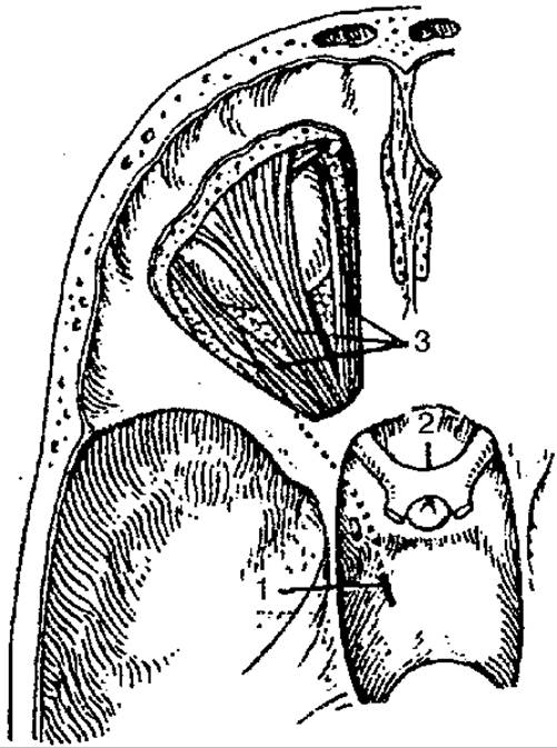
Fig. 126. Abducens nerve:
1 — abducens nerve; 2 — optic nerve; 3 — Muscles of the eye
The Facial Nerve (n. facialis), cranial nerve VII, is a mixed nerve comprising two parts: the facial nerve proper and the intermediate nerve (Fig. 127). The nuclei of the facial nerve lie within the pons. After emerging from the brainstem in the groove between the pons and the Medulla Oblongata, the facial nerve enters the internal acoustic meatus and, passing through the facial canal, exits via the stylomastoid foramen.
Within the facial canal, the nerve divides into several branches:
1) the greater petrosal nerve, which carries parasympathetic fibers to the pterygopalatine ganglion; it exits the canal through an opening on the superior surface of the petrous part of the temporal bone;
2) the chorda tympani — a mixed nerve that branches off the facial nerve, exits through the petrotympanic fissure, and runs forward and downward to join the lingual nerve. It contains afferent taste fibers from the anterior part of the tongue and parasympathetic secretomotor fibers to the sublingual and submandibular Salivary Glands; 3) the stapedius nerve — a motor nerve that innervates the stapedius muscle of the tympanic cavity.
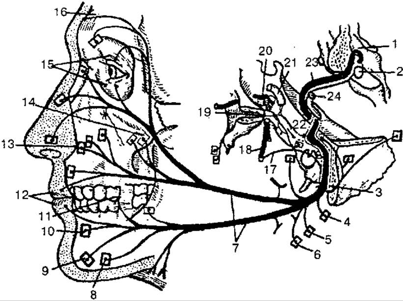
Fig. 127. Facial nerve (diagram):
1 — floor of the IV ventricle; 2 — nucleus of facial nerve; 3 — stylomastoid foramen; 4 — branch to posterior auricular muscle; 5 — branch to posterior belly of digastric muscle; 6 — branch to stylohyoid muscle; 7 — branches of facial nerve to Muscles of facial expression and platysma; 8 — branch to depressor anguli oris muscle; 9 — branch to mentalis muscle; 10 — branch to depressor labii inferioris muscle; 11 — branch to buccinator muscle; 12 — branch to orbicularis oris muscle; 13 — branch to levator labii superioris muscle; 14 — branch to zygomatic muscle; 15 — branches to orbicularis oculi muscle; 16 — branches to frontal belly of epicranius muscle; 17 — chorda tympani; 18 — lingual nerve; 19 — pterygopalatine ganglion; 20 — trigeminal ganglion; 21 — Internal Carotid Artery; 22 — intermediate nerve; 23 — facial nerve; 24 — Vestibulocochlear nerve
Upon exiting its canal through the stylomastoid foramen, the facial nerve gives off branches to the epicranius, posterior auricular, digastric, and stylohyoid muscles. Within the parotid gland, the facial nerve fans out to form the parotid plexus (pes anserinus major). Only motor fibers emerge from this plexus, forming the temporal, zygomatic, buccal, marginal mandibular, and cervical branches. All of these participate in the Innervation of the muscles of facial expression and the platysma muscle of the neck.
The vestibulocochlear nerve (n. vestibulocochlearis), cranial nerve VIII, is formed by sensory nerve fibers originating from the Organ of Hearing and Balance (Fig. 128). It emerges from the brainstem behind the pons, lateral to the facial nerve, and divides into the vestibular and cochlear parts, which innervate the Organ of Hearing and balance.

Fig. 128. Vestibulocochlear nerve (diagram):
1 — semicircular canals; 2 — lateral ampullary nerve; 3 — anterior ampullary nerve; 4 — utricular nerve; 5 — utriculoampullary nerve; 6 — vestibular ganglion; 7 — vestibular nerve; 8 — cochlear nerve; 9 — saccular nerve; 10 — cochlear ganglion (spiral ganglion of the cochlea); 11 — posterior ampullary nerve
The cell bodies of the vestibular part of the nerve lie in the vestibular ganglion, located at the bottom of the internal acoustic meatus. The peripheral processes of these cells form a series of nerves that terminate in receptors within the semicircular canals of the membranous labyrinth of the Inner ear, while the central processes project to the corresponding nuclei of the Rhomboid fossa. The vestibular part participates in regulating THE POSITION OF the HEAD, trunk, and limbs in space, as well as in The system of motor coordination.
The cochlear part of the nerve is formed by the central processes of neurons in the cochlear ganglion, which lies within the cochlea of the labyrinth. The peripheral processes of these cells terminate in the Spiral organ of the cochlear duct, while the central processes reach the corresponding nuclei located in the rhomboid fossa. The cochlear part participates in The formation of The Organ of hearing.
The Glossopharyngeal nerve (n. glossopharyngeus), cranial nerve IX, is a mixed nerve that emerges from the medulla oblongata by 4–5 rootlets and heads toward the jugular foramen (Fig. 129). Upon exiting the cranial cavity, the nerve forms two ganglia: superior and inferior. These ganglia contain the cell bodies of sensory neurons. Beyond the jugular foramen, the nerve descends, courses toward the root of the tongue, and divides into terminal lingual branches that end in the mucous membrane of the dorsum of the tongue. The glossopharyngeal nerve gives off collateral branches that provide sensory innervation to the mucous membrane of the tympanic cavity and the auditory tube (tympanic nerve), the palatine arches and Tonsils (tonsillar branches), the parotid gland (lesser petrosal nerve), the carotid sinus and carotid body (carotid sinus branch), and motor innervation to the stylopharyngeus muscle (stylopharyngeal branch). In addition, branches of the glossopharyngeal nerve join with branches of the Vagus nerve and the Sympathetic trunk to form the pharyngeal plexus.

Fig. 129. Glossopharyngeal nerve:
1 — glossopharyngeal nerve; 2 — superior ganglion; 3 — communicating branch; 4 — inferior ganglion; 5 — branch to stylopharyngeus muscle; 6 — tonsillar branches; 7 — lingual branches; 8 — pharyngeal branches; 9 — carotid sinus branch
The vagus nerve (n. vagus), cranial nerve X, is a mixed nerve (Fig. 130) containing sensory, motor, and autonomic fibers. It is the longest of the cranial nerves, with fibers reaching the Organs of the neck, Thorax, and abdomen. Impulses traveling along the vagus nerve slow The Heart rate, dilate Blood Vessels, constrict the Bronchi, increase intestinal peristalsis, relax intestinal sphincters, and stimulate the secretion of gastric and intestinal glands. The vagus nerve emerges from the medulla oblongata in the posterior sulcus via several rootlets, which unite to form a single trunk and head toward the jugular foramen. Below the jugular foramen, the nerve has two enlargements: the superior and inferior ganglia, formed by the cell bodies of sensory neurons. The peripheral processes of these neurons arise from Internal Organs, the dura mater of the brain, and the skin of the external acoustic meatus, while their central processes project to the Nucleus of the solitary tract in the medulla oblongata.
The vagus nerve is divided into four parts: cranial, cervical, thoracic, and abdominal.

Fig. 130. Vagus nerve:
1 — vagus nerve; 2 — superior ganglion; 3 — inferior ganglion; 4 — meningeal branch; 5 — auricular branch; 6 — communicating branch; 7 — pharyngeal branches; 8 — pharyngeal plexus; 9 — superior cervical cardiac branches; 10 — superior laryngeal nerve; 11 — external branch; 12 — internal branch; 13 — communicating branch with recurrent laryngeal nerve; 14 — inferior cervical cardiac branches; 15 — recurrent laryngeal nerve; 16 — tracheal branches; 17 — esophageal branches; 18 — inferior laryngeal nerve; 19 — communicating branch with internal laryngeal branch; 20 — thoracic cardiac branches; 21 — bronchial branches; 22 — pulmonary plexus; 23 — esophageal plexus; 24 — anterior vagal trunk; 25 — posterior vagal trunk; 26 — anterior gastric branches; 27 — posterior gastric branches; 28 — hepatic branches; 29 — celiac branches; 30 — renal branches
The cranial part lies between the Water/144.html">Origin of the nerve and the superior ganglion; it gives off branches to the dura mater of the brain, the walls of the transverse and occipital sinuses, the skin of the external acoustic meatus, and the external surface of the auricle.
The cervical part includes the portion located between the inferior ganglion and THE ORIGIN OF the recurrent laryngeal nerve. The branches of the cervical part are: 1) pharyngeal branches, which innervate the pharyngeal mucosa, the Constrictor muscles, and the muscles of the soft palate; 2) superior cervical cardiac branches, which, together with branches of the sympathetic trunk, enter the cardiac plexuses; 3) the superior laryngeal nerve, which innervates the mucosa of the Larynx and the root of the tongue, as well as the cricothyroid muscle of the larynx; 4) the recurrent laryngeal nerve, which gives off branches to the Trachea, Esophagus, and heart, and innervates the mucosa and muscles of the larynx, except for the cricothyroid.
The thoracic part extends from the level of origin of the recurrent laryngeal nerve to the level of the esophageal hiatus of the Diaphragm; it gives off several branches to the heart, Lungs, and esophagus, and participates in the Formation of the cardiac, pulmonary, and esophageal plexuses.
The abdominal part consists of the anterior and posterior vagal trunks. They give off branches to The Stomach, Liver, Pancreas, Spleen, Kidneys, and intestines.
The Accessory nerve (n. accessorius), cranial nerve XI, is a motor nerve (Fig. 131). It consists of several cranial and spinal roots and innervates the sternocleidomastoid and trapezius muscles. It has two nuclei: one located in the medulla oblongata, and the other in the Cells of the anterior horns of the cervical Spinal Cord.

Fig. 131. Accessory nerve (diagram):
1 — spinal roots; 2 — cranial roots (vagal part); 3 — trunk of the accessory nerve; 4 — internal branch; 5 — external branch; 6 — muscular branches
The hypoglossal nerve (n. hypoglossus), cranial nerve XII (Fig. 132), is a motor nerve formed by the processes of Nerve Cells of the homonymous nucleus located in the medulla oblongata. The nerve exits the skull through the hypoglossal canal of the Occipital bone, innervating the muscles of the tongue and, partially, some muscles of the neck.
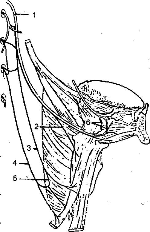
Fig. 132. Hypoglossal nerve and cervical (hypoglossal) loop:
1 — hypoglossal nerve; 2 — thyrohyoid branch; 3 — anterior root; 4 — posterior root; 5 — cervical (hypoglossal) loop; 6 — lingual branches
Last update: 08/08/2026
Editorial and Educational Adaptation: This material has been compiled based on the primary/original source text. The project team performed an editorial review, corrected technical inaccuracies, structured sections, and adapted the content for an educational format.
What was processed:
- elimination of formatting defects (OCR errors, structural breaks, corrupted characters);
- editorial organization of content;
- standardization of terminology in accordance with academic sources;
- verification of factual statements against the original source text.
All mentions of the author, publication year, and origin of the primary text have been preserved in accordance with the source.