Human Anatomy and Physiology - N. I. Fedyukovich 2003
Nervous system
Peripheral nervous system
Spinal nerves
The Spinal Nerves (nn. spinales) are paired, metamerically arranged nerve trunks formed by the fusion of two Spinal Cord roots: the posterior (sensory) and anterior (motor) roots (Fig. 133). At the level of the intervertebral foramen, they unite and exit, dividing into three or four branches: the anterior, posterior, meningeal, and white communicating branches; the latter connect with the ganglia of the Sympathetic trunk. In humans, there are 31 pairs of spinal nerves, which correspond to the 31 pairs of spinal cord segments (8 cervical, 12 thoracic, 5 lumbar, 5 sacral, and 1 pair of coccygeal nerves). Each pair of spinal nerves innervates a specific area of Muscle (myotome), Skin (dermatome), and bone (sclerotome). Based on this, segmental innervation of Muscles, skin, and bones is distinguished.
Class="center">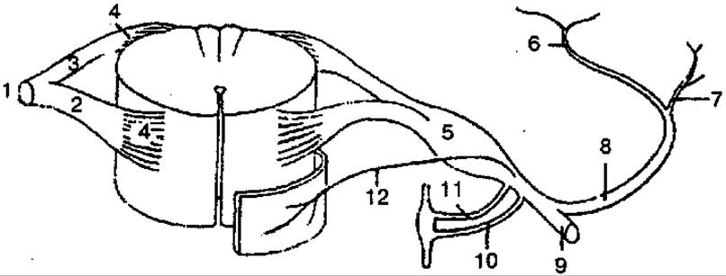
Fig. 133. Diagram of spinal nerve formation:
1 — trunk of spinal nerve; 2 — anterior (motor) ROOT; 3 — posterior (sensory) root; 4 — rootlets; 5 — spinal (sensory) ganglion; 6 — medial part of the posterior ramus; 7 — lateral part of the posterior ramus; 8 — posterior ramus; 9 — anterior ramus; 10 — white ramus; 11 — gray ramus; 12 — meningeal branch
The posterior rami of the spinal nerves innervate the Deep Muscles of the back and occiput, as well as the skin of the posterior surface of the HEAD and trunk. There are posterior rami of the cervical, thoracic, lumbar, sacral, and coccygeal nerves.
The posterior ramus of the first cervical spinal nerve (СІ) is called the suboccipital nerve. It innervates the rectus capitis posterior Major and minor, the obliquus capitis superior and inferior, and the semispinalis capitis.
The posterior ramus of the second cervical spinal nerve (СІІ) is called the greater occipital nerve; it divides into short muscular branches and a long cutaneous branch, innervating the Muscles of the head and the skin of the occipital region.
The anterior rami of the spinal nerves are significantly thicker and longer than the posterior rami. They innervate the skin and muscles of the neck, chest, abdomen, and upper and lower limbs. Unlike the posterior rami, only the anterior rami of the thoracic spinal nerves retain a metameric (segmental) Structure. The anterior rami of the cervical, lumbar, sacral, and coccygeal spinal nerves form plexuses (plexus). These include the cervical, brachial, lumbar, sacral, and coccygeal nerve plexuses.
The Cervical plexus is formed by the anterior rami of the upper four cervical (СІ — CIV) spinal nerves, which are connected by three loop-like arches, and lies on the Deep Muscles of the Neck. The cervical plexus communicates with the accessory and hypoglossal nerves. It contains motor (muscular), cutaneous, and mixed nerves and branches. The muscular nerves innervate the trapezius and sternocleidomastoid muscles, send branches to the deep muscles of the neck, and the Infrahyoid muscles receive innervation from the ansa cervicalis. The cutaneous (sensory) NERVES OF THE cervical plexus give rise to the great auricular nerve, lesser occipital nerve, transverse cervical nerve, and supraclavicular nerves. The great auricular nerve innervates the skin of the auricle and external acoustic meatus; the lesser occipital nerve innervates the skin of the lateral occipital region; the transverse cervical nerve innervates the skin of the anterior and lateral Regions of the neck; and the supraclavicular nerves innervate the skin above and below the clavicle.
The largest nerve of the cervical plexus is the phrenic nerve. It is a mixed nerve, formed by the anterior rami of the III–V cervical spinal nerves, which passes into the thoracic cavity and ends within the substance of the Diaphragm.
The motor fibers of the phrenic nerve innervate the diaphragm, while its sensory fibers innervate the Pericardium and Pleura.
The Brachial Plexus (Fig. 134) is formed by the anterior rami of the lower four cervical (CV — CVIII) nerves, a portion of the anterior ramus of the first cervical (СIV) and the first thoracic (ThI) spinal nerves.
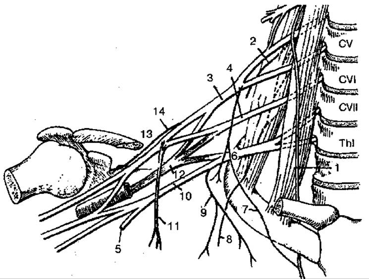
Fig. 134. Brachial plexus (diagram):
1 — phrenic nerve; 2 — dorsal scapular nerve; 3 — superior trunk of the brachial plexus; 4 — middle trunk of the brachial plexus; 5 — subclavian trunk; 6 — inferior trunk of the brachial plexus; 7 — accessory phrenic nerves; 8 — long thoracic nerve; 9 — medial pectoral nerve; 10 — lateral pectoral nerve; 11 — medial cord; 12 — posterior cord; 13 — lateral cord; 14 — suprascapular nerve
In the interscalene space, the anterior rami form three trunks: superior, middle, and inferior. These trunks divide into several branches and proceed to the axillary fossa, where they form three cords (lateral, medial, and posterior) and surround the axillary artery on three sides. The trunks of the brachial plexus, along with their branches lying above the clavicle, are called the suviclavicular part, while those with branches lying below the clavicle are called the infraclavicular part. The branches arising from the brachial plexus are divided into short and long branches. The short branches mainly innervate the bones and soft Tissues of the shoulder girdle, while the long branches innervate the free upper limb.
The Short Branches of the brachial plexus include the dorsal scapular nerve, which innervates the levator scapulae and the rhomboid major and minor muscles; the long thoracic nerve, which innervates the serratus anterior muscle; the subclavian nerve, which innervates the subclavius muscle; the suprascapular nerve, which innervates the supraspinatus and infraspinatus muscles and the shoulder Joint Capsule; the subscapular nerve, which innervates the subscapularis and teres major muscles; the thoracodorsal nerve, which innervates the latissimus dorsi muscle; the lateral and medial pectoral nerves, which innervate the pectoral muscles of the same name; and the axillary nerve, which innervates the deltoid and teres minor muscles, the shoulder joint capsule, and the skin of the upper lateral aspect of the arm.
The Long branches of the brachial plexus arise from the lateral, medial, and posterior cords of the infraclavicular part of the brachial plexus (Fig. 135, A, B).
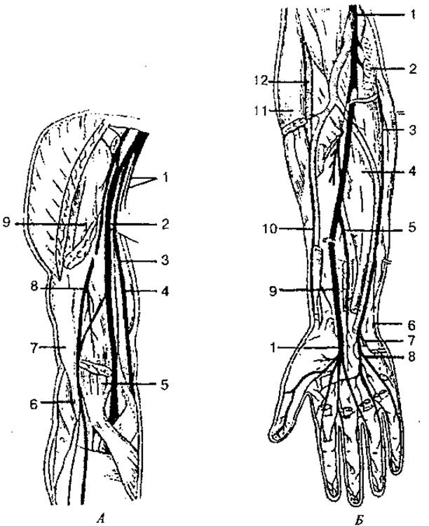
Fig. 135. Nerves of the arm, forearm, and hand:
A — nerves of the arm: 1 — medial cutaneous nerve of the arm and medial cutaneous nerve of the forearm; 2 — median nerve; 3 — brachial artery; 4 — ulnar nerve; 5 — biceps brachii muscle (distal end); 6 — radial nerve; 7 — brachialis muscle; 8 — musculocutaneous nerve; 9 — biceps brachii muscle (proximal end); B — nerves of the forearm and hand: 1 — median nerve; 2 — pronator teres (transected); 3 — ulnar nerve; 4 — flexor digitorum profundus; 5 — anterior interosseous nerve; 6 — dorsal branch of the ulnar nerve; 7 — deep branch of the ulnar nerve; 8 — superficial branch of the ulnar nerve; 9 — pronator quadratus (transected); 10 — superficial branch of the radial nerve; 11 — brachioradialis muscle (transected); 12 — radial nerve
The musculocutaneous nerve arises from the lateral cord and supplies branches to the coracobrachialis, biceps brachii, and brachialis muscles. After sending branches to the elbow joint, the nerve descends as the lateral cutaneous nerve of the forearm. It innervates a portion of the skin of the forearm.
The median nerve is formed by the fusion of two roots from the lateral and medial cords on the anterior surface of the axillary artery. The nerve gives off its first branches to the elbow joint, and then, descending further, to the anterior muscles of the forearm. In the palm, beneath the palmar aponeurosis, the median nerve divides into terminal branches that innervate the muscles of the thumb, except for the adductor pollicis muscle. The median nerve also innervates the JOINTS OF THE wrist, the first four digits, some of the lumbrical muscles, and the skin of both the dorsal and palmar surfaces.
The ulnar nerve originates from the medial cord of the brachial plexus and runs alongside the brachial artery along the medial aspect of the arm, where it gives off no branches. It then courses behind the medial epicondyle of the humerus and passes into the forearm, where it runs with the ulnar artery in the ulnar groove. In the forearm, it innervates the flexor carpi ulnaris and a portion of the flexor digitorum profundus. In the lower third of the forearm, the ulnar nerve divides into dorsal and palmar branches, which then pass into the hand. In the hand, the Branches of the ulnar nerve innervate the adductor pollicis muscle, all interosseous muscles, two lumbrical muscles, the muscles of the little finger, the skin of the palmar surface at the level of the V digit and the ulnar side of the IV digit, and the skin of the dorsal surface at the level of the V, IV, and the ulnar side of the III digits.
The medial cutaneous nerve of the arm arises from the medial cord, supplies branches to the skin of the arm, accompanies the brachial artery, and communicates in the axillary fossa with the lateral branch of the II, and sometimes III intercostal nerves.
The medial cutaneous nerve of the forearm is also a branch of the medial cord and innervates the skin of the forearm.
The radial nerve originates from the posterior cord of the brachial plexus and is the thickest nerve. In the arm, it passes through the radial canal between the humerus and the heads of the triceps brachii muscle, giving off muscular branches to this muscle and cutaneous branches to the posterior surface of the arm and forearm. In the lateral sulcus of the cubital fossa, it divides into deep and superficial branches. The deep branch innervates all the muscles of the posterior compartment of the forearm (extensors), while the superficial branch runs in the sulcus alongside the radial artery, passes to the dorsum of the hand, where it innervates the skin of the lateral 2 1/2 digits, starting from the thumb.
The anterior rami of the thoracic spinal nerves (ThI— ThXII), 12 pairs, run in the intercostal spaces and are called intercostal nerves. The exception is the anterior ramus of the XII thoracic nerve, which passes below the XII rib and is called the subcostal nerve. The intercostal nerves run in the intercostal spaces between the internal and external intercostal muscles and do not form plexuses. The upper six intercostal nerves on both sides reach the Sternum, while the lower five intercostal nerves and the subcostal nerve continue onto the anterior abdominal wall.
The anterior rami innervate the intrinsic muscles of the chest, participate in the Innervation of the muscles of the anterior abdominal wall, and give off anterior and lateral cutaneous branches, innervating the skin of the chest and abdomen.
The Lumbosacral plexus (Fig. 136) is formed by the anterior rami of the lumbar and sacral spinal nerves, which join together to form the lumbar and sacral plexuses. The lumbosacral trunk serves as the connecting link between these plexuses.
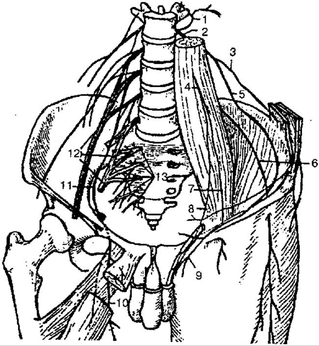
Fig. 136. Lumbosacral plexus:
1 - posterior rami of lumbar nerves; 2 - anterior rami of lumbar nerves; 3 - iliohypogastric nerve; 4 - genitofemoral nerve; 5 - ilioinguinal nerve; 6 - lateral femoral cutaneous nerve; 7 - femoral branch; 8 - genital branch; 9 - anterior scrotal nerves; 10 - anterior branch of obturator nerve; 11 - obturator nerve; 12 - lumbosacral plexus; 13 - anterior rami of Sacral Plexus
The Lumbar plexus is formed by the anterior rami of the upper three lumbar spinal nerves and partially by the anterior rami of the XII thoracic and IV lumbar spinal nerves. It lies anterior to the transverse processes of the lumbar vertebrae within the substance of the psoas major muscle and on the anterior surface of the quadratus lumborum muscle. Short muscular branches arise from all anterior rami of the lumbar nerves, innervating the psoas major and psoas minor muscles, the quadratus lumborum muscle, and the lumbar intertransverse lateral muscles.
The largest branches of the lumbar plexus are the femoral and obturator nerves.
The femoral nerve is formed by three roots that initially run deep into the psoas major muscle and unite at the level of the V lumbar vertebra to form the trunk of the femoral nerve. Heading downward, the femoral nerve lies in the groove between the psoas major and iliacus muscles. It enters the thigh through the muscular lacuna, where it gives off branches to the anterior muscles of the thigh and the skin of the anteromedial surface of the thigh. The longest branch of the femoral nerve is the saphenous nerve. The latter enters the adductor canal along with the femoral artery, then follows the descending genicular artery along the Medial surface of the leg to the FOOT. Along its course, it innervates the skin of the knee joint, the Patella, and partially the skin of the leg and foot.
The obturator nerve is the second largest branch of the lumbar plexus. From the lumbar region, the nerve descends along the medial border of the psoas major muscle into the lesser pelvis, where, together with the obturator artery and vein, it passes through the obturator canal into the thigh, gives off muscular branches to the adductor muscles of the thigh, and divides into two terminal branches: the anterior (innervating the skin of the medial surface of the thigh) and the posterior (innervating the obturator externus, adductor magnus muscles, and the hip joint).
In addition, other major branches arise from the lumbar plexus: 1) the iliohypogastric nerve — innervates the muscles and skin of the anterior abdominal wall, part of the gluteal region and thigh; 2) the ilioinguinal nerve — innervates the skin of the pubis, inguinal region, root of the Penis, and the Scrotum (skin of the Labia Majora); 3) the genitofemoral nerve — divides into two branches: genital and femoral. The first branch innervates part of the skin of the thigh, in men — the cremaster muscle, the skin of the scrotum, and the dartos tunic; in women — the round ligament of the Uterus and the skin of the labia majora. The femoral branch passes through the vascular lacuna to the thigh, where it innervates the skin over the inguinal ligament and the femoral canal area; 4) the lateral femoral cutaneous nerve — exits the pelvic cavity into the thigh, innervating the skin of the lateral surface of the thigh down to the knee joint.
The sacral plexus is formed by the anterior rami of the upper four sacral, the V lumbar, and partially the IV lumbar spinal nerves. The anterior rami of the latter form the lumbosacral trunk. It descends into the cavity of the lesser pelvis, joining the anterior rami of the I - IV sacral spinal nerves. The branches of the sacral plexus are divided into short and long.
The short branches of the sacral plexus include the superior and inferior gluteal nerves (Fig. 137), the pudendal nerve, the nerve to the obturator internus, the nerve to the piriformis, and the nerve to the quadratus femoris. The latter three nerves are motor nerves and innervate the corresponding muscles, passing through the infrapiriform foramen.
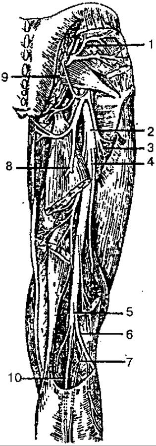
Fig. 137. Nerves of the gluteal region and the posterior surface of the thigh:
1 — superior gluteal nerve; 2 — sciatic nerve; 3, 4 — muscular branches of the sciatic nerve; 5 — tibial nerve; 6 — common fibular nerve; 7 — lateral sural cutaneous nerve; 8 — posterior femoral cutaneous nerve; 9 — inferior gluteal nerve; 10 — medial dorsal cutaneous nerve
The superior gluteal nerve exits the pelvic cavity through the suprapiriform foramen along with the superior gluteal artery and vein, passing between the gluteus minimus and gluteus medius muscles. It innervates these gluteal muscles, as well as the tensor fasciae latae muscle.
The inferior gluteal nerve exits the pelvic cavity through the infrapiriform foramen and innervates the gluteus maximus muscle.
The long branches of the sacral plexus are represented by the posterior femoral cutaneous nerve, which innervates the skin of the gluteal region and partially the skin of the Perineum, and the sciatic nerve (Fig. 138).
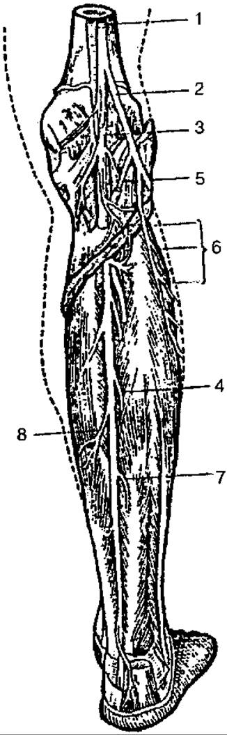
Fig 138. Nerves of the leg (posterior surface):
1 — sciatic nerve; 2 — common fibular nerve; 3 — tibial nerve; 4, 7, 8 — muscular branches of the tibial nerve; 5 — lateral sural cutaneous nerve; 6 — muscular branches of the fibular nerve
The sciatic nerve is the largest nerve in The Human Body. It exits the pelvic cavity through the infrapiriform foramen, runs downward, and at the level of the lower third of the thigh, divides into the tibial and common fibular nerves. They innervate the posterior compartment muscles of the thigh.
The tibial nerve passes through the popliteal fossa, giving off branches along its course to the knee joint, the triceps surae, plantaris, and popliteus muscles. Descending further, the nerve enters the leg, supplying branches to the muscles. Within the popliteal fossa, the tibial nerve gives rise to the medial sural cutaneous nerve, which joins a branch of the lateral sural cutaneous nerve in the lower third of the leg to form the sural nerve. The sural nerve innervates the skin of the lateral aspect of the foot and ankle. In the region of the medial malleolus, the tibial nerve divides into the medial and lateral plantar nerves. These nerves innervate the muscles, skin, and joints of the foot.
The common fibular nerve, branching off from the sciatic nerve, runs laterally and downward into the leg, where it divides into the superficial and deep fibular nerves. The superficial fibular nerve passes through the superior musculofibular canal, supplies branches to the fibularis longus and brevis muscles, and emerges onto the dorsum of the foot, where it innervates the skin of the dorsum of the foot and toes. The deep fibular nerve runs along the anterior surface of the interosseous membrane and emerges onto the dorsum of the foot; it innervates the anterior Muscles of the Leg and foot, the ankle joint, and a portion of the skin of the I - II toes.
The Coccygeal plexus is formed by the anterior rami of the last sacral and the coccygeal spinal nerves. It lies on the coccygeus muscle and sends branches to the skin in the region of the coccyx and the anus.
Last update: 08/08/2026
Editorial and Educational Adaptation: This material has been compiled based on the primary/original source text. The project team performed an editorial review, corrected technical inaccuracies, structured sections, and adapted the content for an educational format.
What was processed:
- elimination of formatting defects (OCR errors, structural breaks, corrupted characters);
- editorial organization of content;
- standardization of terminology in accordance with academic sources;
- verification of factual statements against the original source text.
All mentions of the author, publication year, and origin of the primary text have been preserved in accordance with the source.