BOTANY VOLUME 2 - PLANT PHYSIOLOGY - 2007
9. ALLELOPHYSIOLOGY
9.2. Symbiosis
Alongside the three widespread types of Symbiosis — nitrogen-fixing symbiosis (see 9.2.1), mycorrhiza (see 9.2.3), and Lichens (see 9.2.4) — numerous other living communities of a symbiotic nature have been discovered. Endosymbiosis is characterized by one of the partners penetrating the Cells of the other, either partially or completely. In this case, the invading Structure remains enveloped by a host membrane derived from the Plasmalemma, known as the symbiosomal membrane. This membrane is essential for Metabolic exchange between the two partners and also ensures the suppression of the host's defense reactions against the "immigrant." Parasitic phytopathogenic Fungi (such as the obligate biotrophic oomycete Peronospora or the powdery mildew fungus Erysiphe graminis) penetrate the host's cells using specialized hyphae called haustoria (see 9.3.2), which are likewise surrounded by a host Cell membrane that exhibits all the features of a symbiosomal membrane. Here, the close Structural and functional links between parasitism and symbiosis are also clearly evident.
Symbioses between Algae and invertebrates are particularly noteworthy. For instance, each gastrodermal cell of *Chlorohydra viridissima* harbors 15 to 25 *Chlorella* cells (amounting to a total of 1.5 • 105 algal cells per *Chlorohydra* Organism), while a single cell of *Paramecium bursaria* contains about 1,000 *Chlorella* cells. Surrounded by the host cell's vacuolar membrane, they export approximately 30 — 40% of their photosynthetic products to the animal, likely in the form of glucose and maltose. A similarly high export of photoassimilates from symbiotic dinoflagellates occurs in marine invertebrates, such as the coral *Pocillopora damicornis* and the sea anemone *Anthopleura elegantissima* (with glycerol and organic acids being exported in these cases). The Skeleton-forming calcium carbonate of corals is a direct product of symbiosis. Corals frequently "house" cyanobacteria capable of MOLECULAR Nitrogen Fixation (see 9.2.1). In some Coelenterates, the Nutrition provided by the symbiosis is so abundant that the polyp's Mouth becomes entirely reduced. In the marine flatworm *Convoluta roscoffensis*, larvae must "capture" green algae (*Platymonas convoluta*) to reach maturity. The alga produces mannitol as its primary photosynthetic product, but likely exports predominantly Amino Acids, amides, Fatty acids, and Steroids to the animal host, while receiving uric acid in return. The copepod *Acanthocyclops vernalis* can pass ingested algae through its gut undigested; these algae remain capable of Photosynthesis, supplying the host with O2 and potentially photosynthetic products as well.
Especially remarkable is the symbiont of the colonial ascidian *Didemnum*: it is a unicellular alga with a prokaryotic cellular structure but containing chlorophylls a and b, which is classified within the independent division Prochlorophyta (see 11.2).
In some instances, animal cells acquire only METABOLISM/14.html">Chloroplasts from algae, which can remain photosynthetically active (at least for a certain period). This is characteristic, for example, of cells near the digestive tract of certain transparent marine Mollusks that retain the chloroplasts of their algal diet (siphonaceous green algae). *Elysia viridis* harboring *Codium* chloroplasts achieves a photosynthetic productivity (relative to 1 mg of chlorophyll) comparable to that of the free-living alga *Codium fragile*. These surviving symbioses with algae and chloroplasts (while still little is understood about their benefits to the algae) are regarded as potential models for the symbiotic origin of The Introduction/5.html">Eukaryotic Cell (see 2.4).
9.2.1. Symbiotic Fixation of Atmospheric Nitrogen
The ability to reduce atmospheric nitrogen (N2) to ammonia (NH3) (nitrogen fixation) is found in A number of prokaryotes within the eubacteria and cyanobacteria groups and is mediated by the enzyme Nitrogenase (see below). Biological Nitrogen Fixation replenishes the nitrogen lost annually from the biosphere due to denitrification (see Table 6.21; Fig. 6.87), making it an indispensable component of the global nitrogen cycle. Free-living nitrogen fixers fix 15 — 20 kg of nitrogen per hectare annually. Symbiotic Nitrogen fixation is much more productive, yielding up to 50 — 200 kg of nitrogen per hectare per year (for instance, the *Anabaena-Azolla* symbiosis yields up to 95 kg/ha/yr, the *Frankia-Alnus* symbiosis up to 200, and the *Rhizobium*-legume symbiosis 55 — 140 kg N/ha/yr).
Certain nitrogen-fixing Bacteria live entirely free-living lives (e.g., *Azotobacter vinelandii*, *Clostridium pasteurianum*, and *Rhodospirillum rubrum*), others exist in associations (e.g., *Klebsiella pneumoniae* with plants, animals, and even humans), or in symbiosis with animals incapable of fixing nitrogen (e.g., *Citrobacter freundii*, see below) or with plants (e.g., *Rhizobium* species, see below)—though they may also occur as free-living organisms, in which state they fix less nitrogen or none at all.
Nitrogen fixation is widespread among the Hormogoneae—free-living cyanobacteria that form heterocysts (such as species of the genera *Anabaena*, *Anabaenopsis*, *Cylindrospermum*, *Nostoc*, *Aulosira*, *Calothrix*, *Tolypothrix*, *Trichodesmium*, and *Mastigocladus*)—and takes place specifically within the heterocysts. Some Hormogoneae that do not form
heterocysts (see 11.2, Fig. 11.15) fix N2 only under anaerobic or microaerobic conditions; among unicellular cyanobacteria, this occurs only in isolated cases (*Gloeocapsa*). In symbiotic associations, cyanobacteria live with fungi, diatoms, mosses, ferns, gymnosperms, and angiosperms, as well as with Protozoans and Multicellular animals. In doing so, cyanobacteria (primarily referring to genera of the order Nostocales, especially species of *Nostoc*, *Anabaena*, *Calothrix*, and *Scytonema*) colonize host structures that develop even in the absence of symbionts. The ESTABLISHMENT OF THE symbiosis is largely regulated by the hosts, presumably via secreted substances of as-yet-unknown chemical nature.
For instance, *Geosiphon pyriforme*, a relative of the genus *Glomus* (a lower fungus) whose mycelium permeates the upper soil layers and forms vesicles ~1 mm in size there (Fig. 9.3), phagocytoses cyanobacteria (*Nostoc punctiforme*) from the environment. Within the Cytoplasm, these are surrounded by the host cell's symbiosomal membrane and function much like Plastids, supplying the host with photosynthetic products and reduced nitrogen compounds.
Class="center">Fig. 9.3. *Geosiphon pyriforme*: mycelium with two older and several young vesicles containing *Nostoc* endosymbionts. Inset: confocal laser scanning micrograph of a young *Geosiphon* vesicle 5 days after the uptake of endosymbionts (*Nostoc* appears lighter, while *Geosiphon* has darker cells)

Associations between cyanobacteria and diatoms—such as the benthic species *Rhopalodia gibba* and marine planktonic species (e.g., of the genera *Rhizosolenia* and *Hemiaulus*)—also possess an endosymbiotic nature. Consequently, these diatoms do not require an external nitrogen source when cultured.
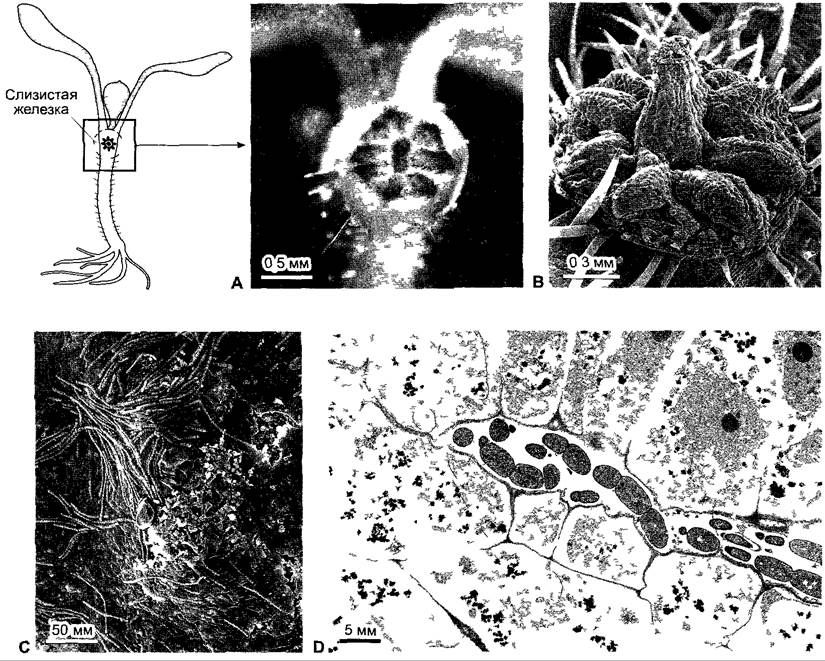
Intracellularly (or enclosed by a symbiosomal membrane), symbiotic cyanobacteria (*Nostoc*) are also found in the mucus-rich glands of tropical *Gunnera* species (Gunneraceae), which develop at the Base of the leaf petiole (Fig. 9.4). The symbionts migrate in the stage of hormogonia—filaments of cells capable of slow gliding movement and division (see 11.2). Presumably attracted by host factors via chemotaxis (see 8.2.1.1), the cyanobacterial cells enter through canals into the mucilaginous glands and, at the base of the glandular channels, are incorporated via phagocytosis by glandular cells whose walls are partially dissolved. Inside, the *Nostoc* symbionts intensively develop nitrogen-fixing heterocysts.
Fig. 9.4. Symbiosis between *Gunnera* and *Nostoc*. A, B — one of two mucilage-secreting glands on the hypocotyl of a *Gunnera* seedling, positioned crosswise relative to the cotyledons; C — hormogonia on the gland surface; D — longitudinal section through a gland showing *Nostoc* hormogonia within a glandular canal lined with mucilage-secreting cells
In all other cases, symbiotic cyanobacteria remain extracellular within their hosts, such as *Anabaena azollae*, which resides in the intercellular spaces of the leaves of the aquatic fern *Azolla* and enters via the apical meristem, meaning it is already present during leaf development; in the coralloid roots of *Macrozamia* species of the cycad family (*Nostoc*); and in the mucus-filled cavities of gametophytes (but not sporophytes) of hornworts (e.g., *Anthoceros punctatus*, *Nostoc* species, Fig. 9.5) and liverworts (e.g., *Blasia pusilla*, *Nostoc* species). The thalli of *Anthoceros punctatus* secrete a factor that induces hormogonium formation while simultaneously attracting these motile filaments of dividing cells through chemotaxis. The invading cyanobacteria, seemingly also under the control of the host plant, intensively form heterocysts that provide the host plant with
fixed nitrogen largely in the form of ammonia (NH3). In this state, photosynthetic CO2 fixation by the cyanobacteria is strongly suppressed, making the symbionts dependent on the host plant for their supply of Organic compounds (including amino acids!) and causing them to grow very slowly. Therefore, the benefit that cyanobacteria derive from such a symbiosis hardly lies in their *in planta* presence; rather, it likely pertains to free-living cells in the immediate vicinity (On the surface) of the host plants, which are induced to form hormogonia and presumably stimulated by substances excreted by the host plants.
Fig. 9.5. *Anthoceros-Nostoc* symbiosis. A — general appearance of *Anthoceros punctatus* thalli; each gametophyte produces a stem-like sporophyte. B — underside of the gametophyte with *Nostoc* colonies appearing dark in the photograph

Symbioses between nitrogen-fixing bacteria and animals (as well as humans) and angiosperms are also known. For instance, termites "house" nitrogen-fixing
bacteria in their guts (*Citrobacter freundii*, *Enterobacter agglomerans*), thereby supplementing their nitrogen-deficient diet. The gut flora of Papuans (indigenous people of New Guinea) likewise contains nitrogen-fixing bacteria. Despite a monotonous diet consisting primarily of protein-poor sweet potatoes, Papuans do not suffer from amino acid deficiencies.
Unlike cyanobacterial symbioses, plant nitrogen-fixing symbioses with bacteria involve The formation of specialized symbiotic structures—ROOT nodules. These occur, for example, in alders and contain streptomycetes of the genus Frankia alni. More than 140 other species from 9 families form nitrogen-fixing root nodules with actinomycetes as symbiotic partners (Table 9.1). Fixation is efficient, ranging from 50 to 200 kg of nitrogen per hectare annually in Alnus species. However, root nodules in members of the Fabales (legumes) are particularly widespread and well-studied; they represent a symbiosis with nitrogen-fixing bacteria from very closely related genera: Rhizobium, Bradyrhizobium, Azorhizobium, Mesorhizobium, and Sinorhizobium. Within the family Fabaceae, root nodules are found in less than half of Caesalpinioideae, the majority of Mimosoideae, and nearly all investigated genera of Faboideae. Legumes are among the earliest cultivated plants of the Stone Age and remain vital crops second only to Poaceae. Their soil-improving properties were already recognized in antiquity (Theophrastus, 4th century BC).
Table 9.1 Genera including species with root nodules containing actinomycetes
Genus |
Family |
Casuanna |
Casuannaceae |
Муriса |
Myncaceae |
Comptonia |
Myncaceae |
Ainus |
Betulaceae |
Dryas |
Rosaceae |
Cercocarpus |
Rosaceae |
Chamaebatia |
Rosaceae |
Cowania |
Rosaceae |
Purshia |
Rosaceae |
Rubus |
Rosaceae |
Conaria |
Coriariaceae |
Ceanothus |
Rhamaaceae |
Colîetia |
Rhamnaceae |
Discaria |
Rhamnaceae |
Retamila |
Rhamnaceae |
Talguenea |
Rhamnaceae |
Trevoa |
Rhamnaceae |
Eîaeagnus |
Eleagnaceae |
Eippophae |
Eleagnaceae |
Shepherdia |
Eleagnaceae |
Paraspoma |
Ulmaceae |
Daiisca |
Datiscaceae |
Nodule bacteria are widely distributed in the soil. Near the host plant, they move toward the root surface via chemotaxis. The substances attracting the bacteria are Flavonoids (see 6.16.1). In Rhizobium meliloti, for example, positive chemotaxis is triggered by luteolin (Fig. 9.6). The bacteria attach to the tips of young root hairs through an interaction mediated by plant Lectins (carbohydrate-binding Proteins; see 6.17.4), which bind to surface structures on the bacteria. This binding induces root Hair curling and the formation of an infection thread (which can be viewed as an inverted, inward-directed tip growth of the root hair); this thread is lined by a Cell wall and grows inward through the root hair. The nodule bacteria reside within the infection thread. It grows through several layers of cortical parenchyma cells until it reaches the developing nodule primordium,
which originates from outer or deeper-lying (see below) cortical parenchyma cells positioned above the protoxylem strands, while already differentiated parenchyma cells re-enter the Cell Cycle (see 7.3.2) and undergo polyploidization. The resumption of active divisions in polyploid cells is induced by Nod factors secreted by the nodule bacteria (Nod stands for Nodulation). These factors are lipochitooligosaccharides (see Fig. 9.6) whose core consists of 3 to 5 N-acetylglucosamine molecules linked by (β1 > 4)-glycosidic bonds, similar to Chitin. This oligosaccharide also bears a number of other characteristic substituents. The Biosynthesis of Nod factors in rhizobial cells is likewise induced by host-plant flavonoids. The necessary biosynthetic Enzymes are encoded by nod genes, which are frequently located on the symbiosis-essential Sym plasmid. The structure of Nod factors determines the host range of the bacteria and regulates the formation of nodule primordia from the outer or inner Regions of the cortical parenchyma. For instance, to exert its inducing effect on alfalfa, the Nod factor must carry a complex sulfate ester at the C-6 position of the N-acetylglucosamine residue at the reducing end (see Fig. 9.6). The presence of medium- to long-chain (often rare) fatty acids in place of the acetyl residue at the first glucosamine unit is also important for biological activity. Nod factors bearing polyunsaturated fatty acids diffuse deeper into the cortical parenchyma and influence the formation of indeterminate nodules, which develop their own meristem at the tip and continue to grow indefinitely (e.g., in pea and alfalfa). Nod factors with saturated fatty acids diffuse less deeply into the cortical parenchyma and direct the formation of determinate nodules lacking their own meristem, which typically cease functioning after a few weeks and are subsequently resorbed by the plant (e.g., in bean and soybean). Nodule bacteria with a narrow host range synthesize only one or a few Nod factors, whereas those with a broad host range produce a larger set of different factors. Through Genetic Engineering combinations of Nod factor genes, nodule bacteria with altered host ranges can be generated purposefully.
Fig. 9.6. Schematic representation of the stages of Rhizobium-legume symbiosis formation. (1) In response to nitrogen deficiency, the root secretes flavonoids, which trigger positive chemotaxis in soil-dwelling flagellated rod-shaped nodule bacteria and activate nodulation genes (nod genes); (2) nodule bacteria attach to the tip of young root hairs via plant lectins; (3) the root hair invaginates at the tip to form an infection thread, inside which the Nod factor-secreting nodule bacteria are harbored and multiply. Their biosynthesis is carried out using enzymes encoded by several of the activated nod genes. Nod factors diffuse into the cortical parenchyma and induce cell divisions, forming a nodule primordium; (4) after the infection thread reaches the nodule primordium, the nodule bacteria are phagocytosed by these cells; (5) upon a significant increase in host cell volume, the nodule bacteria differentiate into bacteroids, while also increasing in volume (about tenfold). Bacteroids no longer divide and fix N2. If the Nod factor structures (bottom fragment) specific for nodulation in alfalfa are absent, nodule primordium induction is suppressed. If the sulfate group is missing, the Nod factor is inactive on alfalfa but remains active on Vicia or Pisum. If the fatty acid is absent, the factor fails to function entirely. For further details, see the text.

Within the nodule primordium, The Cell walls of both the polyploid cells and the infection thread are partially hydrolyzed, and the nodule bacteria are phagocytosed by the plant cells, with the rhizobia initially undergoing further division as the plant cells swell. At this stage, the nodule bacteria secrete abundant auxin. Finally, the cell shape changes, The cell wall is reorganized, the protoplast swells, and the rhizobia transition into so-called bacteroids, which no longer divide and instead fix nitrogen. This process requires bacterial nif or fix genes (short for nitrogen fixation). Among other Functions, they encode the subunits of the enzyme nitrogenase (see below). The bacteroids are permanently surrounded by a plant-derived symbiosomal membrane, also referred to as the peribacteroid membrane. A single membrane vesicle may enclose several bacteroids, numbering approximately 1011 — 1012 per gram of tissue. The peribacteroid membrane, together with the enclosed bacteroids and the intervening space, is termed a symbiosome (Fig. 9.7).
Fig. 9.7. Root nodules of alfalfa (A) and Lotus prescii (B). An electron micrograph of host soybean cells (C) clearly shows symbiosomes containing bacteroids derived from nodule bacteria.

Nitrogen-fixing Tissues inside the root nodule can be recognized by their red coloration. This is due to the presence of leghemoglobin, which is produced as a joint product of the symbiotic partners (the plant synthesizes the Myoglobin-like protein, while the bacteroids appear to provide the heme group). Leghemoglobin binds molecular oxygen similarly to vertebrate Hemoglobin, but with an affinity roughly 10 times higher. This maintains a low partial pressure of oxygen at the site of nitrogen fixation, as nitrogenase is extremely oxygen-sensitive and its genes are repressed in the presence of excessive O2. Simultaneously, leghemoglobin efficiently supplies oxygen to the bacterial Respiratory Chain, which drives ATP synthesis. Indeterminate nodules continuously generate new symbiosome-containing cells at their tips, while cells at the base eventually degenerate. Determinate nodules cease nitrogen fixation after 4 to 6 weeks. The plant resorbs valuable organic substances (particularly compounds containing N, S, and P) from the dying cells. Although this process kills the bacteroids, a greater number of nodule bacteria are released into the soil from the senescing cells than were initially taken up; furthermore, the nodule bacteria proliferate abundantly near The surface of the host plant's root, making the mutual benefit of the process evident.
In soils well-supplied with nitrogen (NO-3 or NH+4), root nodules form in only negligible quantities. However, under nitrogen scarcity, the root begins to exude flavonoids, prompting the formation of numerous root nodules. Older root nodules, however, suppress the formation of new ones through a mechanism that is not yet fully understood, preventing the number of nodules from increasing uncontrollably even under nitrogen-deficient conditions. Nitrogen fixation is sufficient to allow legumes to thrive in soils very poor in nitrogen, though their growth does not reach its maximum potential. Consequently, agricultural legumes require supplementary Fertilization, albeit in small doses. Legumes are also widely grown in crop rotations to improve soil fertility before being plowed under ("green manure").
9.2.2. Biochemistry and Physiology of Nitrogen Fixation
The conversion of molecular nitrogen into ammonia is an exergonic process:
N2 + 3H2 > 2NH3 (ΔG0' = -33.5 kJ/mol);
however, due to the high activation energy of the reaction, it proceeds only under very high pressures and temperatures of 400 — 500 °C in the presence of a catalyst—reduced iron (Haber–Bosch process). This method, used to produce synthetic fertilizers, represents one of the most crucial global technological syntheses (see Table 6.21).
The nitrogenase-catalyzed reaction is likewise very energy-demanding:
N2 + 4NADH + 4H+ + 16ATP > 2NH3 + H2 + 4NAD+ + 16ADP + 16Pi.
The reaction requires 8 electrons, 6 of which are used to reduce N2 and 2 to reduce 2 H+ to H2 (a side reaction whose significance remains unclear)1. NADH + H+ and ATP are supplied by the Krebs cycle and the respiratory chain (see 6.10.3). Electrons are transferred from NADH first to ferredoxin. Reduced ferredoxin then serves as the electron donor for nitrogenase (Fig. 9.8).
1 According to one hypothesis, hydrogen reduction occurs when molecular nitrogen is scarce and represents one of the side Reactions Catalyzed by nitrogenase. It has been proposed that this property of nitrogenase could be harnessed for industrial-scale hydrogen production from Water using cyanobacteria. — Ed. note
Fig. 9.8. Metabolism of symbiosomes and host cells in legume root nodules. Stoichiometry is shown for the nitrogenase reaction only. For further explanations, see the text.

Nitrogenase is a complex enzyme system consisting of two components: dinitrogenase proper and dinitrogenase reductase. The latter is a dimer containing a single [Fe4S4] center shared by both subunits (iron-sulfur centers; see Fig. 6.56). This single-electron carrier accepts one electron from reduced ferredoxin and transfers it to dinitrogenase coupled with the binding and Hydrolysis of two ATP molecules (consecutive
transfer of 6 electrons to N2 thus requires 12 ATP; the obligatorily coupled reduction of 2H+ > H2 requires 2 electrons and thereby consumes an additional 4 ATP).1
1 The ratio between reduced N2 and H2 produced is actually not always equimolar. It varies depending on the availability of nitrogen from the atmosphere. — Ed. note
Nitrogenase is a tetrameric complex with an α2β2-structure, wherein the α and β subunits are highly similar. This tetrameric complex features two catalytic centers that operate independently of each other. Each center consists of a single iron-molybdenum cofactor (FeMoCo) bound by 4 protein subunits. The iron-molybdenum cofactor comprises an Fe4S3 cluster and an Fe3MoS3 cluster. N2 is presumed to bind via 3 iron atoms from each such cluster and is subsequently reduced to 2NH3 without the release of intermediate products.
Nitrogenase is not strictly substrate-specific and, In addition to N2 and H+, can reduce other substrates in vitro (e.g., N2O —> N2 + H2O; C2H2 —> C2H4). The reduction of acetylene (C2H2) to Ethylene (C2H4) is commonly used to assay nitrogenase activity via gas Chromatography.
Under molybdenum deficiency, certain nitrogen fixers (such as Azotobacter vinelandii) express alternative nitrogenases containing vanadium or iron, which also exhibit a distinct structural Organization.
Symbiosomes are characterized by active metabolic exchange across the bacteroid and peribacteroid membranes (Fig. 9.8). Bacteroids export fixed nitrogen predominantly as ammonium ions (NH+4), as they lack the ability to produce Glutamine Synthetase and thus cannot convert ammonia into glutamine (for glutamine formation, see Fig. 6.88). Rhizobia obtain amino acids for their own Protein Synthesis from the host cells. Root nodule cells export excess fixed nitrogen primarily in the form of the amino acids glutamine and asparagine. Certain root nodules, such as those in soybeans, initially convert nitrogen from glutamine and asparagine via Purine Biosynthesis (see 6.14) into inosine monophosphate, and subsequently form allantoin and allantoic acid via xanthine and uric acid, which serve as transport molecules for nitrogen. Export from the root nodules to the host plant, as well as the delivery of nutrients to the nodules, occurs via the vascular bundle located at the periphery of the nodules (export proceeds via the xylem, while import occurs via the phloem).
Bacteroids receive reduced carbon in the form of malate, which host cells produce from imported sucrose (via the glycolytic breakdown of hexoses to phosphoenolpyruvate (see 6.10.1), the carboxylation of phosphoenolpyruvate to oxaloacetate by PEP carboxylase—present at high concentrations in root nodules (for the reaction, see Fig. 6.79)—and the subsequent reduction of oxaloacetate to malate). The oxidation of malate in the Krebs cycle (see Fig. 6.94) supplies NADH+H+ and FADH2. A portion of the NADH and the generated FADH2 is channeled through the bacteroid respiratory chain to produce ATP, while another portion of NADH serves to reduce ferredoxin and supplies electrons for the nitrogenase complex. Leghemoglobin (see 9.2.1), present at high concentrations (~3 mM) in the cytoplasm of host cells, efficiently binds O2 and thereby lowers the concentration of free oxygen sufficiently to prevent damage to nitrogenase. The bacteroid cytochrome $aa_3$-type terminal oxidase (see 6.10.3.3) exhibits a very high affinity for oxygen; low oxygen concentrations are entirely sufficient for the needs of the respiratory chain, especially since consumed oxygen is rapidly replenished by the O2-buffering capacity of leghemoglobin.
Free-living nitrogen fixers employ various mechanisms to protect oxygen-sensitive nitrogenase. Many species synthesize the enzyme exclusively under anaerobic or microaerobic conditions. Obligately aerobic nitrogen fixers (such as Azotobacter) possess specialized protective proteins that bind directly to nitrogenase. Filamentous cyanobacteria form numerous heterocysts, within which nitrogen fixation takes place. These cells feature thick, lipid-rich cell walls that impede O2 entry and do not evolve O2 due to the absence of Photosystem II (see 6.4.5).
Nitrogen fixation is an energetically demanding process. Root nodules typically consume 5–20 mg of CARBOHYDRATES per 1 mg of fixed nitrogen. The fixation yield ranges from 30–100 mg of nitrogen per gram of fresh nodule mass per day; in other words, a single nodule can process approximately 3 to 10 times its own weight in nitrogen daily.
The mutualistic association between plant roots and fungi within the rhizosphere—an ecologically vital symbiosis—is termed mycorrhiza. Such associations already existed during the Devonian period, some 400 million years ago. Approximately 90% of all land plants and around 6,000 species of fungi are capable of forming mycorrhizae (see Box 11.4).
Based on their structural Morphology, distinct types of mycorrhizae are distinguished.
The most widespread type is arbuscular mycorrhiza (AM). It is named after the intracellular structures formed by fungal hyphae within the cortical cells, which swell into vesicles or branch out into treelike structures known as arbuscules (Fig. 9.9). AM fungi and other mycorrhizal fungi do not colonize the epidermis, apical meristem, or root cap.
Fig. 9.9. Arbuscular mycorrhiza (AM) (A — courtesy of S. Dickson; B — courtesy of S. Smith): A — arbuscules of Glomus coronatum in root cells of Allium porrum (captured using a laser scanning confocal Microscope; Plant Cell Walls are not visible); B — vesicles of Glomus mosseae in root cells of Allium porrum (approx. 45×)

In AM, all fungal symbionts belong to the order Endogonales within the class Zygomycetes (see 11.2), predominantly to the genus Glomus; they are obligate symbionts. The host partners comprise species from almost all families of angiosperms. AM is either absent or very poorly developed in families such as Cyperaceae, Amaranthaceae, and Brassicaceae, for instance. In temperate-zone trees, ectomycorrhiza predominantly forms (see below), whereas tropical trees form AM in the vast majority of investigated cases. Among gymnosperms, AM has been observed only in Taxus baccata, Sequoia sempervirens, Sequoiadendron giganteum, and Ginkgo biloba.
In AM, the fungus supplies mineral nutrients (primarily phosphate and Trace Elements) much more efficiently than the root hairs they replace. The plant partner provides carbohydrates in return. The establishment of AM enhances the growth of crop plants, for instance; alongside improved nutrient supply, this can also confer increased resistance to pathogenic fungi and nematodes. The assimilation of plant carbohydrates enhances the strength of sinks (the "source–sink" relationship, see 6.8) for the fungal partner within the root zone. This leads to an increase in the net photosynthetic output of the plant, contributing to the enhanced growth of mycorrhizal plants. The improved resistance of such plants to pests is presumably due, among other factors, to the fact that the establishment of the mycorrhizal symbiosis activates the initially weak pathogen defense mechanisms of the host plant (see
9.3.4).
AM shares certain similarities with the root nodule symbiosis (see 9.2.1, 9.2.2). The arbuscules are separated from the plant cytoplasm by a symbiosome-like membrane derived from the host cell's Plasma Membrane—the periarbuscular membrane—which is structurally and functionally similar to the peribacteroid membrane. Further evidence for similarities in at least certain Developmental Stages of both symbioses comes from the observation that all known root nodule-deficient mutants also fail to undergo mycorrhization.
Meanwhile, there is abundant evidence that intensive signaling molecule exchange takes place between both partners during AM formation, although their precise chemical nature remains to be elucidated.
In ectomycorrhiza, a mantle of fungal hyphae envelops the short and thick lateral roots of the second and third orders (Fig. 9.10) and functionally replaces the missing root hairs. The spreading hyphae of mycorrhizal fungi permeate the surrounding soil much more intensively. The fungi form a dense network predominantly extracellularly between the cells of the primary cortex—known as the Hartig net.
Fig. 9.10. Scanning electron micrograph of a silver fir (Abies alba) root with ectomycorrhiza: A — Overview; B — individual lateral roots

Ectomycorrhizae occur in ~3% of all seed plants. Among them are many forest trees (partly obligately), such as pine, spruce, larch, oak, and beech (Fig.
9.11). In the absence of fungi, these trees typically exhibit stunted growth.
Fig. 9.11. Ectomycorrhiza: A — cross-sectional fragment through a young beech root (approx. 50×); B — electron micrograph of a Hartig net fragment from the mycorrhiza between Lactarius decipiens and silver fir (Abies alba)

The ability to form ectomycorrhizas has already been demonstrated in approximately 65 fungal genera, predominantly Ascomycota and Basidiomycota. Fungi of certain genera—such as Russula, Amanita, Boletaceae, and Lactarius—live almost exclusively symbiotically and form fruit bodies only in association with tree roots. (Because of this, unlike the saprophytic cultivated mushroom, the penny bun/porcini mushroom cannot be induced to form fruit bodies in culture.) Some fungi show a more or less strict Specificity toward particular hosts. Trees, by contrast, do not appear to specialize in specific fungi (Pinus sylvestris, for example, can form ectomycorrhizas with at least 25 different fungi), though certain fungal species may be stimulated by pine more strongly than others. Introduced tree species, such as Pinus strobus or Pseudotsuga taxifolia, form normal mycorrhizas with native fungal species in Europe.
The benefits that trees derive from ectomycorrhizas are considered to include improved mineral salt nutrition and water supply, enhanced delivery of nitrogen and phosphates through the fungal breakdown of humus, provision of growth Hormones by the fungi, and protection against pathogen invasion, which is more effective than in VA-mycorrhizas. In return, the fungi receive carbohydrates and possibly other organic compounds from the host. Because large amounts of substances are specifically required for fruit body formation, their development frequently begins only after intensive SHOOT growth has ceased, during the tree's nutrient-storage phase (August–October).
In Representatives of the genera Picea and Pinus, ectomycorrhiza-endomycorrhiza intermediates (ecto-endomycorrhizas) are widespread, where intracellular structures are super-imposed on the normal ectomycorrhizal morphology. Transitions ranging from ecto- through ecto-endo- to pure endomycorrhizas are found in various members of the order Ericales. Thanks to their highly developed mycotrophy, these plants are able to grow on soils poor in P and N; this adaptation is a prerequisite for the further expansion of heather plants into peatlands, raised bogs, and coniferous forests.
The final link in this evolutionary line within the Ericales are members of the family Monotropaceae (e.g., Monotropa hypopitys, yellow bird's-nest), which are achlorophyllous parasites. Through the hyphae of obligate ecto-endomycorrhizas, these plants are directly linked to ectomycorrhizal forest trees (conifers, beeches). The transfer of 14C-labeled sugars from trees via fungi to Monotropa, and of 32P-labeled phosphate ions back from Monotropa to trees, has been demonstrated experimentally. Substantial amounts of carbon compounds are exchanged between plants linked by mycorrhizal fungi, depending on source-sink relationships. However, these compounds apparently remain within the fungus and are not transferred to the host plants.
Endomycorrhizas occur, for instance, in almost all orchids (Fig. 9.12). Their minute seeds (weighing 0.3–15 µg per seed) contain few intrinsic reserves and require symbiotic fungi (Basidiomycetes) for germination and development into independent autotrophic plants; these fungi supply them with organic material, water, mineral salts, and in part also hormones ("nurse fungi"). Fungal hyphae are present in the outer cortical cells throughout the adult plant as well (with the exception of aerial roots). However, in deeper tissue layers, the hyphae are digested or ruptured. In orchids that are incapable or barely capable of photosynthesis even after germination—such as bird's-nest orchid (Neottia), coralroot (Corallorhiza), and ghost orchid (Epipogium)—the higher plant must parasitize the fungus to obtain all essential nutrients and hormones.
Fig. 9.12. Endomycorrhiza of the orchid Platanthera chlorantha: A — fragment of a tangential section through the cortical parenchyma containing intracellular fungal hyphae, as well as two mucilage cells with raphides (115×); B — an invading infection hypha on the outside of a root hair tip. Penetration of The plant cell wall occurs via the formation of an appressorium (penetration hypha). The process closely resembles the infection of plant cells by a phytopathogenic fungus, but the plant's defense response is suppressed; C — infection hyphae growing from the root hair toward the cortical parenchyma (B, C — 235×)

A lichen is a symbiosis in which fungi combine with algae or cyanobacteria to form a novel organism that externally generally functions as a single entity. Partner recognition is mediated by lectins (see 6.17.1.2). The fungus — the mycobiont — associates with the photobiont1 (alga or cyanobacterium, respectively) in various ways, occasionally utilizing haustoria. The photobiont retains, and in some cases even enhances, its specific metabolism—photosynthesis, and in Nostoc also nitrogen fixation (see 9.2.1).
1 In Russian-language literature, the term phycobiont is more commonly used to emphasize the algal Nature of the symbiotic partner. — Ed. note.
Approximately 25% of the roughly 65,000 known fungal species participate in lichen formation. Lichen-forming fungi are found across all major taxonomic groups of the fungal kingdom.
Given that lichens can harbor 28 different cyanobacteria or algal genera, it is unsurprising that The Nature of the assimilates transferred from the photobiont to the mycobiont varies. To date, glucose (in all cyanobacterial photobionts) and sugar alcohols (in all green algal photobionts) have been identified as transport metabolites. If a lichen contains both green algae and cyanobacteria (e.g., in cephalodia, as in Peltigera aphthosa), the fungus receives sugar alcohols from the former and glucose from the latter; the mycobiont converts both groups of substances into mannitol, the primary fungal reserve substance. Exchange is abundant and rapid: within just 2 min of the onset of photosynthesis in an atmosphere of labeled 14CO2, detectable quantities of labeled assimilates are present in the fungus.
The export of organic nitrogen compounds from the nitrogen-fixing Nostoc symbiont to the mycobiont also occurs rapidly; moreover, in Peltigera aphthosa, for instance, the cyanobacteria within the cephalodia supply nitrogen to the fungus but not to the green algae within the lichen. There is evidence that the fungal partner stimulates Nitrogen fixation by the symbiotic cyanobacteria.
Presumably, photobionts in lichens also receive vital substances from the fungi, such as mineral salts and water; otherwise, lichens could not be regarded as symbiotic systems. Little is known about the specifics of how algae are supplied by the fungus. The relationship between lichen partners is sometimes interpreted as a moderate parasitism of the fungus on the photobionts.
Last update: 07/08/2026
Editorial and Educational Adaptation: This material has been compiled based on the primary/original source text. The project team performed an editorial review, corrected technical inaccuracies, structured sections, and adapted the content for an educational format.
What was processed:
- elimination of formatting defects (OCR errors, structural breaks, corrupted characters);
- editorial organization of content;
- standardization of terminology in accordance with academic sources;
- verification of factual statements against the original source text.
All mentions of the author, publication year, and origin of the primary text have been preserved in accordance with the source.