Peptide Self-Regulation of Living Systems (Facts and Hypotheses) - Shataeva L. K. 2003
Peptide Interactions with the Cell Membrane
Interaction of Regulatory Peptides with Receptors and Phospholipids
METABOLISM/2.html">THE CONCEPT OF specific Cell-surface receptors was introduced by Paul Ehrlich in the early 20th century. To explain receptor Specificity, he used the lock-and-key model previously developed by Emil Fischer for selective enzyme catalysis. It is worth noting that the spatial matching implied by the lock-and-key concept is a remarkably persistent mental model rooted in everyday experience. Vast electroencephalographic data have contributed virtually nothing to this mechanical model. When modeling the interaction of regulatory Peptides with Membrane Receptors, it is generally assumed that a specific region of the receptor mirrors and complements The Structure of the Ligand (Govyrin & Zhorov, 1994). The outer portions of certain receptors bear a partial resemblance to the variable region of y-globulin molecules, which is why diagrams often depict them as fork-like structures. Based on this same model, it is hypothesized that certain synthetic peptides may partially match the Fc region of IMMUNOGLOBULINS in a complementary fashion, thereby triggering an allergic response in the body.
The specificity of regulatory peptides that induce histamine release from intestinal mast Cells has been studied in the greatest detail (Jasani et al., 1979). A comparison of peptide activities across a broad range of sizes and Amino Acid Sequence variations (including various ACTH fragments) with The activity of the degranulating peptide merely demonstrated that activity requires the presence of a block of four basic amino acid residues and an amidated C-terminus in the peptide chain. However, all variants were 2 to 4 orders of magnitude less active than the natural degranulator. In other words, a wide spectrum of regulatory peptides can stimulate histamine release from mast cells, but each acts only within a specific concentration range.
A more modern approach to studying peptide regulation involves identifying a cascade of sequential interactions that alters the Conformations of both the lipid and peptide Components of the membrane, ultimately delivering the corresponding information signal to The Cell. It has been found that certain peptide regulators either inhibit or activate Enzymes located on or near The surface of The cell membrane.
Specifically, A number of Proline-containing peptides derived from food Proteins (IY, VW, IW, VAP, IKP, LRP, 1RP, etc.) inhibit the membrane enzyme that converts angiotensin I into angiotensin II and inactivates bradykinin (Yamamoto, 1997). This zinc-containing peptidase is located on the membranes of vascular endothelial cells and neuroepithelial cells. Its most potent inhibitor has been discovered among snake venom peptides—pyrEKWAP. Dietary peptides act as Competitive Inhibitors and account for the pronounced antihypertensive effect of fermented dairy products, as lactic acid Bacteria release these inhibitors through the Hydrolysis of casein. Thus, the regulatory action of these peptides is driven by the inhibition of this membrane enzyme.
Another example is the fibroblast growth factor (a 140-amino-acid polypeptide), which activates phospholipase Cβ, resulting in a sharp surge in the cytosolic concentration of Inositol 1,4,5-trisphosphate (a secondary messenger). This is accompanied by an inotropic response of the myocardium. Consequently, The Effect of the growth factor boils down to a temporary increase in the activity of several Hydrolases and an alteration in the charge and potential of The Plasma Membrane caused by phospholipid hydrolysis (Tappia et al., 1999).
These observations corroborate findings on the vital role of Phospholipids in cellular autoregulation, as they serve as precursors for messenger molecules (diacylglycerol, phosphatidic acid, ceramides) that function both at the cell surface and in the Cytosol, activating the G-protein system and subsequent signaling cascades (Morris, 1999).
Among the regulatory enzymatic reactions that modify membrane phospholipids, researchers have noted not only hydrolysis, but also the phosphorylation of inosine by a GMP-dependent kinase associated with troponin T (Yuasa et al., 1999). It is well established that the phosphatidylinositol "HEAD" can bind up to three phosphate groups, which dramatically increases the local density of negative charges. Alterations in charge distribution across the mosaic surface of the plasma membrane can, in their own right, act as a signal transmitted from the membrane into the cell interior or to adjacent cells at tight junctions.
It is crucial to highlight the important role of sialic acid, which serves as the terminal moiety of the glycosidic portion of numerous Membrane receptors and determinants, carrying a permanent negative charge. Furthermore, it is a constituent of glycosphingolipids in the plasma membrane of Neurons and the synaptic membrane, where it participates in neurotransmitter release. Brain excitation processes also unfold with the involvement of sialic acids. A broad spectrum of age-related brain pathologies, including Alzheimer's disease (AD), is accompanied by a general decline in the levels of sialic acid-containing gangliosides and cerebrosides (Reutter et al., 1982).
To account for the specific mechanisms by which Neuropeptides and venoms trigger histamine release from intestinal mast cells, a model of receptor-independent activation of the G-protein system was proposed (Mousli et al., 1990). It was observed that the activity of the studied amphiphilic peptides and neurotoxins (all containing blocks of basic amino acid residues) was suppressed by low calcium concentrations and hydrophobic quaternary amines, and failed to manifest if the cells were pre-desialylated with neuraminidase. All of this points to the direct involvement of negatively charged groups on the mast cell surface in the binding of regulatory peptides.
As noted previously, preserving the native Structure and function of Membrane Proteins requires the presence of membrane phospholipids. Consequently, substances that alter the COMPOSITION AND PROPERTIES of the phospholipid bilayer thereby affect the Functional Properties of membrane proteins. For instance, local anesthetics disrupt the structure of The Lipid Bilayer, leading to impaired sodium channel function. Opioid peptides presumably exert a similar effect on the cell membrane.
To a certain extent, these facts place membrane peptide receptors and phospholipids on an equal footing regarding their ability to selectively perceive environmental changes, undergo corresponding conformational shifts, and transmit this information not only into the cell interior but also to neighboring cells via bilayer membrane contact systems. For this reason, fluorescent and colorimetric Methods FOR STUDYING the intermolecular interactions of regulatory peptides with phospholipid membranes are currently undergoing intensive development (Dobretsov, 1989; Kolusheva et al., 2000).
Class="center">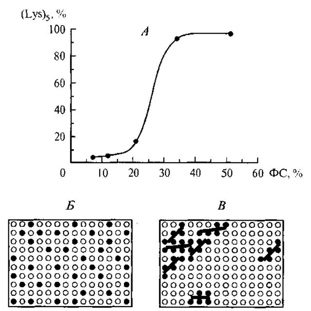
Fig. 10. Dependence of pentalysine binding to the phospholipid membrane surface on its phosphatidylserine content (A), and the transition from a uniform distribution of phosphatidylserine (B) to pentalysine–phosphatidylserine clusters (C).
Relatively recently, researchers investigated the Interaction of a basic oligopeptide (pentalysine) with a model phospholipid (PC/PS) membrane containing varying concentrations of acidic phosphatidylserine (Denisov et al., 1998), as illustrated in Fig. 10. The non-linear Nature of the plot is governed by Changes in the self-association of charged PS groups on the membrane surface and cooperative effects during peptide adsorption. In the absence of the peptide, the negatively charged phosphatidylserine groups are distributed uniformly across the surface due to mutual electrostatic repulsion; however, the sorption of the positively charged oligopeptide gathers them into clusters, disrupting this uniform surface distribution. At low phosphatidylserine concentrations, this disruption is minimal (the initial segment of the curve), but as the concentration rises, the selectivity of oligopeptide binding increases. Obviously, this is accompanied by A change in the local membrane polarization within the cluster regions.
During The formation of "peptide–polar phospholipid layer" intermolecular bonds, a polar (charge–charge) complementarity between the participants is observed, which is uncharacteristic of specific interactions between peptide chains themselves.
Studies on the binding of higher-molecular-weight peptides to Liposomes containing phosphatidylglycerol have demonstrated that cytochrome c and the basic polypeptide histone H1 compete for binding to the phospholipid membrane, whereas polylysine K19 binds most strongly to acidic phospholipids (Rytomaa & Kinnunen, 1996).
The liquid-crystalline STRUCTURE OF THE membrane allows individual phospholipids to move freely within the layer and arrange themselves into a conformation that complements the charge distribution of the regulatory peptide. The working hypothesis posits that in certain cases, there is no need for the regulatory peptide to cross into the cell interior; rather, it is sufficient for it to reorganize and stabilize a charge-complementary phospholipid cluster On the surface of the membrane bilayer. Furthermore, given that the phospholipid composition of cell membranes varies significantly across different Tissues (see Table 10), the structure of the regulatory peptide must likewise be tissue-specific.
It should be noted that regardless of the mechanism by which a ligand acts on the membrane—whether through binding to receptors or phospholipid groups that alter membrane properties—the process is described by identical equations. The simplest case relating peptide concentration (C) to the system's response to the received signal (R) can be represented by a cooperative isotherm:
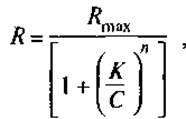
where Rmax is the maximum response corresponding to receptor saturation; K is the binding constant; and n is the cooperativity parameter.
Under this approach, cellular receptors are likened, to some extent, to enzymes, and ligands to substrates, since their binding triggers a specific reaction, i.e., a response (Volkenstein, 1981). In the absence of cooperativity—that is, when n = 1 and ligand interaction occurs with individual receptors—this equation becomes identical to the Michaelis–Menten Equation for enzyme kinetics. In cases of positive cooperative interaction (n > 1), the resulting system response to the signal is amplified. This is generally believed to stem from interactions among membrane receptors themselves. A prime example is found in the highly sensitive chemoreceptors of crustaceans, which discriminate between the scents of Amino Acids and peptides (Belli & Rechnitz, 1989). These receptors are located at the dendritic terminals of Nerve Cells. The sensitivity of the blue crab's chemoreceptors to amino acid odors becomes apparent at a concentration of 10-5 M, dictating the crab's subsequent behavioral response. It is hypothesized that once at least a single receptor interacts with a single ligand, the cooperative response of all receptors induces local depolarization of the nerve Cell Membrane and the corresponding Action Potential. The sensitivity of a membrane receptor to changes in the state of its neighbors is determined by the integrative Properties of the bilayer lipid membrane housing the hydrophobic domains of the receptors; under these conditions, the exponent n can reach values of several units.
The receptors of certain sensory cells located within the epithelium of a sensory organ operate at the boundary with the external environment, particularly the air (e.g., olfactory cells, Hair Cells of the auditory system). They possess a significantly more complex architecture than chemoreceptors operating in a humoral medium. Receptor cells proper are invariably oriented normal to the outer boundary of the organ. They are surrounded by supporting cells, and their apical regions feature specialized membrane structures, such as cilia and microvilli. For instance, olfactory neurons bear 6 to 8 modified cilia, and it is hypothesized that their tips house odor chemoreceptors.
However, a defining feature of Olfaction is its vectorial nature—meaning the direction of the signal source is perceived. The Morphology of cell membranes in the apical region of the sensory cell undoubtedly plays a role in this process. Cilia, microvilli, and hairs at the terminals of olfactory, gustatory, and vestibular sensory cells resemble an antenna array composed of vibrators and slits (Principles of Physiology, 2001). It is known that the phase difference of oscillations excited in identical elements combined in a specific manner enables the perception of a signal as a directional beam. In addition, an extra spatial orientation effect arises from the interaction of two Different types of oscillations at the interface between two media: the air and the substantially denser medium of the sensory organ. Mechanical vibrations, particularly sound waves, propagate through the gas phase as longitudinal waves and through liquids as transverse waves. Their interaction at the sensory organ boundary can generate mechanical jolts and beats (Ivanytskyi & Deyev, 2001). The cilia on the Hair cells of the Inner ear perceive not only the sound frequency but also these beats, which force them to pivot toward the sound source. This shift in ciliary orientation is instantly detected by the neuron, which relays an electrical signal to the effector organ. The electrophysical properties of the cell membrane enable it to transmit local—and even point-like—potential changes across the lipid liquid-crystalline structure at high speed and with significant Amplification. This is contingent upon the periodic capacitance fluctuations of the lipid bilayer (the leading parameter) being commensurate with the frequency of the excitation signal (Rabinovich & Trubetskov, 1984).
Presumably due to this intricate Organization of sensory receptors, many sensory cells are incapable of division: their degradation or destruction is replenished by stem cells (situated close to the sensory cells) which, when necessary, differentiate into new sensory cells (Alberts et al., 1994).
It is plausible that Sensory Organs employ this mechanism to achieve cellular adaptation. If the external environment serves as a source of overly intense stimulation that damages cellular receptors, new sensory cells develop from the sensory organ's stem cells. The receptors of these new cells retain their sensitivity to minute variations in the irritating factor against a backdrop of a persistently high level of ambient stimulation (Richardson, 1993).
For most differentiated tissues whose cells are capable of division, receptor turnover occurs not only during Cell Division but also through the intracellular recycling of chemoreceptors within vesicles. Consequently, a cell adapted to high concentrations of a specific ligand can generate chemoreceptors with heightened sensitivity to very minor fluctuations in those high concentrations.
Thus, the effects of ultra-low doses are by no means paradoxical in cooperative systems. In traditional pharmacology, these patterns are described by the dose-response saturation function. However, the biochemical interpretation of this relationship in the ultra-low concentration range demands a certain degree of caution, as the concentration of receptors interacting with the ligand is not accounted for in the equation given on page 130. It is traditionally assumed that the receptor concentration is significantly lower than that of the specific ligands, meaning the reaction can be described by a first-order equation. In systems where ligand concentration or activity can be measured independently by both biochemical and biological methods simultaneously, a linear correlation between them is typically absent. Specifically, when comparing the neuraminidase activity in the hydrolysis of an oligomeric substrate with its activity in hydrolyzing erythrocyte receptors, a non-linear relationship is observed. In this case, as in virus-induced erythrocyte hemagglutination and many other biological interactions measured over a specific time interval, the reaction titer (response) is logarithmically related to the analytical concentration of the reactant (Shatayeva et al., 1978). This indicates that the reactions triggered by the reactant upon acting on the cell may be cascading (chain) reactions rather than cooperative ones.
The lateral mobility of the phospholipid components within the plasma membrane and their sensitivity to the distribution of positively charged groups in the surrounding environment make them inherently vulnerable to external molecular aggression. Most peptide Antibiotics and toxins exploit these exact mechanisms to disrupt cell permeability. Notably, streptolysin O (a peptide toxin) binds to plasma membrane Cholesterol at picomolar concentrations, forming a ring-shaped structure (pore) with a diameter of 30 nm (Palmer et al., 1998). This process culminates in the leakage of cellular contents and subsequent cell death. The antibiotic peptides defensins and protegrins, which feature a structural excess of Lysine and Arginine, operate in a similar fashion (Kokryakov, 1999).
The activity of peptide antibiotics depends in specific ways on the compositional makeup of the cytoplasmic membrane and the transmembrane electrical potential of the target cell. Bacterial membranes contain a high proportion of acidic phospholipids (phosphatidylglycerol and cardiolipin), which are virtually absent in Introduction/5.html">Eukaryotic Cell membranes. These phospholipids serve not only as markers but also as binding sites for animal-derived peptide antibiotics, particularly defensins and protegrins, via strong ion-ion bonds established through a cooperative mechanism similar to the concentration dependence shown in Fig. 10A. Furthermore, the Membrane Potential of bacteria is known to be 1.5–2 times higher than that of eukaryotic cell membranes. Consequently, peptide antibiotics with a high content of lysine and arginine can enter the cell through the membrane via Electrophoresis, much like what has been described for the microbial antibiotics polymyxin B and gramicidins (Franklin, Snow, 1984).
Interestingly, a number of protegrins and cecropins contain Tryptophan near the N-terminus: KW, RW, GW, SW, KW, RRW. At the same time, it has been found that a structurally defined peptide attached to a liposome—consisting of 11 amino acid residues with a tryptophan residue at the N-terminus—can "pull" the liposome into the Cytoplasm via a membrane fusion mechanism. The Mechanism of viral particle entry into the cell is organized in a similarly analogous way (Pecheur et al., 1998).
Thus, the interaction of various classes of peptides with the cytoplasmic membrane occurs through multiple mechanisms and Supports diverse cellular Functions, yet reveals a number of underlying structural regularities. Depending on their structure, regulatory peptides can exhibit a wide range of activities:
1) directly acting on the cellular receptor by establishing intermolecular bonds with segments of the receptor's peptide chain, which is thereby activated, undergoes a conformational change, and transmits the signal of the established bond into the cytoplasmic space;
2) acting on the membrane bilayer structures by forming ion-ion bonds with the polar heads of phospholipids, thereby altering their mutual arrangement, polarization, and membrane potential, which serves as a signal for intracellular messengers;
3) embedding into the structure of the lipid bilayer with the help of aromatic and basic amino acid residues near the N-terminus, altering membrane permeability to ions and solvent, and compromising membrane integrity;
4) activating membrane enzymes—hydrolases and transferases—that alter The ratio of positive and negative charges on the cell membrane surface, thereby initiating a cascade of chemical and electrochemical transmembrane reactions;
5) acting as agonists (synergizing) in the activation of membrane proteins (enzymes and receptors) by other metabolic substances; it is entirely possible that the tissue-specific action of regulatory peptides belonging to the cytomedin class is determined precisely by their role as agonists in modulating the specific functions of differentiated tissues.
Last update: 06/08/2026
Editorial and Educational Adaptation: This material has been compiled based on the primary/original source text. The project team performed an editorial review, corrected technical inaccuracies, structured sections, and adapted the content for an educational format.
What was processed:
- elimination of formatting defects (OCR errors, structural breaks, corrupted characters);
- editorial organization of content;
- standardization of terminology in accordance with academic sources;
- verification of factual statements against the original source text.
All mentions of the author, publication year, and origin of the primary text have been preserved in accordance with the source.